Reading...
![]()
Play button
![]()
Play button
![]()
Use LEFT and RIGHT arrow keys to navigate between flashcards;
Use UP and DOWN arrow keys to flip the card;
H to show hint;
A reads text to speech;
56 Cards in this Set
- Front
- Back
|
The sciatic nerve innervates the motor and sensory parts of the distal lower extremity except for which part innervated by which nerve?
|
The medial calf and foot, which are innervated by the saphenous, which is formed from the continuation of the femoral.
|
|
|
Give the origin of the sural nerve
|
Formed from the tibial nerve (medial branch) which is given off before the tibial nerve goes deep to the gastrocnemius, and from the peroneal nerve (lateral branch) by the biceps femoris before it reaches the fibular head. They anastamose, go posterior to the lateral malleolus, and give off branching rami
|
|
|
Give the borders of the femoral triangle, the positioning of the thigh to where it is most easily seen, and its contents from lateral to medial
|
Borders:
Medial: adductor longus Lateral: sartorius Superior: inguinal ligament Floor: pectineus Roof: Fascia lata except for cribiform fascia by saphenous opening It is most easily seen in the FABER position. Contents are NAVEL: femoral nerve, femoral branch of the genitofemoral nerve, femoral artery, femoral vein (all in the femoral sheath), extra space, lymphatics/lacunar ligament. |
|
|
What are the main motor branches of the femoral nerve?
|
Iliacus, pectineus, quadriceps femoris, sartorius
|
|
|
Name the muscles inserted into the pes anserine area from anterior to posterior. What collagenous structure is this insertion superficial to?
|
Sartorius, gracilis, semitendinosis. Superior to the insertion of the MCL.
|
|
|
What divisions of what nerve roots form the lateral femoral cutaneous nerve?
|
The posterior divisions of L2 and L3
|
|
|
Describe the course of the LFCN from the root level
|
From L2 and L3 nerve roots to the lumbar plexus. Posterior divisions go just lateral to the ilioinguinal nerve. Passes under the inguinal ligament just medial to the anterior superior iliac spine. Stays under the fascia lata. Lateral branch goes just anterior to the greater trochanter and down to the knee. Medial branch goes to the anterior thigh.
|
|
|
What is LFCN neuropathy called?
|
meralgia paresthetica
|
|
|
Describe the anatomy behind the greater and lesser sciatic foramina
|
The greater sciatic foramen is formed from the greater sciatic notch and the sacrospinous ligament going from the ischial spine to the sacrum. The lesser sciatic foramen is formed from the lesser sciatic notch, the sacrospinous ligament superiorly, and the sacrotuberous ligament inferomedially.
|
|
|
Describe the origin and course of the ilioinguinal nerve
|
From L1, sometimes T12. Travels along the concavity of the ilium, then perforates the transverse abdominis, wrapping medially to innervate the area around the medial inguinal crease. May join up with the iliohypogastric. Goes through the superficial inguinal ring to the inguinal canal. Does not go through the deep inguinal ring. Innervates the upper scrotum in men and the mons pubis in women.
|
|
|
Describe the origin and course of the iliohypogastric nerve
|
Like the ilioinguinal nerve, it originates from T12 and L1, goes along the concavity of the ilium and pierces the transverse abdominis. There a posterior branch goes and innervates the lateral gluteal area and an anterior branch passes through the external oblique muscle and innervates the inferior aspect of the abdomen/inguinal areas.
|
|
|
Describe the origin and course of the genitofemoral nerve.
|
Originates at L1/2, goes along the convexity of the ilium, runs anteriorly to the psoas behind the ureter. Just above the inguinal ligament it divides into a femoral and genital branch. The femoral branch travels next to the femoral nerve through the inguinal canal. The genital branch goes through the inguinal canal and deep inguinal ring to innervate the cremaster and the scrotum in men. In women, it passses through the round ligament to innervate the ipsilateral mons pubis and labia majora.
|
|
|
What nerve primarily innervates the hip joint?
|
The obturator nerve.
|
|
|
What nerves form the peripatellar plexus?
|
Branches from the anterior division of the lateral femoral cutaneous nerve and the saphenous nerve
|
|
|
Describe the course of the obturator nerve and the muscles it innervates. What muscle does it confusingly NOT innervate, given it's name (and what nerve innervates it)?
|
Start with ventral roots of L2-4. Pass along the psoas to the medial border of the pelvic brim. Runs lateral to the internal iliac artery and the ureter. Lies anterior to the obturator vessels and goes to the thigh through the obturator foramen/obturator canal. It turns into an anterior and posterior branch, separated by the obturator externus and then the adductor brevis. The posterior branch innervates the obturator externus, adductor brevis, sometimes the adductor magnus, and it sometimes gives an articular branch to the knee. The anterior division gives off a branch to the articulation of the hip joint. It sometimes innervates the pectineus. It also innervates the adductor longus, sometimes brevis, gracilis. The obturator internus is not innervated by the obturator nerve, rather by the "nerve to the obturator internus".
|
|
|
Describe the formation and course of the superior hypogastric nerves and inferior hypogastric plexus
|
Start as a sympathetic/parasympathetic plexus from a combination of an extension of the lumbar plexus and sacral parasympathetics. Travel around the aorta. At around L4 level, just inferior to the formation of the common iliac arteries, it forms the superior hypogastric plexus. At L5, they divide into the superior hypogastric nerves and follow the iliac vessels to around the bladder and viscera. Here they form the inferior hypogastric plexus.
|
|
|
Where does the ganglion of impar (aka ganglion of Walther) lie?
|
Just in front of the sacrococcygeal joint.
|
|
|
What is the terminal ganglion of the sacral sympathetic and parasympathetic chains?
|
The ganglion imapr
|
|
|
Where does the tibial nerve lie in relation to the soleus? Tibialis posterior? FHL and FDL?
|
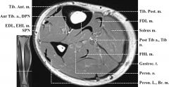
deep to the soleus, directly superficial to the tibialis posterior, equal depth and medial to the FHL, equal depth and lateral to the FDL.
|
|
|
Where does the peroneus longus insert?
|
The first metatarsal and medial cuneiform
|
|
|
Where does the peroneus brevis insert?
|
the fifth metatarsal
|
|
|
What part of the femoral head is not covered in hyaline cartilage? What is attached to this area?
|
The fovea. The ligamentum teres.
|
|
|
What lies inside the ligamentum teres?
|
The central branch of the obturator artery.
|
|
|
What major muscle groups attach to the greater trochanter? The lesser trochanter?
|
The greater trochanter receives insertions from the gluteal group, the lesser from the adductor group.
|
|
|
Give femoral shaft to head angles for coxa valga and coxa vara
|
Coxa valga: greater than 135 degrees
Coxa vara: less than 125 degrees |
|
|
Name the three major ligaments supporting the hip joint and their location
|
anterior: iliofemoral
posterior: ischiofemoral and pubofemoral |
|
|
What muscle(s) are the main hip flexor?
|
iliopsoas.
|
|
|
What muscle(s) are the main hip extensor?
|
gluteus maximus and hamstrings
|
|
|
What muscle(s) are the main hip abductors?
|
Gluteus medius and minimus
|
|
|
What muscle(s) are the main hip adductors?
|
Adductor brevis and longus
|
|
|
What muscle(s) are the main hip internal rotators?
|
tensor fascia lata. When hip flexed, gluteus medius and minimus (anterior fibers).
|
|
|
What muscle(s) are the main hip external rotators?
|
Gluteus medius and minimus (posterior fibers) with hip in extension, quadratus femoris, gamelli, piriformis, obturator internus and externus
|
|
|
What two structures does the ischial bursa lie between?
|
The ischial tuberosity and the gluteus maximus
|
|
|
What three structures does the greater trochanteric bursa lie between?
|
The greater trochanter, the iliotibial tract, and the tendon of the gluteus medius.
|
|
|
What nerve innervates the gluteus medius?
|
The superior gluteal nerve
|
|
|
What is another name for the inominate bones?
|
The iliac wings.
|
|
|
List the major ligamentous/fascial stabilizers of the sacroiliac joint
|
Thoracolumbar fascia from the 12th rib, lumbar spinous and transverse processes, pelvic brim. Sacrotuberous ligament, iliolumbar ligament. Interosseus ligament. Dorsal sacroiliac ligament. Anterior sacroiliac ligament. Sacrospinous ligament. Pubic symphysis.
|
|
|
List the major muscles attaching to the sacroiliac joint
|
gluteus maximus, gluteus medius, latissimus dorsi, multifidi, biceps femoris, psoas, piriformis, transverse abdominis, abdominal obliques.
|
|
|
What is the largest joint as regards to articular surface and joint volume?
|
the knee
|
|
|
How do the menisci of the knee receive the bulk of their nutrition?
|
Via the synovial fluid
|
|
|
In extension, what part of the patella is in contact with the articular groove of the femur?
|
The superior pole
|
|
|
What are the main flexor muscles of the knee? The secondary flexor muscles?
|
The hamstrings. Secondary muscles include the gracilis, sartorius, and gastrocnemius
|
|
|
The orientation of the superficial and deep inferior patellar bursa are in relation to what structure?
|
The patellar ligament.
|
|
|
List the main components of the hindfoot, midfoot, and forefoot
|
Hindfoot: talus and calcaneus
Midfoot: tarsal bones (navicular, cuboid, cuneiform) Forefoot: metatarsals and phalanges |
|
|
What joint is responsible for ankle flexion and extension? For inversion and eversion? What degree of eversion and inversion?
|
The junction of the fibula, tibia, and talus creates a mortise that allows for dorsi- and plantarflexion. The talocalcaneal joint allows for 15 degrees of eversion and 30 degrees of inversion.
|
|
|
What is thought to be the main function of the calcaneocuboid and talonavicular joints? How do they do this?
|
Possibly climbing. They allow about 20 degrees of adduction and 10 degrees of abduction.
|
|
|
Where do the deep layers of of the deltoid ligament attach? The superficial layers?
|
The deep layers of the deltoid ligament attach to the medial talus. The superficial layers attach to the talus, the sustenaculum tali, and the navicular tuberosity.
|
|
|
What nerves innervate the ankle joint?
|
The deep peroneal and the tibial.
|
|
|
Where does the anterior talofibular ligament run?
|
From the lateral malleolus to the lateral talus.
|
|
|
Name the four major ligaments of the ankle.
|
Deltoid medially. Laterally, the anterior talofibular ligament, the calcaneofibular ligament, and the posterior talofibular ligament.
|
|
|
What is prone to being entraped in the anterior tarsal tunnel?
|
The deep peroneal nerve.
|
|
|
Where does the deep peroneal nerve lie in the leg as it descends to the ankle?
|
Right next to the anterior tibial artery in front of the interosseous membrane.
|
|
|
What are the three components of the tarsal tunnel? What structures pass through it?
|
The medial malleolus, the flexor retinaculum, and the lacunate ligament. The tibial nerve, tibial artery, tibial vein, tibialis posterior, flexor digitorum longus, flexor hallucis longus.
|
|
|
Where does the achilles bursa lie?
|
Between the base of the tibia, the posterior calcaneus, and the achilles tendon.
|
|
|
What is the most common reason for coxa valga? For coxa vera? |
Coxa valga: slipped femoral epiphysis Coxa vara: femoral neck fracture or other proximal femoral fracture. |
|
|
What is the strongest ligament in the human body? |
The iliofemoral ligament (of Bigelow) |