![]()
![]()
![]()
Use LEFT and RIGHT arrow keys to navigate between flashcards;
Use UP and DOWN arrow keys to flip the card;
H to show hint;
A reads text to speech;
109 Cards in this Set
- Front
- Back
|
Give a brief description of workup and empiric treatment of vulvodynia
|
MRI of the pelvis and lumbar spine to rule out malignancy, lumbar radic, abnormality of the plexus, etc. Urinalysis and culture, STD testing including herpes. If all these ruled out, consider COX2 inhibitors, empiric doxycycline 100mg po BID x14 days, TCA or gabapentin, and eventually caudal ESI. Many need psych eval.
|
|
|
Describe the symptoms of proctalgia fugax
|
Intermittent pain of the rectum often associated with depression, more common in women. Sometimes made better with insertion of something in the rectum.
|
|
|
What is the typical workup for proctalgia fugax?
|
MRI pelvis, colonoscopy, hemeoccult, O and P (not mentioned but I think this would be beneficial). Essentially, rule out neoplastic, infectious, and anatomic pathology. May need psych eval.
|
|
|
Differentiate the symptoms of proctodynia and proctalgia fugax
|
proctodynia is dull and constant. Proctalgia fugax is more severe and intermittent.
|
|
|
What is thought to be the etiology of proctalgia fugax?
|
It is also known as levator ani syndrome or a disorder of the internal anal sphincter. It is characterized by spasms of the levator ani muscle. There may also be a dysfunction of the pudendal nerve
|
|
|
What are some treatments of proctalgia fugax?
|
botox of the levator ani or internal anal sphincter, calcium channel blockers, pudendal nerve block. Hyosciamine (levo-atropine, an antimuscarinic anticholinergic) lozenge under the tongue at night, warm baths, low dose benzodiazepine at night, drotaverine (phosphodiasterase 4 antispasmodic) as a breakthrough.
|
|
|
What are common radiographic findings and symptoms of osteitis pubis? What is its typical etiology? |
Waddling gait, groin pain radiating to the thighs, tenderness over the pubis. The typical etiology is hematogenous spread of infection following bladder, inguinal, or prostate surgery. |
|
|
What is the treatment of osteitis pubis? |
NSAIDs, PT, topicals, local injection |
|
|
What is the innervation of the piriformis muscle? |
The sacral plexus (nerve to the piriformis, L5-S2) |
|
|
What is the "double crush" syndrome in the context of piriformis syndrome? |
Concomitant piriformis syndrome and lumbar radiculopathy. |
|
|
What is the one most common cause of chronic hip joint pain? What are the four lesser common causes of chronic hip joint pain? |
Osteoarthritis 2) Post-traumatic arthritis 2) Villonodular arthritis 3) Collagen vascular diseases (usually a polyarthropathy) 4) Infection |
|
|
What injury and what systemic illness predispose to Baker's cysts? |
Injury: tear of the medial meniscus or tendinitis of the medial hamstring. |
|
|
Where is the location of the suprapatellar bursa? |
Beneath the patella, extending superiorly to the undersurface of the quadriceps
|
|
|
What holds the suprapatellar bursa in place? |
A small portion of the vastus intermedius called the articularis genus muscle. |
|
|
What is the old, colloquial name for prepatellar bursitis? |
"Housemaid's knee" |
|
|
Describe the locations of the infrapatellar bursae. |
The superficial infrapatellar bursa sits between the patellar ligament and the subcutaneous tissue. The deep infrapatellar bursa sits between the patellar ligament and the tibia. |
|
|
If the medial knee has been subjected to trauma, the pes anserine bursa and what other non-bony structure are often injured? |
the medial collateral ligament. |
|
|
What is the etiology of anterior tarsal tunnel syndrome? |
Compression of the deep peroneal nerve by the superficial fascia of the ankle. |
|
|
What is the etiology of posterior tarsal tunnel syndrome? |
Entrapment of the tibial nerve at the flexor retinaculum and the lacunata ligament. |
|
|
Where are the two places along the Achilles tendon where tendonitis is most common? |
At the site of insertion to the calcaneus and five centimeters proximally, where the tendon is the thinnest. |
|
|
Where are callouses most common with metatarsalgia? |
second and third metatarsal heads. |
|
|
How do you distinguish sesmoiditis from metatarsalgia on physical exam? |
The pain of metatarsalgia is centered over the metatarsal heads and does not move with foot/digit flexion. |
|
|
What is the prevalence of plantar fasciitis between men and women? |
twice as common in women |
|
|
What is the main difference between CRPS types one and two? |
Type one has no appreciable nerve damage. Type two does. |
|
|
Describe bone scan findings with CRPS |
homogenous unilateral hyperperfusion in the affected part at 30 seconds post injection during perfusion phase and at 2 minutes during the blood pool phase. At three hours post injection, there is unilateral periarticular isotope uptake. |
|
|
What is the most common connective tissue disorder? |
Rheumatoid arthritis |
|
|
List the American College of Rheumatology's classification criteria for RA |
4/7 must be present. 1-4 must last at least six weeks. 2) arthritis of 3 or more of the following: PIP, MCP, wrist, elbow, knee, ankle, MTP 3) Arthritis of the wrist, MCP, PIP 4) Symmetric joint involvement 5) rheumatoid nodules. Usually over bony prominences, extensor surfaces, juxta-articular regions 6) Positive RF 7) Radiographic changes including erosions or bony decalcification of the involved joints. |
|
|
Which way do the fingers drift with RA? |
Ulnar drift |
|
|
What cells make rheumatoid factor? |
plasma cells |
|
|
What does the CBC look like with somebody with RA? |
1) Erythrocytes: mild normocytic, normochromic anemia, with hemoglobin typically above 10 g/dL. |
|
|
What does synovial fluid analysis look like with those with RF? |
leukocytosis with predominantly PMNs. Decreased viscosity. Increased proteins. No crystals. |
|
|
What are typical early radiographic findings of RA? |
Ulnar drift, soft tissue changes indicating synovitis, second and third MCP joints. |
|
|
What are the boutonniere and swan neck deformities? |
Boutonniere deformity: the PIP is flexed and DIP is extended. From the PIP slipping through the extensor tendons like a button. |
|
|
Distinguish where OA tends to affect first versus RA. |
OA: first MCP, Heberden's and Bouchard's (DIP, PIP), knee, shoulder spine. |
|
|
What disease-modifying drug is becoming first line treatment for RA? |
methotrexate. Folic acid. LFTs. |
|
|
What TNF-alpha blocking drugs are now frequently given as stand alones or in combination (sometimes with methotrexate) for RA? |
entanercept (SQ twice weekly) and infliximab (IV infusion). |
|
|
What is the second most common collagen vascular disease? |
systemic lupus erythematosis |
|
|
What percentage of people with SLE have polyarthralgias? How destructive do these tend to be in comparison to RA? |
90%. Less destructive. |
|
|
What is Jaccoud's arthritis? What is the common clinical presentation? |
Severe arthritis with SLE. These patients tend to present with an acute constellation of symptoms similar to rheumatic fever. Heart issues: pericarditis and myocarditis Subcutaneous nodules: over bones or tendons (like rheumatoid nodules) Erythema marginatum Syndeham's chorea |
|
|
What is the common cutaneous lesion with SLE? |
Butterfly/malar rash. Discoid rash. This indicates that the disease will be milder |
|
|
How frequent is ANA positive with SLE? |
98% |
|
|
What are two common instances when ANA is falsely positive? |
syphillis and some drug-induced lupus like states (hydralazine, procainamide, and several beta blockers frequently causes this) |
|
|
What is a less sensitive but more specific lab test for SLE? |
anti double stranded DNA antibody. |
|
|
What's the deal with CRP with SLE? |
In contradistinction to RA, they are frequently normal. However, other studies have shown that hsCRP does have a good correlation with disease severity. |
|
|
In the context of SLE, what should elevated anticardiolipin antibodies alert you to? |
elevated coagulability. |
|
|
What is the main philosophy of treating mild SLE? |
Monitor for renal damage and a hypercoagulable state (anticardiolipin). Also monitor for becoming "severe" such as with cardiomegaly, pleural effusions. |
|
|
What is the main philosophy for treating severe SLE? |
Look out for system failure. Start prednisone 60 mg/d. Higher dose IV doses may be given as well. If comorbid renal disease, add cyclophosphamide and azathioprine. Prophylactically anticoagulate. |
|
|
What is the deal with pregnancy and SLE? |
it is associated with flaring of the symptoms. |
|
|
What is the most common demographic of systemic scleroderma? |
Women between ages 30 and 50 |
|
|
What are common initial features of scleroderma? |
Sclerodactyly (decreased movement of fingers), Raynaud's phenomenon, esophageal dysmotility. |
|
|
What is the appearance of skin with scleroderma? |
shiny and atrophic with a swollen, taut appearance. Telangiectasias, hyperpigmentation, and mask-like facies may occur. Subcutaneous calcifications may occur. |
|
|
What organ is typically affected worst with scleroderma? |
The kidneys, with rapid sclerosis of the small arteries. |
|
|
What percentage of those with scleroderma have elevated ANA? |
90% |
|
|
How does ANA differ with scleroderma versus CREST syndrome? |
ANA has an antinuclear pattern with scleroderma and an anti-centromere pattern with CREST syndrome (calcinosis, Raynaud's, esophageal dysmotility, sclerodactyly, telangiectasias - a milder disease than systemic sclerosis). |
|
|
What medications are useful for systemic sclerosis? |
1) Renal manifestations: ACE inhibitors and vasodilators like minoxidil. 2) Musculoskeletal: steroids, NSAIDs 3) CCBs Raynaud's (also vasodilators) 4) Methotrexate and penicillamine to slow the progression of fibrosis. 5) PPIs/H2 blockers for esophageal dismotility. 6) Potential antibiotics for small intestinal bacterial overgrowth. |
|
|
What is the most important part of the workup of polymyositis? |
Workup for malignancy, as this has a very high comorbidity. |
|
|
What usually precedes polymyositis? |
a viral infection |
|
|
What type of rash is pathognomonic for dermatomyositis? |
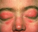
A heliotrope periorbital rash. Sometimes a generalized maculopapular rash may appear |
|
|
What lab is good to follow for progression of polymyositis? |
creatine kinase |
|
|
What are the five things to look for in diagnosis polymyositis or dermatomyositis? |
1) Proximal muscle weakness 2) Elevated CK, oftentimes CRP/ESR 3) Heliotrope rash (dermatomyositis) 4) proximal EMG changes 5) Muscle biopsy |
|
|
What medications should be considered with dermato/polymyositis? |
Start with corticosteroids like prednisone 60 mg/day, with a taper. Consider methotrexate, cyclosporine, azathioprine, cyclophosphamide. |
|
|
Distinguish polymyalgia rheumatic and polymyositis by EMG findings and weakness. |
With polymyalgia rheumatica, there is no focal proximal muscle weakness, but rather muscle aches, particularly around the neck/pecs, and pelvis. Also, PMR should have a normal EMG. |
|
|
What is a consistent laboratory finding with polymyalgia rheumatica? |
ESR over 100. CRP is also usually very high. |
|
|
If PMR is diagnosed (and temporal arteritis ruled out - remember 50% comorbidity), what is the treatment? |
Lower dose corticosteroids, like prednisone 20 mg/day. Following ESR is not helpful as it often remains high. |
|
|
Where is the most common lesion causing "central pain"? Technically, any lesion from where to where can cause central pain? |
the ventral posteriolateral thalamus, spinothalamic tract From the dorsal horns to the cortex |
|
|
Why is fentanyl so strong in comparison to morphine? |
Very high mu affinity. Full agonist. Most of its potency is because it is very lipophilic, thus easily crossing the blood brain barrier. |
|
|
Distinguish Freud's theory of conversion disorder to the more commonly accepted theory. |
Freud thought conversion disorder was a subconscious process that occurred as a defense against unacceptable impulses. Modern thought is that it is a pathological learned behavior that has more to do with secondary gain, social support, etc. |
|
|
Conversion disorder is relatively rare in the U.S. Why does Waldman think this is? |
Because Freud made it so famous that it is usually immediately recognized. |
|
|
Is conversion disorder intentional? |
No. That would make it malingering or factitious disorder. |
|
|
What CNS pathology is commonly seen with "la belle indifference"? |
Right cerebral hemisphere strokes. It is frequently seen in conversion disorder too (not an organic pathology). |
|
|
Describe Munchausen Syndrome |
going to frequent doctors presenting textbook perfect diseases and life threatening maladies. Often accompanied by self-injury so as to try to validate those complaints. The most notorious is injection of fecal matter in the bloodstream to induce a septic reaction. |
|
|
Regarding primary and secondary gains, what's the main difference between Munchausen syndrome and conversion disorder? |
Conversion disorder typically has clear primary or secondary gains. Strangely, Munchausen syndrome doesn't. Though Munchausen syndrome is consciously done and conversion disorder is unconsciously done. |
|
|
Give the six degrees of thermal injury |
1) First degree: erythema and mild pain. Only in the epidermis. 2) Second degree: fluid extravasation and blisters. Papillary layer of the dermis. 3) Third degree: charring of the skin and subcutaneous tissues. Eschar formation. Destruction of pain receptors. 4) Fourth degree burn: the dermis is destroyed and muscle/bone are exposed. No pain. 5) Fifth degree: Muscle has been destroyed. 6) Thermal injury to bone. |
|
|
Give the adult rule of nines for assessing total body surface area. |
Head 9% Anterior torso 18% posterior torso 18% one lower extremity 18% one upper extremity 9% perineum 1% |
|
|
What is the Parkland Formula for fluid replacement in a burn? |
4 ml x TBSA% x weight in kilograms
Fluid given in lactated ringers (LR is superior to NS over the short term as it can is alkalinizing in regards to acidosis associated with acute blood loss - however, it is not good over the long term as it only has a Na of 130 and K of 4). |
|
|
What are the six determinants associated with severity of electrical injury? |
1) Voltage 2) Amperage type (DC versus AC) 3) Path of current 4) Resistance/conductance of tissue 5) Surface area contact 6) Duration of exposure |
|
|
Which is more dangerous: direct or alternating current? Why? |
Alternating current is more dangerous (three times more) because of rapid changes in current flow. This more easily induces tetany, making it impossible for the person to withdrawal the body part. AC also can penetrate the epidermis better than DC. |
|
|
What is joule heating? |
The more resistant the tissue/substance, the more the electricity is converted to heat. |
|
|
Describe the differences in the appearance of the skin with high voltage, low voltage, and lightening electrical injuries. |
High voltage: deceptively minor skin damage with perhaps a small, leathery entrance/exit area with some hyperemia. Low voltage: skin and subcutaneous tissue is edematous with dry, shriveled skin around it. Lightening: "Lichtenberg figure": a christmas tree like burn. |
|
|
Differentiate nociception, pain, and suffering. |
Nociception: the nervous conduction of pain signals (free nerve endings via A delta and C fibers to the spinothalamic tract, VLP thalamus, etc). |
|
|
What is the female to male incidence of multiple sclerosis? |
2:1 |
|
|
Where do plaques associated with multiple sclerosis tend to aggregate? |
Periventricular white matter, the basal ganglia, the long tracts of the cord and brainstem, the optic nerves |
|
|
What are the most common presentations of MS? |
Optic neuritis, transverse myelitis, internuclear opthalmoplegia (medial longitudinal fasciculus lesion), pain/paresthesias. |
|
|
What type of MS do more than 70% of those suffering from it have? |
The relapsing/remitting form with near complete remission during remissions. |
|
|
What is the most common cause of optic neuritis? |
MS |
|
|
Besides MS, what is in the differential diagnosis for transverse myelitis? |
Post infectious etiologies, particularly EBV and Lyme. Of course anything damaging the cord (tumor, infection, aneurysm, etc) may cause it. |
|
|
What is the aphorism regarding fundoscopic exam with acute optic neuritis? |
"The patient sees nothing (acute vision loss) and the physician sees nothing (normal optic disk early on)." |
|
|
What is the Uhthoff phenomenon? |
optic neuritis symptoms worse when doing something warm, like eating warm food or taking a hot shower. |
|
|
What is relapsing/remitting MS typically treated with? |
beta interferon and glatiramer acetate (copaxone) |
|
|
What are the three main theories as to the etiology of "Post Polio Syndrome?" |
1) Polio is healed and the person has their normal deficits with those deficits that normally occur with aging. 2) A recurrence of dormant polio like shingles with zoster. 3) An enterovirus similar to polio causing an autoimmune reaction |
|
|
What anticholinestherase inhibitor is sometimes used for weakness with polio? |
Mestinon/pyridostigmine. |
|
|
What percentage of those with Guillain-Barre Syndrome require ventilation? |
30% tingling in the fingers and toes. |
|
|
What is a common infection prior to GBS? |
mild respiratory prodrome/virus, typically without fever, 2-4 weeks prior. |
|
|
What physical exam finding is thought to be pathognomonic for GBS (and its absence should cause serious doubt that this is GBS)? |
arreflexia. Normal or hyperreflexia is almost never seen with GBS. |
|
|
What are the typical three phases of GBS and their durations? |
1) Evolution: 2 weeks |
|
|
What percentage of those with GBS can expect a nearly full recovery? |
85% |
|
|
Describe the Miller Fischer variant of GBS |
Descending (not ascending) weakness. Ophthalmolplegia. Ataxia, areflexia. |
|
|
What bacteria have been associated with GBS/AIDP? |
Campylobacter, borrelia berdorferi, Haemophilus influenzae, Mycoplasma pneumoniae |
|
|
What tends to be seen with CSF with AIDP? |
normal cytology with increasing protein |
|
|
What is frequently seen with MRI with AIDP? |
Frequently nothing specific. However, often with gadolinium enhancement you can seen hyperintensity along the ventral nerve rootlets. |
|
|
What type of hemoglobin is seen with sickle cell anemia? |
hemoglobin S |
|
|
What two tissues/organs are most susceptible to sickle cell crises? |
the long bones and the spleen |
|
|
What is the cause of an aplastic crises with sickle cell anemia? |
infection with parvovirus b19 causing an acute halt in erythropoeisis. |
|
|
What are three non-analgesic medications that may be beneficial to those with sickle-cell pain? |
Zinc may help stabilize the erythrocyte membrane. Oxygen |
|
|
Define dependence in the context of medications and physiology. |
The physiologic state in which continued intake of a substance is required to maintain homeostasis. |
|
|
Define tolerance in the context of medications and physiology. |
an organism adapts to the effects of the drug over time, with a diminution of one or more of the drug's actions. |
|
|
Define addiction in the context of medications and physiology. |
A disease state characterized by dysfunctional behavior surrounding the use of a substance. |
|
|
What is the main area of the brain often associated with addiction? |
The "mesolimbic pathway". This connects the ventral tegmental area (not ventral trigeminal area as stated in Waldman's) adn the nucleus accumbens (main dopamine releasing area in the basal ganglia). |
|
|
What are the three most common theories of addiction? |
1) Homeostasis theory: disequilibrium between positive reward of drug and negative of withdrawal. 2) Incentive salience theory: sensitization of the dopamine system and dysfunction of modulators cause the patient to seek the drug at the expense of all else. 3) Habit theory: addiction is in part a learned behavior. |