![]()
![]()
![]()
Use LEFT and RIGHT arrow keys to navigate between flashcards;
Use UP and DOWN arrow keys to flip the card;
H to show hint;
A reads text to speech;
12 Cards in this Set
- Front
- Back
|
A 77-year-old woman with frequency, urgency and urge incontinence is able to contract her pelvic muscles appropriately. She has failed to respond to three different anticholinergic medications including one in combination with imipramine. Urinalysis is normal. CMG shows detrusor overactivity. The next step is:
|
sacral neuromodulation.
Explanation of the Answers: In the patient with an overactive bladder who does not achieve satisfactory results with maximum tolerable medical therapy the options are sacral neural modulation, bladder augmentation or diversion. Sacral neural modulation is reversible and therefore the best first step. Patients must be able to accurately fill out a voiding diary prior to the procedure. Pelvic floor rehabilitation does have a role in urge incontinence either alone or in conjunction with medical therapy, but is generally used as the first step in treatment. There is no data to support the use of electrical stimulation in the patient who can perform the exercises appropriately. Phenol injections intended to denervate the bladder have been abandoned. |
|
|
A 24-year-old man has a complete T-10 spinal cord transection after a diving accident. Videourodynamics three weeks after the injury will likely show:
|
Correct Answer:
detrusor areflexia. Explanation of the Answers: A complete spinal cord injury is usually followed by a period of decreased spinal cord activity at and below the level of the lesion, producing a state of "spinal shock". Both autonomic and somatic activity are suppressed, resulting in detrusor areflexia. The bladder neck is generally closed and competent. Striated sphincter tone is decreased, but usually preserved enough to maintain continence. Urinary retention is generally the rule during this stage. |
|

What type of voiding symptoms might this patient have?
|
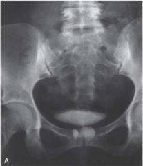
Dyspaurunia, post void dribbling is a big one as this is a urethral diverticulum. Also might have hematuria, chronic UTIs. Also worry about cancer as 5% of urethral cancers in women arise in urethral diverticulums.
|
|
|
What are theories for the development of urethral diverticula in women?
|
*Endoscopic collagen injection
*Trauma during vaginal delivery, from delivery itself or assisted delivery as in with tonsils. Although there are probably other unknown factors that may facilitate the initiation, formation, or propagation of urethral diverticula, *infection of the periurethral glands seems to be the most generally accepted common etiologic factor in most cases |
|
|
Where is the ostia most commonly located in urethral diverticula?
|
*in more than 90% of cases, the ostium is located posterolaterally in the mid or distal urethra, which corresponds to the location of the periurethral glands
*Inflammation was found in most of these patients suggest an infectious etiology. |
|
|
What is the modern hypothesis for the development of acquired urethral diverticula in women?
|
*These authors propose that acquired urethral diverticula result from infection and obstruction of the periurethral glands. These glands are normally found in the submucosal layer of the spongy tissue of the distal two thirds of the urethra.
*Repeated infection and abscess formation in these obstructed glands eventually result in enlargement and expansion. Initially, the expanding mass displaces the spongy tissue of the urethral wall and then enlarges to disrupt the muscle envelope of the urethra. *This results in herniation into the periurethral fascia. The enlarging cavity can then expand and dissect within the leaves of the periurethral fascia and urethropelvic ligament. This expansion occurs most commonly ventrally, resulting in the classic anterior vaginal wall mass palpated on physical examination in some patients with urethral diverticula. However, these may also expand laterally or even dorsally about the urethra. Eventually, the abscess cavity ruptures into the urethral lumen, resulting in the communication between the urethral diverticulum and the urethral lumen |
|
|
What are typical signs and symptoms of urethral diverticula?
|
*Vaginal or pelvic mass, hematuria, pelvic pain, urethral pain, dysuria, frequency, post-void dribbling, dyspaurenia, incontinence, vaginal or urethral discharge, double voiding, sensation of incomplete voiding
* Recurrent UTI, vaginal pain, urethral discharge at exam, vaginal mass |
|
|
What type of PE shoud be done when suspecting a urethral diverticulum?
|
*Thorough pelvic examination, as many of these patients have an anterior vaginal wall mass.
|
|
|
What PE signs might alert you to concern with a urethral diverticulum?
|
*A particularly hard anterior vaginal wall mass may indicate a calculus or malignant neoplasm within the urethral diverticulum and mandates further investigation
|
|
|
Does the characteristic "milking: maneuver for urethral diverticulum usually work and thus can it be used to rule out diverticulum?
|
No, most time don't see any discharge.
|
|
|
The earliest finding in diabetic voiding dysfunction is what?
|
*Delayed detrusor sensation
|
|

|

|

