Reading...
![]()
Play button
![]()
Play button
![]()
Use LEFT and RIGHT arrow keys to navigate between flashcards;
Use UP and DOWN arrow keys to flip the card;
H to show hint;
A reads text to speech;
106 Cards in this Set
- Front
- Back
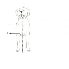
What is the plane in the image? What are the directions indicated in the image?
|
Median plane; also called mid-saggital plane.
1. Medial 2. Lateral |
|
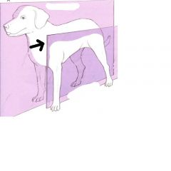
What is the plane indicated in the image?
|
Sagittal
|
|
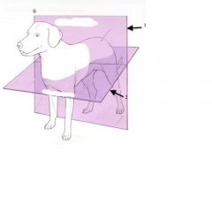
What are the planes indicated in the image?
|
1. Transverse
2. Dorsal |
|
|
Where does directional terminology change on the forelimb and what does it change to?
|
Changes at the antebrachiocarpal joint. Changes from Cranial/Caudal to Dorsal/palmar (on the manus)
|
|
|
How does the directional terminology change on the hind limb?
|
Changes from cranial/caudal to dorsal/plantar
|
|
|
What is the "elbow" joint called?
Describe the joint. |
Cubital joint
Complex joint (composed of humerus, radius, ulna). Resting position is less than 180 degrees in cranial position. |
|
|
What is the joint separating the manus from the antebrachium?
|
Antebrachiocarpal joint
|
|
|
Why are there two distinct bones in the antebrachium?
|
To allow supination and pronation
|
|
|
How are numbered bones counted? (ie metacarpals)
|
Medial to Lateral
|
|
|
Where does the axis run between the metacarpals?
|
Between digits III and IV
|
|
|
What is the difference between a digit and phalanx?
|
Part of a whole; 3 phalanges and the claw compose a digit.
|
|
|
What is unique about the feline distal phalanx?
|
The distal phalanx is held lateral to the middle phalanx by the dorsal elastic ligaments.
|
|
|
What do muscles act on?
|
Joints; muscle must cross joint to act on it
|
|
|
What is flexion?
|
The act of being bent
|
|
|
What is the flexor surface?
|
Area that decreases the lesser resting angle
|
|
|
What is the extensor surface?
|
Area that increases the lesser resting angle
|
|
|
When a muscle crosses two joints where is the major action of the muscle?
|
On the more distal attachment
|
|
|
What are muscle action groups?
|
Intrinsic muscles in similar location which may have similar action/attachments at proximal or distal attachment.
|
|
|
What is the root word for shoulder?
|
Omo
|
|
|
What is the root word for key (clavis)?
|
Cleido
|
|
|
What is the root word for arm?
|
Brachialis
|
|
|
What is the root word for widest?
|
Latissimus
|
|
|
What is the root word for before?
|
Ante
|
|
|
What is the root word for below or underneath?
|
Infra
|
|
|
What is the root word for above or over?
|
Supra
|
|
|
What is the root word for a band?
|
Fascia
|
|
|
What is the root word for elbow?
|
Cubitus
|
|
|
What is the root word for round?
|
Teres
|
|
|
What is the root word for hand?
|
Manus
|
|
|
What does cephalic mean?
|
Cranial; towards the head
|
|
|
What is the root word for wrist?
|
Carpus
|
|
|
What is the root word for toothed?
|
Serrated
|
|
|
What is the root word for bony?
|
Osseous
|
|
|
What is the root word for mouth?
|
os
|
|
|
What is the root word for braid?
|
plexus
|
|
|
What is the root word for armpit?
|
Axilla
|
|
|
What is the root word for a little swelling?
|
Tubercle
|
|
|
What is the root word for something shaped like a crow's beak?
|
Coracoid
|
|
|
What are the subunits of the superficial pectoral muscles?
|
Descending pectoral and transverse pectoral
|
|
|
What are the parts of the brachiocephalicus?
|
Cleidobrachialis
Cleidocephalicus (pars cervicalus) Cleidocephalicus (pars mastoidea) |
|
|
What are the parts of the STERNOCEPHALICUS?
|
Mastoid part, Occipital part
|
|
|
What are the parts of the trapezius?
|
Cervical part and thoracic part
|
|
|
What are the parts of the Rhomboideus?
|
Capitis, Cervicis, Thoracis
|
|
|
What are the parts of the Serratus ventralis?
|
Serratus ventralis cervicis and Serratus ventralis thoracis
|
|
|
What are the parts of the deltoideus?
|
Scapular part and Acromial Part
|
|
|
What are the parts of the Triceps brachii?
|
Long head, Lateral head, Accessory head, Medial Head
|
|
|
What are the parts of the Flexor carpi ulnaris?
|
Ulnar head and humeral head
|
|
|
What are the parts of the deep digital flexor?
|
Humeral head, Ulnar head, Radial head
|
|
|
How does the feline acromion of the scapula differ from that of the canine?
|
It has hamate and suprahamate processes (the later projects over the infranatus m)
|
|
|
What is the supratrochlear foramen known as in the feline humerus?
|
Supracondylar Foramen
|
|
|
What are two fossae found in the feline humerus that are not present in the canine humerus?
Describe them. |
Radial and Coronoid fossae.
Coronoid fossa is medial to radial fossa and accommodates medial coronoid process of ulna during elbow flexion. |
|
|
Where do the brachial a. and medial n. pass through in the feline humerus?
|
The supracondylar foramen
|
|
|
What are the carpal organs?
|
Specialized sensory (tactile) in the caudal forearm of felines. Contain whiskers and pressure points.
|
|
|
What intrinsic thoracic muscle is well developed in the feline but not in the canine?
|
Brachioradialis
|
|
|
What is the brachial plexus?
|
Branch of nerves extending from the C6, C7, C8, T1, T2
|
|
|
What is the key nerve of the forelimb?
|
The radial nerve; innervates the elbow extensors which must work in order to bear weight on limb
|
|
|
What is the autonomous zone of the Radial n. in the dog?
|
Dorsal D2, D3, D4, Axial D5
|
|
|
The dorsal side of the paw has only ________ nerves in the dog and cat.
|
Superficial
|
|
|
Where does the lateral digital extensor terminate in the feline?
|
Digits II-IV
|
|
|
Why is it impossible to test the median nerve of a dog?
|
The radial n. and median n. anastomose on the palmar side making it impossible to distinguish between the two nerves.
|
|
|
What is the autonomous zone of the Radial n. in the cat?
|
Dorsal D2, D3
|
|
|
Where do the palmar metacarpals terminate in the feline?
|
At the distal metacarpals; they do not anastomose with with palmar common digital nerve
|
|
|
How do the radial and ulnar heads of the deep digital flexor differ in the feline compared to the canine?
|
The are similar in size and therefore relatively larger than in the dog
|
|
|
How is it possible to test the median n. in the cat?
|
The palmar superficial and deep set of nerves do not anastomose and each digital nerve are either median or ulnar fibers
|
|
|
What is the autonomous zone of the Ulnar n. in the dog?
|
Dorsal or palmer D5 (Abaxial)
|
|
|
What is an autonomous zone?
|
Area of skin innervated solely by a specific peripheral nerve with no overlap with adjacent nerves
|
|
|
What is the autonomous zone of the Ulnar n. in the cat?
|
Dorsal or palmer D5
|
|
|
What is a cutaneous zone?
|
Entire area of skin innervated by a peripheral nerve
|
|
|
What is the autonomous zone of the median n. in the dog?
|
Can not be determined
|
|
|
What is the autonomous zone of the median n. in the cat?
|
Palmer D1, D2, D3
|
|
|
What are the intrinsic extensors of the shoulder joint?
|
Supraspinatus
Infraspinatus Supscapularis |
|
|
What is the blood supply and innervation of the intrinsic extensors of the shoulder joint?
|
Superficial cervical a.
Suprascapular n. Subscapular n. |
|
|
What are the intrinsic flexors of the shoulder joint?
|
Deltoideus, Teres major, Teres minor
|
|
|
What is the blood supply and innervation of the intrinsic flexors of the shoulder joint?
|
Caudal circumflex humeral a., subscapular a. and the axillary n.
|
|
|
What are the extensors of the elbow?
|
Triceps brachii
Anconeus Tensor fasciae antebrachii |
|
|
What is the blood supply and innervation of the extensors of the elbow?
|
Deep brachial a. and collateral ulna; Radial n.
|
|
|
What are the flexors of the elbow?
|
Biceps brachii, brachialis
|
|
|
What is the blood supply and innervation of the flexors of the elbow?
|
Bicipital a., Cr. Circumflex humeral a., Transverse cubital a., and musculocutaneous n.
|
|
|
What are x-rays a form of?
|
Electromagnetic radiation (ionizing)
|
|
|
What percentage of available energy is transformed to x-rays?
|
1%
|
|
|
What primarily produces the QUANTITY of x-rays produced?
|
Available electrons (mA)
Length of exposure (mAs) |
|
|
As x-rays pass through a patients, what are the possible interactions a ray can have?
|
X -rays can be absorbed, scattered and can pass through
|
|
|
Why is it important to ALWAYS take at least 2 views when making radiograph images?
|
Radiographs are 2D representations or 3D images; to conceive an actual relationship of structures it is necessary to have multiple views.
|
|
|
What are the 5 basic radiograph opacities in increasing whiteness?
|
Air
Fat Water Bone Metal |
|
|
What are the advantages of radiographs?
|
Widely available, Inexpensive, fast, can be portable
|
|
|
What are the disadvantages of radiographs?
|
2D representation of 3D images
Radiation exposure Better for bone and air; limited for soft tissue |
|
|
How is ultrasound imaging produced?
|
Sound waves are produced and bounce off objects differently, creating an image of what is being scanned
|
|
|
What are the variations in ultrasound images called?
|
Echogenecities (hyperechoic=brighter, hypoechoic=darker)
|
|
|
What are the advantages of ultrasound imaging?
|
It is safe, fairly portable and good for soft tissue
|
|
|
What are the disadvantages of ultrasound imaging?
|
It is poor for bone or gas, machine and user dependent, machines are expensive
|
|
|
How are CT images produced?
|
X-ray machine spirals around a patient and acquires multiple images and then computer processes them into a 3D image
|
|
|
What are the variations in CT images called?
|
Attenuations (hyper vs hypoattenuating)
|
|
|
What is MRI and how is it produced?
|
Magnetic Resonance Imaging; produced by high strength magnetic field
|
|
|
What are variations in MRIs called?
|
Intensity (hypo vs hyperintensity)
|
|
|
What signs should be used when evaluating images?
|
Size, Margin, Shape, Opacity, Number, Location
|
|
|
What are orthogonal views?
|
Two projections made at 90 degrees to one another
|
|
|
How are radiograph images named?
|
According to the direction of the beam; from point of entrance to exit. Left to right lateral = right lateral.
|
|
|
What are the 3 components of radiation safety?
|
Time, Distance, Shielding
|
|
|
How does the distance from the source affect the intensity of the primary beam?
|
By the inverse square law
|
|
|
Where are the superficial cervical lymph nodes located and what do they drain?
|
Deep to the omotransversius; drains nodes of the head, superficial neck, most of thoracic limb, craniodorsal thoracic wall
|
|
|
Where is the accessory lymph node located and what does it drain?
|
Between latissimus dorsi and deep pectoral mm. Drains the lateral thoracic wall, cranial mammae, medial proximal forelimb,
|
|
|
Where is the axillary lymph node located and what does it drain?
|
Located at branching of axillary and lateral thoracic vv. Drains medial proximal forelimb, cranial mammae. Efferent to veins.
|
|
|
What is the cutaneous zone of for the axillary nerve?
|
Cranial cutaneous antebrachial n.
|
|
|
What is the cutaneous zone for the radial nerve?
|
Lateral cutaneous antebrachial n.
|
|
|
What is the cutaneous zone for the ulnar nerve?
|
Caudal cutaneous antebrachial n.
|
|
|
What is the cutaneous zone for the musculocutaneous nerve?
|
Medial cutaneous antebrachial n.
|