![]()
![]()
![]()
Use LEFT and RIGHT arrow keys to navigate between flashcards;
Use UP and DOWN arrow keys to flip the card;
H to show hint;
A reads text to speech;
32 Cards in this Set
- Front
- Back
|
Define photoreceptors. |
Cells which function to convert light from waves into visual information. |
|
|
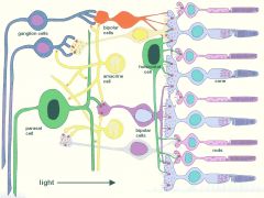
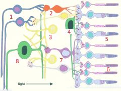
1. ganglion cell 2. bipolar cells 3. amacrine cells 4. horizontal cells 5. cone 6. rods 7. bipolar cells 8. parasol cell |
|
|
What sits behind the rods and cones? |
The pigment epithelium and choroid. |
|
|
What is within the outer segment of rods and cones? |
Stacked with membrane layers called discs. |
|
|
What is the role of discs? |
House molecular machinery which responds to light. |
|
|
What does the inner segment of photoreceptor cells house? |
Mitochondria and other organelles as well as providing a synaptic terminal connect to other neurons. |
|
|
What is difference in the morphology of discs in rods and cones? |
In rods the discs are intracellular structures In cones the discs are membrane folds. |
|
|
What is different about the sensitivity between rods and cones? |
1. Rods are extremely sensitive and respond to very low levels of illumination 2. Cones are less sensitive and only respond to bright light |
|
|
What are muller cells and what is their role? |
Muller cells are glial cells which support the photoreceptors to allow light to pass through all cell layers and stimulate the photoreceptors which are found at the back of the retina. They have been compared to fibre optic cables which deliver light rays directly to the photoreceptors. |
|
|
What is the role of the pigment epithelium and choroid layer? |
Absorb light which bypasses the photoreceptor cells. This prevents refraction and scattering of light which could blur the vision |
|
|
What is the photopigment found in rods? |
Rhodopsin |
|
|
What is the general structure of a photopigment? |
Photopigments consist of membrane bound proteins called opsins which are bound to a chromophore molecule |
|
|
What is the chromophore molecule of all photopigments? |
Retinal |
|
|
What vitamin is retinal derived from? |
Vitamin A |
|
|
What is the light sensitive part of a photopigment? |
The chromophore. |
|
|
What is the role of the bound opsin in a photopigment? |
The opsin bound to the retinal is the reason for the different light absorptions of each of the photopigments. (defines the wavelength specificity) |
|
|
How is the efficiency of photoreceptors increased? Why is it done this way? |
1. Aim is to pack as many photopigments into each photoreceptor cell as possible. 2. Efficiency of photopigments is fixed |
|
|
How is surface area of light receiving areas of the rods and cones increased? |
Discs are stacked perpendicular to the incoming light allowing for maximum surface area to catch light. |
|
|
How many photopigment molecules are in each photoreceptor cell? |
Over 1 billion photopigment molecules in each PR cell. |
|
|
What is special at the membrane potential of a photoreceptor cell? |
The only type of sensory cell that is relatively depolarised at rest --> +35mv When it is stimulated it becomes hyperpolarised |
|
|
Describe in detail the hyperpolarisation mechanism of photoreceptors. |
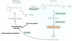
1. (In the action of the light) - Action of the enzyme guanylyl cyclase converts GTP to high concentrations of cyclic GMP 2. cGMP maintains lighand gated ion channels open - allows constant influx of sodium and calcium ions 3. When light hits the photoreceptor cell. The molecules of retinal in the discs assume a new conformation. They convert from an 11-cis conformation to an All-trans conformation. Visually, straightens the molecule. This is induced by the absorption of photon energy 4. All-trans rhodopsin converts to metarhodopsin I -> metarhodopsin II then All-trans retinal dissociates from opsin. 5. Opsin shape altered - Promoting interactions between opsin and transducin (G protein) 6. Transducin activation in turn activates cGMP - phosphodiesterase 7. Activated cGMP-phosphodiesterase rapidly degrades cGMP 8. Decrease of cytoplasmic cGMP concentration allows the cation channels to close 9. The loss of depolarising currents allows the membrane potential to hyperpolarise (-70mv) 10. Retinal molecule returns to original conformation after light activation ends and is reassociated with opsin via an enzyme driven mechanism. |
|
|
Describe the process of dark adaptation. |
1. Bright light, fully activates rhodopsin on the rods - Rods required for light vision 2. Going from light --> dark means that no rhodopsin is available to be activated as it is all activated by the bright light 3. All-trans Retinal and opsin must be reformed back together to allow further stimulation to occur and allow vision in low light a process which can take several minutes. |
|
|
How can vitamin A help with night vision? |
Vitamin A is good for night vision as it provides retinal for rhodopsin in the rods - allowing speedier dark adaptation. |
|
|
Describe the process of light adaptation. |
Going from dark --> light Initially the image is too bright and poor contrast as all of the rods become immediately activated. Over time retinal dissociates from the rhodopsin and only the less sensitive cone cells remain activated so that the light is not overwhelming. |
|
|
What is GCAP, what role does it have in vision? |
GCAP is - Guanylyl cyclase activating protein - it enhances guanylyl cyclase activity and is inhibited by calcium. When it is dark and calcium levels are high GCAP is inactivated. When it is light and Calcium levels are low (due to closure of the Na+/Ca2+ channels) GCAP activated guanylyl cyclase - boosting its efficacy and thus the activation of the cell. |
|
|
What is the role of calmodulin in vision? |
Calmodulin reduces channel affinity for cGMP - channels more likely to be closed (easier hyperpolarisation) this occurs during dark adaptation to |
|
|
Where are cones found? What are they specialized for? |
1. Concentrated in the fovea 2. High acuity day vision. |
|
|
What are the different types of cones? |
1. L-Cones 2. M-Cones 3. S-Cones |
|
|
What is the molecular structure of rhodopsin? |
7-transmembrane spanning alpha helices |
|
|
What is the basis of colour sensitivity in cones? |
Differences in primary sequence of cone opsins. |
|
|
What residue binds retinal to opsin? |
Residue 296. |
|
|
Biochemically, summarise how prolonged exposure to bright light leads to light adaptation. |
1. Ca2+ levels decline due to closure of cation channels and Na+ / Ca2+ exchanger. 2. Guanylyl cyclase activity increases 3. Channel affinity for cGMP increases 4. Lowered Ca2+ increases phosphorylation of metarhodopsin II - speeding up activation 5. Cation channels repoen and photoreceptors depolarise. |