![]()
![]()
![]()
Use LEFT and RIGHT arrow keys to navigate between flashcards;
Use UP and DOWN arrow keys to flip the card;
H to show hint;
A reads text to speech;
116 Cards in this Set
- Front
- Back
|
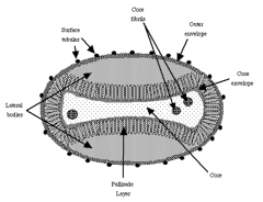
Papovaviridae: ss or ds? DNA or RNA? enveloped or nonenveloped? |
Papovaviridae are dsDNA viruses that are nonenveloped. Include the Papillomaviruses and Polyomaviruses |
|
|
There are both Early and Late genes on the papovaviridae genome. What do the Early genes encode for and when in the viral infection are they expressed? How about for Late genes ? |
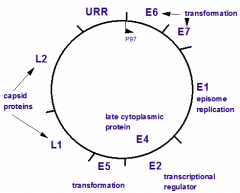
Early (E) genes: encode non structural proteins (DNA replication, transcription, etc.) and are expressed early in a viral infection and are associated with transformation Late (L) genes: encode structural proteins (capsid proteins) and are expressed late in a viral infection |
|
|
Of the three capsid proteins (VP 1-3) for papovaviridae viruses which one is the most important structural protein? Which one is important for receptor binding? |
VP2 is important for structure VP1 is important for receptor-binding |
|
|
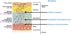
What is the tissue tropism for papovaviridae viruses? |
Surface epithelium: - Keratinized layer: permissive (full replication) - Dermal layer: semi-permissive (transformation and partial replication Mucosal epithelium: - Digestive system - Urogenital system |
|
|
CLASS QUESTION! Is the dermal layer of surface epithelium permissive or semi-permissive to papillomavirus infections? |
Semi-permissive. There is transformation and only partial viral replication |
|
|
Papovaviridae is broken up into two genera. What are they and what types of disease do the cause? |
1. Papillomaviridae is a tumorigenic virus (papilloma = wart) 2. Polyomaviridae causes neurological disease |
|
|
Are papillomaviruses species specific or do they have a broad host specificity? |
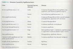
They are species specific and antigenically distinct (wont be getting from touching a dogs papilloma). Each species has multiple types and they are distinguished based on their characteristic restriction endonuclease cleavage, and have to use PCR or VN to do so. |
|
|
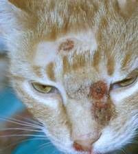
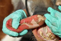
Canine Papillomaviruses There are ____ types in dogs. Papillomas occur commonly in the _______ mucosa, and they usually ___________ spontaneously. |
There are THREE types in dogs. Papillomas occur commonly in the ORAL (mouth, lips, tonguem pharynx) mucosa, and they usually RESOLVE spontaneously. |
|
|
If you were to cut open a papilloma and look at it histologically would you expect to find inflammatory cell infiltrate? |
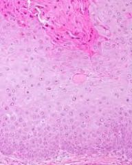
NO, papillomas are just caused by the proliferation of the epithelial cells. |
|
|
How do dog and horse immune systems react once they have experienced a papillomavirus infection? |
They develop a life long immunity! |
|
|
What specific epidermal layer will you see inclusion bodies due to an equine papillomavirus infection? |
Stratum spinosum |
|
|
Do you typically see papillomas in younger or older animals? Why does this make sense as far as the immune system's involvement? |
Predominantly see in younger animals, because once an animal has been exposed to the virus it develops a life long immunity. |
|
|
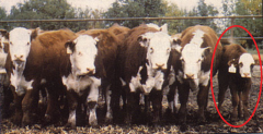
Equine sarcoids are caused by what type of virus? Where do you find the lesions? |

Caused by a papillomavirus and you find the lesions all over the body! |
|
|
CLASS QUESTION! What is the difference between EP (equine papillomavirus) and ES (equine sarcoids)? |
LOCATION of lesions! Papillomavirus = head (muzzle and oral cavity) Sarcoids = all over the place |
|
|
What is the preferred treatment for equine sarcoids? |
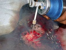
Cryosurgery using liquid nitrogen. Involves two freeze-thaw cycles |
|
|
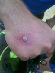
QUIZ QUESTION! What type of infection is associated with papillomavirus infections? |
Benign epithelial tumors (papillomas) |
|
|
Poxviridae: ss or ds? DNA or RNA? enveloped or nonenveloped? |
Poxviruses are enveloped, dsDNA viruses. They are very large, brick shaped viruses. |
|
|
CLASS QUESTION! What is the consequence of a poxvirus binding to the epidermal growth factor receptor? |
It triggers cell proliferation which supplies the virus with a lot of materials for replication in the cytoplasm. |
|
|
Where do poxviruses replicate once they get into the host cell? |
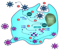
In the cytoplasm, which means that they bring their own mRNA and DNA synthesis machinery for replication purposes. |
|
|
What type of pathology (CPE and grossly) do you see with a poxvirus infection? |
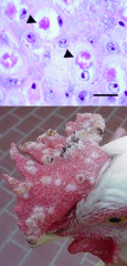
CPE: very large eosinophilic intracytoplasmic inclusion bodies (ballooning) Grossly: epithelial thickening and papule formation |
|
|
What are the two genera within the poxviridae family? |
Orthopoxviruses (cowpox, small pox, and monkeypox viruses) and Parapoxviruses (Orf and psuedocowpox viruses) |
|
|
Cowpox Who is the reservoir host? Where do you see lesions on the cows? How is it transmitted? What are other susceptible hosts? |
Reservoirs: rodents Lesions: teats Transmission: entry through breaks in the skin Susceptible hosts: cows, humans, cats |
|
|
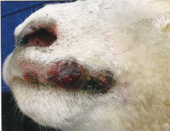
Poxvirus lesions appear on the face and head of cats and can often be mistaken for papillomaviruses. What else should you notice that could help you distinguish between the two? |
Cats with a poxvirus infection will also appear sick (fever, lethargy, depression, etc.) |
|
|
Orf virus is a parapoxvirus that causes lesions where on the body? |
Lesions are located on the gums, tongue, eyelids, feet, and teats. ORF is also referred to as "scabby mouth" which makes sense as far as the lesions. |
|
|
Orf virus commonly infects which species? Does it have zoonotic potential? |
Major hosts are sheep and goats, but it can infect humans. Must be careful and wear gloves when vaccinating with attenuated vaccine. |
|
|
What is the classical sequence of poxvirus lesions? |
Papule -> Vesicle (growing) -> Pustule (neutrophils join) -> Scab (rupture) |
|
|
QUIZ QUESTION! What type of infection is associated with poxvirus infection? |
Epithelial lesions (papule - vesicle - pustule - scab) with lymphocyte infiltration. |
|
|
QUIZ QUESTION! What is the difference in histological pathology between papillomavirus infection and poxvirus infection? |
Papillomavirus infections will result in cell proliferation, and if there are inclusion bodies they will be in the nucleus. Poxvirus infections willresult in cell proliferation along with lymphocyte infiltration, and inclusion bodies they will be in the cytoplasm. |
|
|
Rhabdoviridae: ss or ds? DNA or RNA? enveloped or nonenveloped? |
Rhabdoviridae are enveloped, -ssRNA viruses that are bullet shaped. Have a very short genome so rely on RNA editing to make more proteins. |
|
|
How do rhabdoviruses gain entry into host cells? |
They bind using their G protein spikes and engulfed by pinocytosis/endocytosis |
|
|
Rhabdoviruses replicate in the (cytoplasm or nucleus) of the host cell and their inclusion bodies are called __________. |
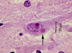
Rhabdoviruses replicate in the CYTOPLASM of the host cell and their inclusion bodies are called NEGRI BODIES. |
|
|
CLASS QUESTION! What is in the Negri Bodies? |
They are cytoplasmic inclusion bodies within nerve cells where you can find rhabdovirus virions. |
|
|
There are many genera of rhabdoviruses but we are most concerned with which two for this class? |
Vesiculoviruses (VSV) and Lyssaviruses (Rabies) |
|
|
What species can contract rabies viruses? |
All mammals, meaning it is zoonotic! |
|
|
What is the cell tropism for rabies viruses? |
Neurons, muscles, salivary glands (virus shedding) |
|
|
Rabies neurological pathogenesis depends on which region of the brain is affected by the virus. If the limbic system is affected then you get _____ rabies, whereas if the neocortex is affected you get _____ rabies. |
Limbic system you get FURIOUS rabies Neocortex you get DUMB rabies. |
|
|
What are the clinical signs that you see with the furious form of rabies? |

Aggression Seizures Hydrophobia - difficulty swallowing, excessive salivation Paralysis and Death |
|
|
What are the clinical signs that you see with the dumb form of rabies? |
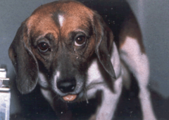
Apathetic Stupor Paralysis Coma Death |
|
|
What is the path of the rabies virus through the body from initial bite wound to when the animal becomes infective? |
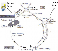
1. Infection through bite wound 2. Virus replicates in local myocytes for weeks or months. 3. Virus travels up peripheral nerve to CNS (Can do this right after bite as well and skip myocyte replication) 4. Virus replicated in the CNS 5. Virus travels back out through the peripheral nerves to salivary glands, skin, and most other organs |
|
|
What other virus causes CNS symptoms that resembles a rabies infection? |
Canine Distemper Virus. CDV is more progressive though and has other sign like diarrhea. |
|
|
When it comes to testing for rabies virus can you just collect samples from an alive animal? Why? |
No they need to be euthanized, because you need to take brain stem and cerebellum samples for FA testing. |
|
|
Can you vaccinate against rabies? Is there a treatment for it? |
Yes! Humans = killed virus vaccine Animal = live and killed virus vaccines Treatment involves rabies immune globulin. |
|
|
QUIZ QUESTION! What type of disease is associated with rabies virus infections? |
Neurological disease in both furious and dumb forms |
|
|
Which domestic species can you find VSV is? |
Horses, Cattle, and Swine |
|
|
Where can you find VSV globally? |
Only found in the US ('Merica!) Three strains are New Jersey, Indiana 2 and Indiana 3. |
|
|
How is VSV transmitted? |
Horizontally through insect vectors (sandflies, midges, blackflies), saliva and exudates |
|
|
What are clinical signs of VSV and what other viral disease does it resemble? |
Excessive salivation Blanched, raised areas Broken vesicles It looks a lot like Foot and Mouth Disease |
|
|
CLASS QUESTION! How many viruses do you know that cause viral vesicular disease of livestock? |
Foot and mouth disease virus (FMDV, Picornar) Swine vesicular disease virus (SVDV, Picornar) Vesicular exanthema of swine virus (VESV,Calici) VSV infects horses (Rhabdo) |
|
|
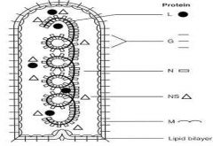
Prions: ss or ds? DNA or RNA? enveloped or nonenveloped? |
TRICK QUESTION! They are infectious protein particles |
|
|
What does the name "prion" mean? |
PRoteinaceous Infectious particle (-ON by analogy to virion) |
|
|
What is PrPC? |
PrPCis a normal surface protein that is present at highest levels in neurons of the brain and spinal cord. It has many alpha-helixes, it's soluble, and it is cleavable by protease. |
|
|
What is PrPSC? |
PrPSC is a mutated form of PrPC that is infectious. It's highly resistant (organic, heat, and protease), its has beta-pleated sheets, and is located on the outside of the cells as well as in cytoplasmic vesicles |
|
|
CLASS QUESTION! What is the difference between normal prions and an abnormal prion? |
PrPC = alpha-helix, susceptible to protease, only on plasma membrane PrPSC = beta-pleated sheets, resistant to protease, found on PM and in cytoplasmic vesicles |
|
|
How do prions "replicate"? |
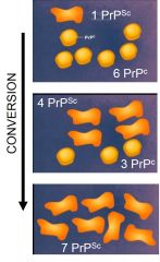
CONVERSION, not actually replicating! The PrPSC can convert the PrPC into other PrPSC, thus creating more |
|
|
CLASS QUESTION! What is the difference between virus replication and prion replication? |
Viruses actually replication, whereas prions multiply through conversion. |
|
|
CLASS QUESTION! Where did the abnormal prion come from? |
It was the result of a mutation of a PrPC |
|
|
How are prions transmitted? |
Ingestion of infective prions They are first taken up at the Peyers Patches, then travel to other lymphoid sites where they multiply, and finally they travel to the CNS via peripheral nerves |
|
|
What type of disease do prions cause? |
Neurological Disease |
|
|
Do prion infections result in an acute or chronic disease? |
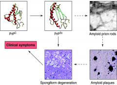
Chronic disease. Progressive infections of the nervous systems where you see amyloid deposits forming plaques. |
|
|
There are many different prion "strains" out there that cause different lesions, have different incubation times, and different mortality rates. Though they all generate the same characteristic _______ after infections. |
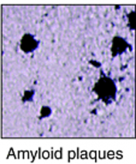
They all generate the same characteristic AMYLOID PLAQUES after infections. |
|
|
Are prions species specific? |
The PrPC amino acid sequence is different between species which makes transmission initially difficult between species. But the barrier will disappear after multiple attempts as passing over. |
|
|
CLASS QUESTION! What does the PrPSC in a new host still have physiochemical properties of the original prion but the amino acid sequence of the new host? |
The physiochemical properties don't change when the prions get transfered, but the amino acid sequence does. |
|
|
What diseases are associated with prions? |
Transmissible Spongiform Encephalopathy (TSE) - Scrapie - Bovine Spongiform Encephalopathy (mad cow) - Transmissible mink encephalopathy - Feline spongiform encephalopathy - Chronic Wasting Disease |
|
|
What are the clinical signs of prion diseases in general? |
Dementia and/or ataxia It's more of a progressive loss of brain function |
|
|
How is bovine spongiform encephalopathy (mad cow) transmitted? |

Ingestion of infected meat (CNS and distal ileum) |
|
|
What diagnostic test do you use to determine if a cow is currently infected with BSE? |
There is no test to detect the disease in a live animal. |
|
|
CLASS QUESTION! What is the difference between PrPC and PrPSC as far as protease digestion? |
PrPSC is resistant to protease digestion and that is why you can find it hanging out is cytoplasmic vesicles. |
|
|
What type of disease is Chronic Wasting Disease (CWD), and what animal species are you going to see it in? |
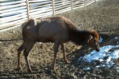
Its a prion disease that you will see in elk and deer. |
|
|
Flaviviridae: ss or ds? DNA or RNA? enveloped or nonenveloped? |
Flaviviridae viruses are enveloped, +ssRNA viruses. Genera include: Flavivirus, Pestivirus, Hepacivirus |
|
|
What two diseases are we most concerned with when talking about Pestiviruses in large animals? |
Bovine Viral Diarrhea (BVD) Hog Cholera |
|
|
What is the cell tropism of Pestiviruses? |
Lymphoid Tissue and Mucosal Epithelium |
|
|
There are two genotypes of BVDV (1 and 2) and within those they each have two biotypes. What are the two biotypes? |
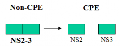
Non-CPE BVDV and CPE BVDV |
|
|
What is the difference between Non-CPE and CPE BVDV? |
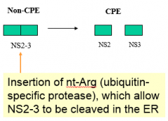
There was an INSERTION of an Arg into the NS2-3 sequence that allowed for protease cleavage, splitting the NS2 and NS3. Together NS2-3 is Non-CPE BVDV Seperate NS2 and NS3 is CPE BVDV |
|
|
What is BVDVs tissue tropism? |
Epithelial cells (arterial walls, oviducts, uterus, mammary gland epi, hair follicles) resulting in mucosal disease Reproductive System Digestive System Lungs Kidneys Adrenal glands |
|
|
What does a BVDV infection look like in healthy, non-pregnant animals? |
Can have subclinical, mild or severe form of the disease. If the animal recovers though it will have a life long resistance to BVDV. |
|
|
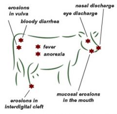
Which biotype of BVDV causes mucosal disease and what are the signs? |
The CPE BVDV causes the mucosal disease. Signs will be high fever, depression, anorexia, bloody diarrhea, hemorrhage, ulcers, and eventually death. |
|
|
How do acute BVDV infections occur? |
Horizontally transferred from either another acutely infected animal or from a PI animal. Pass it through bodily fluids (nasal discharge, tears, saliva, urine,feces, milk and semen) and on contaminated environments and fomites. |
|
|
What is the difference between acute mucosal disease and chronic mucosal disease in reference to BVDV infections? |
Acute mucosal disease you'll see erosions and ulcers in the GI tract, hemorrhage and extensive necrosis. Chronic mucosal disease has very mild symptoms that last for a while. You'll see intermittent diarrhea, gradual wasting, and lameness. |
|
|
How do you get persistently infected BVDV calves? |
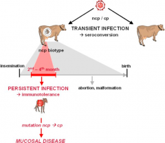
Two ways of getting PI animals: 1. PI cow give it toher fetus 2. Acuteinfection of a pregnant cow during the first 120 to150 days of gestation |
|
|
CLASS QUESTION! Under what conditions does BVDV cause mucosal disease? What is the mechanism behind it? |
When the NonCPE BVDV gets the Arg insertion into the NS2-3 and its becomes the CPE BVDV, then you will see mucosal disease. |
|
|
An acute infection of a pregnant cow during which of these times frames will cause the calf to be persistently infected with BVDV? A. <80 days B. 80-125 days C. >125 days |
A. <80 days - embryonic death B. 80-125 days - PI calf C. >125 days - develops Ab response and clears |
|
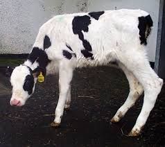
Which one is persistently infected with BVDV? |
Experienced retardation in growth 50% mortality rate in the first year of life |
|
|
What samples can you use for diagnostic testing of BVDV and what are the commonly used tests? |
Antigen capture ELISA (ear notch) RT-PCR (blood or ear notch) |
|
|
What countries has hog cholera been eradicated from? |
Australia, Canada, and the US |
|
|
How contagious is hog cholera? How is it mostly transmitted? |
Its highly contagious to swine and its transmitted orally mostly. |
|
|
What clinical signs will you see with acute hog cholera infections? |
High fever Severe depression Multiple superficial and internal hemorrhages (very high morbidity and mortality rates) |
|
|
The hog cholera virus initially replicated at the entry site but then it quickly spreads to ________. |
The hog cholera virus initially replicated at the entry site but then it quickly spreads to LYMPHOID TISSUE (ex. tonsils and cervical lymph nodes). |
|
|
CLASS QUESTION! What is the histological feature of a HCV infection? |
Hemorrhage and Necrosis |
|
|
Flaviviruses are in the same family (Flaviviridae) as what other genuses? |
Pestiviruses and Hepaciviruses |
|
|
What diseases are we most concerned with when talking about Flaviviruses? |
West Nile Virus Yellow Fever Virus Murray Valley E Virus Dengues |
|
|
How is West Nile Virus transmitted and what species are the reservoir hosts? |
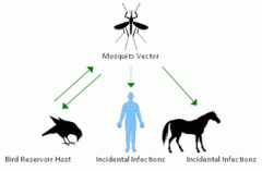
Mosquitoes (vector) transmit it from birds (reservoir) to humans and horses |
|
|
Explain the progression of the West Nile Virus within the mosquito from ingestion to transmission. |
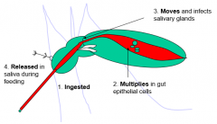
1. Virus is ingested from reservoir host with a blood meal 2. Virus replicates in the gut epithelial cells 3. Virus moves into the salivary glands 4. Virus released in saliva during next blood meal |
|
|
Explain the progression of the West Nile Virus within mammals once bitten by mosquito. |
1. Virus transmitted in mosquitoes saliva 2. Virus replicates in the skin and surrounding lymph nodes 3. Virus is circulating in bloodstream (viremia) and the mammal will experience "Flu-like" symptoms 4a. Virus is cleared by immune system OR 4b. Virus crosses blood-brain barrier 5. Virus causes inflammation and cerebral interstitial edema 6. Damage to the neurons and glial cells results in Neurological Signs |
|
|
How do you control West Nile Virus? |

Very effective, killed vaccine! (And I assume trying to control vectors couldn't hurt) |
|
|
Togaviridae: ss or ds? DNA or RNA? enveloped or nonenveloped? |
Togaviridae viruses are tightly enveloped +ssRNA viruses. Genera include: Alphaviruses and Rubiviruses |
|
|
What general clinical signs do you see with Togaviruses? |
CNS effects. More specifically encephalitis, meningitis, paralysis, fever, headache, seizures, coma and death. Easy to remember since a lot of the virus names include "Encephalitis" in the name. (Ex. Eastern Equine Encephalitis Virus or EEEV) |
|
|
How are Togaviruses transmitted from one host to another? |
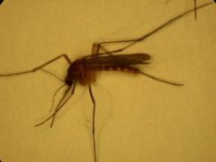
Arthropods like mosquitoes |
|
|
How do you control the spread of Togaviruses? |
You control the vectors! |
|
|
Picornaviridae: ss or ds? DNA or RNA? enveloped or nonenveloped? |
Picornaviridae viruses are nonenveloped +ssRNA viruses. They have the smallest viral genome, so replication is very fast Many genera but the viruses we need to keep track of are Foot and Mouth Disease and Swine Vesicular Disease |
|
|
What type of disease does FMDV cause? How is it transmitted? |
It is a vesicular disease that effects the mucosal epithelium. Transmission is through aerosols, contaminated milk, and oral-fecal. |
|
|
What role do pigs play in the spread of FMD? |
Aerosols produced by infectious animals contain large amounts of virus, but PIGS excrete 1000x-3000x more viruses than cattle. They are amplifying hosts |
|
|
CLASS QUESTION! Why is FMDV so infectious? |
It only takes 10 viral particles to become infected. The virus can become aerosolized which makes spread much easier. |
|
|
(Cattle, Sheep, Pigs, Horses) FMD infects which species? SVD infects which species? |
FMD infects Cattle, Sheep, and Pigs SVD infects Pigs only. |
|
|
Swine vesicular disease exhibits the same clinical signs in pigs as what other (related) disease? |
Foot and Mouth disease. SVD is much less devastating though |
|
|
Arteriviridae: ss or ds? DNA or RNA? enveloped or nonenveloped? |
Arteriviridae viruses are enveloped, +ssRNA viruses. The viruses we need to keep track of are Equine Virual Arteritis (EVA) and Porcine Respiratory and Reproductive Syndrome Virus (PRRSV) |
|
|
Porcine Respiratory and Reproductive Syndrome Virus (PRRSV) has what tissue tropism? What is the result? |
PRRSV affects alveolar macrophages, dendritic cells in the tissues and the vascular system The result being immunosuppression. |
|
|
Equine Viral Arteritis has many clinical signs, but the two most important are what? |
Abortion and Edema |
|
|
The virus that causes Equine Viral Arteritishas what tissue tropism? What is the result? |
The virus affects lymphoid tissue and the vascular system (similar to PRRSV). Immunosuppression and leukopenia are the resulting concerns. |
|
|
CLASS QUESTION! What are the cells that both PRRSV and EVA infect? |
Lymphoid cells and Vasculature |
|
|
CLASS QUESTION! What are the common clinical features from infections of PRRSV and EVA? |
Immunosuppression resulting in respiratory distress, abortions, and hemorrhages |
|
|
Reoviridae: ss or ds? DNA or RNA? enveloped or nonenveloped? |
Reoviridae viruses are nonenveloped dsDNA viruses with a double protein shell. The viruses we need to keep track of are both Orbiviruses and they are Bluetongue and EHD. |
|
|
How are Orbiviruses transmitted between hosts? |
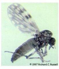
Arthropod vectors: Culicoides spp. |
|
|
What is the tissue tropism of BT viruses? What are the main clinical features? |

The BT virus affects mucosal epithelium, hematopoietic cells, and endothelial cells. Widespread hemorrhages, mucosal ulcers, excessive salivation, and congestion of hoof laminae are the most common clinical signs. |
|
|
Epizootic Hemorrhagic Disease is most commonly found in what animals in the US? |
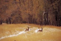
Deer |
|
|
How is EHD transmitted? |
By arthropod vectors: Culicoides spp. |
|
|
What is the tissue tropism of EHDV? What are the main clinical features? |
Hematopoietic and Endothelial Cells Resulting in edema and extensive hemorrhaging |