![]()
![]()
![]()
Use LEFT and RIGHT arrow keys to navigate between flashcards;
Use UP and DOWN arrow keys to flip the card;
H to show hint;
A reads text to speech;
11 Cards in this Set
- Front
- Back
|
From superficial to deep, what are the meningeal layers surrounding the brain & SC?
What are their functions? |
pachymeninx (thick/durable) 1. Dura mater
leptomeninges 2. Arachnoid mater 3. Pia mater
function: 1. protect CNS from mechanical injury (#1) 2. provide blood supply to skull and cerebral hemispheres 3. provide space for CSF flow (via arachnoid in in subarachnoid space) |
|
|
Describe the appearance of and spatial relationships b/w the 3 layers of meninges |
1. dura -appearance: thick; 2 layers-- periosteal & meningeal layers -relationship: fused w/ endosteum of skull w/in cranium, BUT epidural space exists in spinal canal
2. arachnoid -appearance: spiderweb-like sheath -relationship: connected to Pia via arachnoid trabeculae; space b/w arachnoid & pia = subarachnoid space
3. pia -appearance: thin mesh w/ tiny BV; follows all sulci & gyri) -relationship: see arachnoid |
|
|
what are the 3 infoldings of the dura and what is their purpose? |
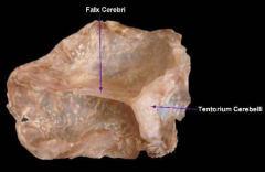
1. Falx cerebri separate cerebral hemis. along midline
2. tentorium cerebelli separate cerebral hemis from cerebellum 3. membrane covering hypophyseal (pituitary) fossa
|
|
|
Where would you do a cisternal CSF tap? |
cerebellomedullary cistern (largest part of subarachnoid space) |
|
|
What produces CSF?
Describe this structure |
choroid plexus
the plexus is a projection of ependymal cells that are derived from the ventricles |
|
|
What's the difference between veins and & sinuses in the venous system of the brain? |
veins: found in & out of the brain; drains into sinuses
sinuses: found in dura mater or bones of skull; formed by the separation of the 2 layers of dura |
|
|
Why is the brain highly dependent on reliable and constant arterial blood supply? |
brain can't store glucose, thus it needs constant influx of glucose from arterial blood |
|
|
What are the 2 main vascular supplies to the brain in the dog?
|
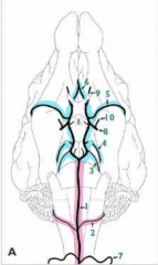
1. basilar artery (pink)
2. cerebral arterial circle (circle of willis) (blue) |
|
|
what are the 2 ways in which blood gets into the brain? |
1. Vertebral artery from C6 and traveling cranially, it passes thru transverse foramina. Then, it branches from intervertebral foramina traveling ventrally to form ventral spinal artery, which becomes the basilar artery
vertebral can also just directy connect to internal carotid, which enters brain to help form cerebral arterial circle
2. common carotid artery splits into Internal and external carotid artery; Internal carotid enters into brain contributing to cerebral arterial circle alongside w/ basilar artery, vertebral artery, & branch of maxillary artery |
|
|
In the dog, which artery is the main supply to the cerebral arterial circle? |
internal carotid artery |
|
|
why would occlusion/severance of common carotids/internal carotid in dog cause PARTIAL/SLOW loss of consciouness? |
despite the occlusion, you still have the vertebral artery pumping blood into brain via basilar artery |