Reading...
![]()
Play button
![]()
Play button
![]()
Use LEFT and RIGHT arrow keys to navigate between flashcards;
Use UP and DOWN arrow keys to flip the card;
H to show hint;
A reads text to speech;
85 Cards in this Set
- Front
- Back
|
What regulates the transitions between different phases of the cell cycle? |
- Cyclins
- CDKs - Tumor Suppressors |
|
|
What are the stages of mitosis?
|
- Prophase
- Metaphase - Anaphase - Telophase |
|
|
What is the shortest phase of the cell cycle?
|
Mitosis
|
|
|
What are CDKs?
|
Cyclin-Dependent Kinases
- Constitutive - Inactive until activated by Cyclin |
|
|
What are Cyclins?
|
- Regulatory proteins
- Control cell cycle events by activating CDKs - Phase specific |
|
|
What do CDKs and Cyclins form / do?
|
- Form Cyclin-CDK complexes
- Must be both activated and inactivated for cell cycle to progress |
|
|
What are the tumor suppressors involved in the cell cycle? Functions?
|
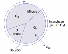
- p53
- Hypophosphorylated Rb - These inhibit G1-to-S progression - Mutations lead to unrestrained cell division |
|
|
What are the types of cells (in terms of cell cycle activity)?
|
- Permanent: remain in Go, regenerate from stem cells
- Stable (Quiescent): enter G1 from Go when stimulated - Labile: never go to Go, divide rapidly w/ a short G1 |
|
|
What kind of cells are "Permanent"? What happens to these cells?
|
- Neurons, skeletal and cardiac muscle, RBCs
- Remain in Go, regenerates from stem cells |
|
|
What kind of cells are "Stable (Quiescent)"? What happens to these cells?
|
- Hepatocytes and lymphocytes
- Enter G1 from Go when stimulated |
|
|
What kind of cells are "Labile"? What happens to these cells?
|
- Bone marrow, gut epithelium, skin, hair follicles, germ cells
- Never go to Go, divide rapidly with a short G1 |
|
|
What are the phases of the cell cycle?
|
- Mitosis
- G1 ←→ Go - S phase (synthesis) - G2 |
|
|
What happens in the Rough Endoplasmic Reticulum (RER)?
|
- Synthesis of secretory (exported) proteins
- N-linked oligosaccharide addition to many proteins |
|
|
What is the name for the RER in neurons? Function?
|
Nissl Bodies
- Synthesizes enzymes (eg, ChAT - choline acetyltransferase makes ACh) - Synthesizes peptide neurotransmitters |
|
|
What happens in Free Ribosomes (unattached to any membrane)?
|
Site of synthesis of cytosolic and organellar proteins
|
|
|
Which cells in the body are rich in RER?
|
- Mucus-secreting goblet cells of the small intestine
- Antibody-secreting plasma cells |
|
|
What happens in the Smooth Endoplasmic Reticulum (SER)?
|
- Steroid synthesis
- Detoxification of drugs and poisons |
|
|
Which cells in the body are rich in SER?
|
- Liver hepatocytes
- Steroid hormone-producing cells of the adrenal cortex |
|
|
Which organelle is the distribution center for proteins and lipids from the ER?
|
Golgi
|
|
|
What happens in the Golgi?
|
- Sends the proteins and lipids to the vesicles and plasma membrane
- Modifies N-oligosaccharies on asparagine - Adds O-oligosaccharides on serine and threonine - Adds mannose-6phosphate to proteins for trafficking to lysosomes |
|
|
What is the "sorting center" for material from outside the cell or from the Golgi?
|
Endosomes
|
|
|
What is the function of Endosomes?
|
Sorting Center
- Receives material from outside the cell or from the Golgi - Sends it to lysosomes for destruction OR back to the membrane / Golgi for further use |
|
|
What happens in I-cell disease?
|
Inclusion Cell Disease
- Inherited lysosomal storage disorder - Failure of addition of mannose-6-phosphate to lysosome proteins - Enzymes are secreted outside the cell instead of being targeted to the lysosome |
|
|
What are the symptoms of Inclusion Cell Disease (I-Cell Disease)?
|
- Coarse facial features
- Clouded corneas - Restricted joint movement - High plasma levels of lysosomal enzymes - Often fatal in childhood |
|
|
What are the vesicular trafficking proteins?
|
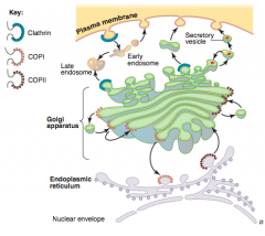
COPI:
- Golgi → Golgi (retrograde) - Golgi → ER COPII: - Golgi → Golgi (anterograde) - ER → Golgi Clathrin: - trans-Golgi → Lysosomes - Plasma Membrane → Endosomes (receptor-mediated endocytosis) |
|
|
What is the peroxisome? Function?
|
- Membrane enclosed organelle
- Involved in catabolism of very long fatty acids and amino acids |
|
|
What is the proteasome? Function?
|
- Barrel-shaped protein complex
- Degrades damaged or unnecessary proteins tagged for destruction with ubiquitin |
|
|
Where do proteins tagged with ubiquitin go?
|
Proteasome, where they are degraded
|
|
|
What is the organization of a microtubule?
|
- Cylindrical structure
- Helical array of polymerized dimers of α and β tubulin - Each dimer has 2 GTP bound - Grows slowly and collapses quickly |
|
|
What are microtubules used for?
|

- Flagella
- Cilia - Mitotic spindles - Slow axoplasmic transport in neurons |
|
|
What is the function of molecular motor proteins?
|
Transport cellular cargo toward opposite ends of microtubule tracks
|
|
|
Which molecular motor protein transports cellular cargo in the retrograde direction along microtubules (+ → -)?
|

Dynein
|
|
|
Which molecular motor protein transports cellular cargo in the anterograde direction along microtubules (- → +)?
|

Kinesin
|
|
|
Which drugs act on microtubules?
|
- Mebendazole / Thiabendazole (anti-helminthic)
- Griseofulvin (anti-fungal) - Vincristine / Vinblastine (anti-cancer) - Paclitaxel (anti-breast cancer) - Colchicine (anti-gout) |
|
|
Which anti-helminthic drugs act on microtubules?
|
Mebendazole and Thiabendazole
|
|
|
Which anti-fungal drug acts on microtubules?
|
Griseofulvin
|
|
|
Which anti-cancer drugs act on microtubules?
|
- Vincristine
- Vinblastine - Paclitaxel (anti-breast cancer) |
|
|
Which anti-gout drugs act on microtubules?
|
Colchicine
|
|
|
What disease is caused by mutations in the lysosomal trafficking regulator gene (LYST)?
|
Chédiak-Higashi Syndrome
|
|
|
What causes Chédiak-Higashi Syndrome?
|
- Mutations in the lysosomal trafficking regulator gene (LYST)
- The product of LYST is required for the microtubule dependent sorting of endosomalproteins into late multi-vesicular endosomes - Results in recurrent pyogenic infections, partial albinism, and peripheral neuropathy |
|
|
What are the symptoms of Chédiak-Higashi Syndrome?
|
- Recurrent pyogenic infections
- Partial albinism - Peripheral neuropathy |
|
|
What is the organization of a cilia?
|
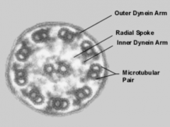
- 9 + 2 arrangement of microtubules
- Anoxemal dynein: ATPase that links peripheral 9 doublets and causes bending of cilium by differential sliding of doublets |
|
|
What disease is signified by immotile cilia?
|
Kartagener's Syndrome (Primary Ciliary Dyskinesia)
|
|
|
What causes Kartagener's Syndrome (Primary Ciliary Dyskinesia)? Symptoms?
|
- Immotile cilia due to a dynein arm defect
Symptoms - Male infertility (immotile sperm) and decreased female fertility - Bronchiectasis - Recurrent sinusitis (bacteria and particles not pushed out) - Associated with situs inversus |
|
|
What structures are actin and myosin part of?
|
- Microvilli
- Muscle contraction - Cytokinesis - Adherens junctions |
|
|
What structures are microtubles part of?
|
Movement:
- Cilia - Flagella - Mitotic spindles - Axonal trafficking - Centrioles |
|
|
What structures are intermediate filaments part of?
|
Structure:
- Vimentin - Desmin - Cytokeratin - Lamins - Glial fibrillary acid proteins (GFAP) - Neurofilaments |
|
|
What is the plasma membrane composed of?
|
- Asymmetric lipid bilayer
- Contains cholesterol, phospholipids, sphingolipids, glycolipids, and proteins |
|
|
What are the intermediate filaments? What kind of cells are they found in?
|
- Vimentin - CT
- Desmin - Muscle - Cytokeratin - Epithelial Cells - GFAP - NeuroGlia (astrocytes) - Neurofilaments - Neurons |
|
|
What is the organization and effect of the Sodium-Potassium Pump?
|
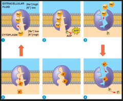
- Na+/K+ ATPase located in plasma membrane
- ATP site is on cytosolic side - For each ATP consumed, 3 Na+ go OUT and 2 K+ go IN - During cycle, pump is phosphorylated |
|
|
What drugs / toxins affect the Na+/K+ ATPase?
|
- Ouabain
- Cardiac glycosides (Digoxin and Digitoxin) |
|
|
What is the action of Ouabain?
|
Inhibits the Na+/K+ ATPase by binding to the K+ site
|
|
|
What is the action of Cardiac Glycosides (Digoxin and Digitoxin)?
|
Directly inhibit the Na+/K+ ATPase, which leads to indirect inhibition of Na+/Ca2+ exchange → ↑ [Ca2+]i → ↑ Cardiac contractility
|
|
|
What is the most abundant protein in the human body?
|
Collagen
|
|
|
What are the characteristics of Collagen?
|
- Most abundant protein in humans
- Extensively modified by post-translational modifications - Organizes and strengthens ECM |
|
|
What are the types of Collagen? Location? (Mnemonic?)
|
Be (So Totally) Cool, Read Books
- Type I: Bone, Skin, Tendons - Type II: Cartilage - Type III: Reticulin - Type IV: Basement membrane or Basal lamina |
|
|
What is the most common type of Collagen?
|
Type I
|
|
|
Where is Collagen Type I found?
|
- Bone, Skin, Tendon [Be (So Totally) Cool, Read Books]
- Also in Dentin, Fascia, Cornea, and involved in late wound repair |
|
|
In what disease is Type I Collagen defective?
|
Osteogenesis Imperfecta
|
|
|
Where is Collagen Type II found?
|
- Cartilage (including hyaline)
[Be (So Totally) Cool, Read Books] - Also in Vitreous Body, Nucleus Polposus |
|
|
Where is Collagen Type III found?
|
- Reticulin
[Be (So Totally) Cool, Read Books] - Reticulin is a part of skin, blood vessels, uterus, fetal tissue, and granulation tissue |
|
|
In what disease is Type III Collagen defective?
|
Ehlers-Danlos (ThreE-D)
|
|
|
Where is Collagen Type IV found?
|
Basement membrane or Basal lamina
[Be (So Totally) Cool, Read Books] Type Four under the Floor (BM) |
|
|
In what disease is Type IV Collagen defective?
|
Alport Syndrome
|
|
|
What diseases does a defect in each of the types of collagen cause?
|
- Type I: Osteogenesis Imperfecta (found in bone)
- Type II: N/A - Type III: Ehlers-Danlos (threE-D) - Type IV: Alport Syndrome (found in BM) |
|
|
What are the steps of collagen synthesis? Location?
|
Inside Fibroblasts:
1. Synthesis (RER) 2. Hydroxylation (ER) 3. Glycosylation (ER) 4. Exocytosis Outside Fibroblasts 5 Proteolytic processing 6. Cross-linking |
|
|
What happens during the first step of collagen synthesis? Location?
|
Synthesis (RER of fibroblast)
- Translation of collagen α chains (PRE-PROCOLLAGEN) - Usually Gly-X-Y (X and Y are proline or lysine) |
|
|
What happens during the second step of collagen synthesis, after synthesis? Location?
|
Hydroxylation (ER of fibroblast)
- Specific proline and lysine residues - Requires vitamin C (deficiency leads to scurvy) |
|
|
What happens during the third step of collagen synthesis, after hydroxylation? Location?
|
Glycosylation (ER of fibroblast)
- Pro-α-chain hydroxylysine residues are glycosylated - Formation of PROCOLLAGEN via hydrogen and disulfide bonds (triple helix of 3 collagen α chains) - Problems forming triple helix → osteogenesis imperfecta |
|
|
What happens during the fourth step of collagen synthesis, after glycosylation? Location?
|
Exocytosis (fibroblast to outside fibroblast)
- Exocytosis of PROCOLLAGEN into extracellular space |
|
|
What happens during the fifth step of collagen synthesis, after exocytosis? Location?
|
Proteolytic Processing (outside fibroblasts)
- Cleavage of disulfide-rich terminal regions of procollagen - Transforms into insoluble TROPOCOLLAGEN |
|
|
What happens during the sixth step of collagen synthesis, after proteolytic processing? Location?
|
Cross-Linking
- Reinforcement of many staggered TROPOCOLLAGEN molecules by covalent lysine-hydroxylysine cross-linking (by Cu2+ containing lysyl oxidase) - Forms collagen fibrils - Problems with cross-linking → Ehlers-Danlos |
|
|
What causes Osteogenesis Imperfecta?
|
Genetic bone disorder
- Causes brittle bone disease - Most common cause is an Autosomal Dominant Type I collagen defect |
|
|
What are the symptoms of Osteogenesis Imperfecta?
|

1:10,000
- Multiple fractures with minimal trauma; may occur during birth process - Blue sclerae d/t translucency of CT over choroidal veins - Hearing loss (abnormal middle ear bones) - Dental imperfections d/t lack of dentin - May be confused with child abuse |
|
|
What causes Ehlers-Danlos Syndrome?
|
Faulty collagen synthesis (many types)
- Can be autosomal or recessive - Type I or Type V collagen most frequently affected in severe classic Ehlers-Danlos Syndrome |
|
|
What are the symptoms of Ehlers-Danlos Syndrome?
|
- Hyperextensible skin
- Tendency to bleed (easy bruising) - Hypermobile joints - May be associated with joint dislocation, berry aneurysms, organ rupture |
|
|
What causes Alport Syndrome?
|
Variety of gene defects resulting in abnormal type IV collagen
- Most common form is X-linked recessive |
|
|
What are the symptoms of Alport Syndrome?
|
- Progressive, hereditary
- Nephritis - Deafness - May be associated with ocular disturbances (Remember Type IV collagen is an important structural component of the BM of the kidney, ears, and eyes) |
|
|
Where is elastin found?
|
- Skin
- Lungs - Large arteries - Elastic ligaments - Vocal cords - Ligament flava (connects vertebrae → relaxed and stretched conformations) |
|
|
What is elastin made of? Organization?
|
- Rich in proline and glycine, nonhydroxylated forms
- Tropoelastin with fibrillin scaffolding - Cross-linking takes place extracellularly |
|
|
What gives elastin its elastic properties?
|
Cross-linking that takes place extracellularly
|
|
|
What can break down Elastin?
|
Elastase - normally inhibited by α1-antitrypsin
|
|
|
What causes Marfan's Syndrome?
|
Defect in Fibrillin
|
|
|
What is the genetic cause of emphysema?
|
- Can be caused by α1-antitrypsin deficiency
- Results in excess elastase activity |
|
|
What causes wrinkles with aging?
|
Reduced collagen and elastin production
|