Reading...
![]()
Play button
![]()
Play button
![]()
Use LEFT and RIGHT arrow keys to navigate between flashcards;
Use UP and DOWN arrow keys to flip the card;
H to show hint;
A reads text to speech;
27 Cards in this Set
- Front
- Back
|
peritrichial cells
|
innervate hair follicles
|
|
|
merkel cells
|
neuroendocrine cells
|
|
|
interpapillary peg
|
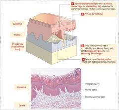
useful for resisting shear stress along the dermis
several rows of dermal papillae work up into epidermis (secondary dermal ridge demaracation) |
|
|
layers from dermis out to stratum corneum
|
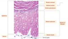
point: a continual differentiation process constantly occuring from a set of stem cells located in the stratum vasal; undergo mitosis; differentiate as they transit to surface; die by apoptosis leaving the keratin layer (protein/lipid layer)
stratum basal (stem cells); at granulosum layer you show granular elements, lamellar bodies, nucleoli flatten out; |
|
|
stratum lucidum
|

only present in thicker skin
|
|
|
stratum spinosum
|

lots of desmosome junctions along the stratum spinosum
|
|

|
thick skin (back, palmsl soles of feet); convuluted interface; lack of hair, sweat glands are deep in dermis;
|
|

|
thin skin
dense irregular connective tissue; thinner epidermis; dermis contaisn dense connective tissue; shallow epidermal ridges; thin keratin layer; hair follicles present (not in thick skin); sebaceous and sweat glands present |
|
|
keratinocyte
|

dominant cell composing stratified squamous epithelium itself;
|
|
|
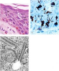
melanocytes
|
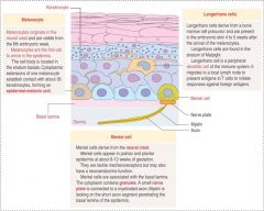
sit down on basal laminae, generate melanin, have dendritic projections out through spinosum layers; communicate with keratinocytes (epidermal-melanin unit); keratinocytes take up melanin secreted by melanocytes = mechanism of skin pigmentation
|
|
|
merkel cells
|

neural crest derived; form a nerve plate w myelinated axon at the basal lamina; form mechanoreceptors
|
|
|
langerhans cells
|

dendritic APC that survey keratinocyte layers and are focused on the basal layer; can migrate out and make it down to a lymph organ to stimulate T cells
|
|

|
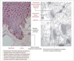
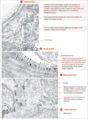
two adjacent cells linked by desmosomes showing tonofilaments projecting in towards the cell; the intracellular space expresses cadherin proteins
INTEGRINS used to attach the basal layer to the basement membrane Cadherins are higher up "spines" o fstratum spinosum are cytoplasmic extensions with desmosomes Note melanocytes at basal level, melanin granules in keratinocytes |
|

|
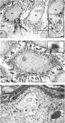
note nucleus in basal cell vs granulosum layer (bottom); cell in granulosum layer is in final stage of differentiation producing lamellar bodies and apoptotic mechanisms
|
|

|
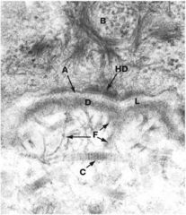
basal keratinocyte with anchoring filament; hemidesmosomes tie down basal layer to the dermis
|
|
|
bullous pemphigold proteins
|
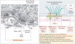
form an aggregate structure that sits down through membrane and projects into extracellular space; in combo w integrins into basal laminae;
autoimmune dz; antibodies target BP180 and BP230 disrupts hemidesmosomes -> loss of epidermal integrity = complemtn activation, mast cell & eosinophil degranulation |
|
|
L-DOPA pathway is precursor for..
|
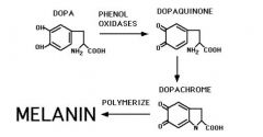
melanin & catecholamine synthesis
|
|
|
UVB
|
causes sunstan or sunburn; bleaches pre-existing melanin in keratinocytes by inducing synthesis of new melanin by melanocytes
|
|
|
induction of melanin in melanocytes via melaoncyte stimulating hormone
|

GPCR binds to stimulating hormone -> transcription factor activated (kreb) -> passed our of nucleus -> activates genes important for melanin synthesis
MITF; key player in inducing melanin synthesis; lack of functioning MITF = albinism and premature aging; also found in excess in melanoma |
|
|
Turning on MITF
|

Turns on genes to synthesis tyrosine hydroxylase important for the synthesis of melanin and catecholamines
|
|
|
Gricellae Syndrome
|
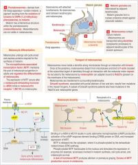
mutation in myosin 5a gene important for final transport step
|
|
Langerhans cells
|
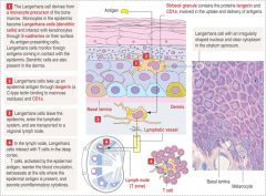
monocyte derived; no desmosomes; can leave epidermis to lymphatic system;
|
|
|
merkel cells
|
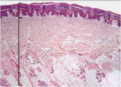
nerve "twig" cut in section at the bottom of the dermis;
|
|
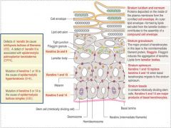
point: express different kinds of keratin as you go from the basal layer to keratin layer
|

along basal level of stratum; mutations in keratin 5 and 14 is a cause of epidermolysis bullosa simplex; in spiny layer stratum spinosum keratin 1 and 10 (mutations here = epidermolyitc hyperkeratosis); in stratum granulosum keratin 2e and 9 + protein filaggrin (= aggregation of keratin) - lemellar bodies express lipids responsible for cell envelope; defects in2e and 9 (epidermolytic palmoplantar keratoderma)
|
|
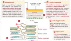
point: different components fall together as cells move upwards; retain claudin-1 and claudin-4;
|

structure of cornified cell envelope is a lipid layer on outersurface generated by lamellar bodies; beneath is involucrin; beneath in small proline rich proteins and loricrin; these are all cross linked together into a weave called transmutaminase; mutations in that causes specific diseases
|
|
|
defects in loricrin
|

vohwinkel syndrome and progressive symmetric erythrokeratodermia; lead to structural disruption of corneal layer
|
|
|

in granulosum layer lamellar bodies and keratinohyaline granules = conc aggregates of granules that are secreted and cross linked
|