Reading...
![]()
Play button
![]()
Play button
![]()
Use LEFT and RIGHT arrow keys to navigate between flashcards;
Use UP and DOWN arrow keys to flip the card;
H to show hint;
A reads text to speech;
37 Cards in this Set
- Front
- Back
|
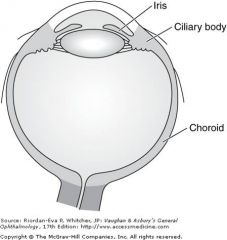
Uvea
|
Iris, ciliary body, choroid
|
|
|
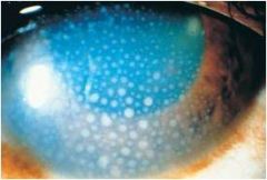
Keratic Precipitates secondary to uveitis; inflammation = increased vessel permeability = cells//exudate accumulate in anterior chamber -> inflammatory cells adhere to corneal endothelium and become visible keratic precipitates
|
|
|
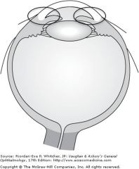
Normal uve tract (iris, ciliary bdy, choroid
|
|
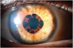
Posterior synechiae; adhesions between iris and lens
|
Anterior synechiae; adhesion between iris and cornea
|
|
|
Posterior synechiae (iris stuck to anterior surface of the lens)
|
|
|
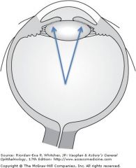
Panuveitis
|
Combo of intermediate an dposterior uveitis
|
|
|
Pt w pain, redness, photophobia, dereased vision with autoimmune dz
|
Uveitis; dx w slit lamp and indirect opthalmoscopy; can lead to vision loss if inadequately tx
|
|
|
mc secondary intraocular malignancy in adults
|
metastasis to uvea (iris, ciliary body, choroid)
|
|
|
mc primary intraocular malignancy
|
uveal melanoma no lymphatic within the eye; primary uveal melanoma tends to metastasize to liver
|
|
|
risks of uv light
|
uveal melanoma no lymphatic within the eye; primary uveal melanoma tends to metastasize to liver
|
|
|
mc spread for eye neoplasms
|
hematogenous; no lymphatic within the eye; primary uveal melanoma tends to metastasize to liver
|
|
|
uveal melanoma no lymphatic within the eye; primary uveal melanoma tends to metastasize to liver
|
|
|
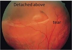
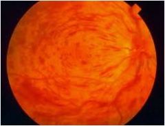
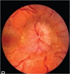
rhetamotgenous retinal detachmnet; The detached retina appears blurred in the top of the photo – notice how the blood vessels are not in focus in the picture. This is because the retina is bowed forward and the picture is two dimensional. The tear is shaped like a “horseshoe” or a “V” due to pull from the vitreous.
|
|
|
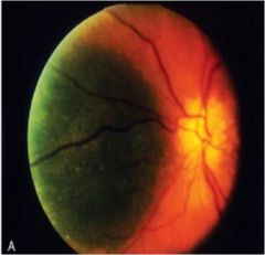
exudative retinal detachment; fluid build up behind macula
|
|
|
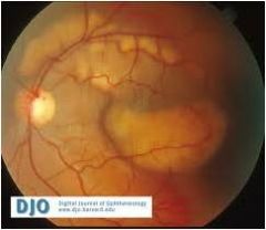
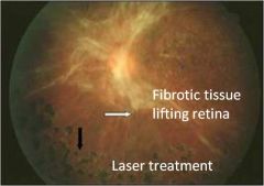
Traction retinal detachment; fibrosis pulls retina off underlying tissue
|
|
|
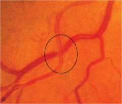
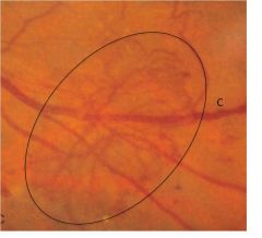
AV nicking secondary to hypertensive retinopathy -> artery compresses vein due to high pressure; venous stasis distal to this crossing may cause occlusion of the retinal vein branches
|
|
|
copper wiring secondary to htn retinopathy; thickened vessel wall alters light reflex = orange metallic appeaance; note some AV nicking
|
|
|
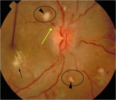
cotton wool spots, flame hemorrhage, macular star from exudates in the outer plexiform retinal layer all secondary to hypertensive retinopathy
|
|
|
hypertensive retinopathy w flame hemorrhages as well as exudates
|
|
|
Neovascularization secondary to diabetic retinopathy; microcirculatory changes lead to lack of perfusion which can cause upregulation of VEGF and retinal angiogenesis
|
|
|
Why don't you give too much oxygen to newborns?
|
Increased risk for retinopathy of prematurity (nasal perfused/temporal avascular); by administering oxygen temporal blood vessels constrict causing ischemia of the distal tissue; ischemia upreg pro-angiogenic factors such as VEGF which can result in angiogensis; signifiant contraction of the neovascular membrane = detachment of the retina
|
|
|
Sickle cell retinopathy
|
Low 02 tension in retinal periphery = sickling of rbc and vascular occlusion; can cause hemorrhages = traction/detachment or neovascularization = traction/retinal detachment
|
|
|
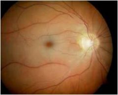
retinal artery occlusion
|
acute painless monocular loss of vision; pale retina w cherry red macula
|
|
|
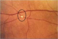
Hollenhorst plaque (fragment of atherosclerotic plaque) present in an arteriole
|
|
why the cherry red spot?
|
CRAO with “cherry red spot” – this occurs due to ischemia, edema, and whitening of the retinal layers surrounding the fovea. In the fovea, the retina is thin and consists only of photoreceptors allowing the redness of the underlying choroid to remain visible (macula has its own blood supply = choroid artery)
|
|
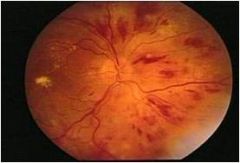
retinal vein occlusion
|
dilated/tortuous vessels in addition to diffuse flame hemorrhages; could be secondary to a thrombus or comrpression of vein at sites of AV nicking (due to atheriosclerosiss)
|
|
|
Black spots and distortion in central vision
|
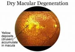
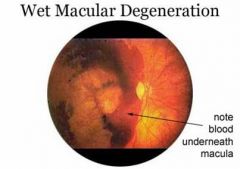
Macular degeneration
|
|
|
dry maular degeneration; black spot in vision
|
|
|
wet macular degeneration; black spot in center of vision
|
|
|
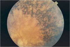
Retinitis pigmentosa is characterized by bilateral degeneration of the retina as well as the pigmented epithelium of the choroid; poor NIGHT VISION
|
|
|
Immunocompromised + aids + vision problems
|
CMV retinitis
|
|
|
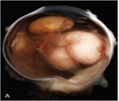
mc primary intraocular malignancy affecting children
|
retinoblastoma; neuronal cell of origin; malignancy arising from retina
60% sporadic 40% familial two hit hypothesis |
|
|
retinoblastoma
|
|
|

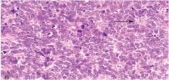
retinoblastoma histology; "flexner-wintersteiner rosettes" reflecting photoreceptor differentiation
areas of dystrophic calcifications also seen |
|
|
Papilledema; optic nerve is hyperemic and swollen; can be secondary to nerve compression of elevation of CSF pressure surrounding the nerve; pressure elevation in the nerve = venous stasis at head = swelling
bilateral papilledema = elevated intracranial pressure |
|
|
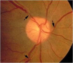
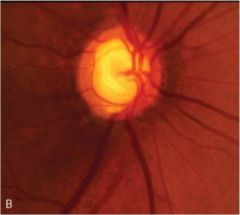
glaucomatous optic nerve damage; note increased risk ratio compared with the normal eye due to loss of ganglion cells and thinning of the retinal nerve fiber layer (disc gets bigger)
|
|
|
optic neuritis
|
usually secondary MS
|