Reading...
![]()
Play button
![]()
Play button
![]()
Use LEFT and RIGHT arrow keys to navigate between flashcards;
Use UP and DOWN arrow keys to flip the card;
H to show hint;
A reads text to speech;
16 Cards in this Set
- Front
- Back
|
Ora serrata
|
Border between the posterior 2/3 light sensitive zone (pars optica) and anterior 1/3 light-nonsensitive zone (pars ciliaris and iridica)
|
|
|
Muller cell
|
suporting neuroglial cell; supports cones, rods, bipolar and ganglion cells in the reti
|
|

|

cornea - scleral layer
|
|
|
Drusen
|
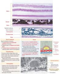
Accumulation of amyloid on the inner side of Bruch's membrane; forms an inward bulging region called drusen; early indication of macular degeneration (pushes photoreceptors away from blood supply)
|
|

|
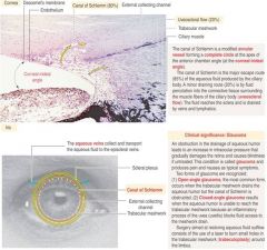
Ciliary body normal
|
|
|
Relationship of the ciliary body to glaucoma
|
|
|
|
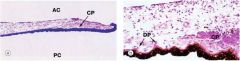
Iris; DP = dilator pupillae (radially oriented - sympathetic innervation), CP = constrictor pupillae (circumferentially oriented - parasympathetic innervation)
|
|
|

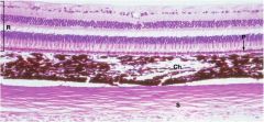
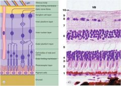
R = retina, P = pigmented epithelium, Ch = choroid, S = sclera
|
|
|
Retinal layers
|
|
|
Iodopsin
|
Photopigment in cones disks
|
|
|
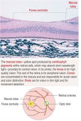
Fovea centralis
|
|
|
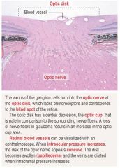
Optic disk
|
|
|
Conjunctiva; epithelium covering exposed portion of sclera; stratified columnar with goblet cells; melanocytes in basal layer
|
|
|
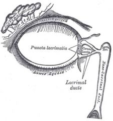
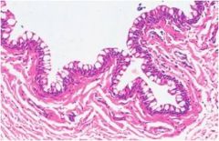
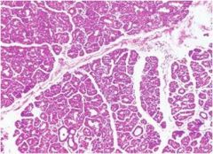
Lacrimal gland; secretion of tears which drain into nasal cavity via naso-lacrimal duct
|
|
|
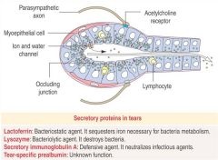
Structure of the lacrimal gland
|
|
|
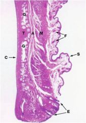
Eyelid; F = hair follicles, C = conjunctiva
|