Reading...
![]()
Play button
![]()
Play button
![]()
Use LEFT and RIGHT arrow keys to navigate between flashcards;
Use UP and DOWN arrow keys to flip the card;
H to show hint;
A reads text to speech;
67 Cards in this Set
- Front
- Back
|
Is it true that the sternal angle is part of the boundary of the thoracic inlet?
|
False
No, you are right - the thoracic inlet is actually bounded by T1 posteriorly, the superior border of the manubrium anteriorly and the 1st ribs laterally. |
|
|
Is it true that the right lung is larger than the left?
|
True
Yes. The right lung weighs about 620g and left 560g - in the average adult. |
|
|
Are the eleventh and twelfth ribs referred to as the floating ribs?
|
True
Yes - they are called floating ribs because they are short and have no attachment to the sternum. In truth, these ribs are not really floating, as they articulate with the body of their own numbered vertebra. |
|
|
Does the surface marking of the pleura extend above the clavicle?
|
True
Yes, in fact the surface marking of the pleura reaches 2-3cm above the medial third of the clavicle. |
|
|
Does the origin of the right brachiocephalic (innominate) vein lie behind the right sternoclavicular joint?
|
True
Yes. Both brachiocephalic veins are formed by the union of the internal jugular and subclavian veins each behind their respective sternoclavicular joint. |
|
|
Does the tubercle of a typical rib articulate with the transverse process of the vertebra numerically above?
|
False
Yes. The tubercle actually articulates with the corresponding transverse process. However, the head of each typical rib has two facets: one for articulation with the numerically corresponding vertebra, and the other for the vertebra above. |
|
|
Does the twelfth rib have a single facet on its head?
|
True
Yes. This is because the 12th rib, like the 10th and 11th ribs, articulates only wih its own vertebral number. Note that the 10th rib occasionally has two facets. |
|
|
Does the 12th rib have a tubercle?
|
False
Yes you are correct, the 12th rib has no angle, tubercle, or subcostal groove. In fact, it's the smallest of all the ribs. |
|
|
Does the right vagus nerve pass in front of the right lung root?
|
False
That's right - it passes at the back of the right main bronchus and gives branches to the pulmonary plexus before entering into the formation of the oesophageal plexus. |
|
|
Does the inferior angle of the scapula lie at the level of the fourth rib?
|
False
Very good! The inferior angle of the scapula actually lies at the level of the seventh rib, and is a fairly good guide to the seventh intercostal space at the back of the chest. |
|
|
Do you agree that the right upper lobe usually contains three bronchopulmonary segments?
|
True
Yes - the right lung has three upper lobe segments (apical or superior, anterior and posterior). In contrast, there are two segments in the middle lobe and five in the lower lobe. |
|
|
In a typical intercostal space, is the intercostal nerve the highest of the neurovascular structures?
|
False
Well done. From the above downwards, it is the vein, artery and then nerve - hence the intercostal nerve is the lowest neurovascular structure in an intercostal space. Note also that the bundle is sheltered under the subcostal groove for most of its course. |
|
|
Does the sinu-atrial node lie in the wall of the right atrium near the superior vena cava?
|
True
Yes. It lies in this position at the upper end of the crista terminalis, and impulses are transmitted from it through the atrial wall to the atrioventricular node. |
|
|
Does the thoracic trachea have complete fibrocartilaginous rings in its walls?
|
False
Excellent, the tracheal walls are strengthened by incomplete rings of hyaline cartilage deficient posteriorly. |
|

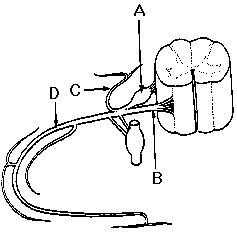
This diagram (Figure 7) shows a typical spinal nerve and the spinal cord.
Is the part of the diagram labelled "B" a spinal nerve? |
False
"B" is a ventral root, which unites with a dorsal root to form a spinal nerve. |
|

This diagram (Figure 7) shows a typical spinal nerve and the spinal cord.
Does the ventral root contain only efferent fibres? |
True
These are all efferent fibres coming from motor neurons in the ventral horn of the spinal cord. |
|

This diagram (Figure 7) shows a typical spinal nerve and the spinal cord.
Are there synapses in the swelling with the label "A"? |
True
That's right - since "A" is the spinal or dorsal root ganglion, it is made up of sensory cell bodies, not synapses. |
|

Is the structure "D" called an intercostal nerve in the thoracic region?
Is the structure "D" called an intercostal nerve in the thoracic region? |
True
" D" is the ventral ramus of a spinal nerve, running under the subcostal groove as an intercostal nerve. |
|
This diagram (Figure 7) shows a typical spinal nerve and the spinal cord.
Is "C" covered with a sheath of dura mater? |
False
Dura covers the dorsal and ventral roots only as far as the formation of the mixed spinal nerve. |
|
|
Does a grey ramus contain preganglionic sympathetic fibres?
|
False
That's right - it's the white ramus that transmits pre-ganglionic sympathetic fibres from the spinal nerve to the sympathetic trunk. Remember that after synapse, the grey ramus transmits postganglionic fibres back into the spinal nerve. |
|
|
Is it true that all spinal nerves have white rami communicantes?
|
False
Well done - the point to remember is that pre-ganglionic sympathetic fibres leave the spinal cord in spinal nerves T1-L2 only, and therefore only these nerves have white rami communicantes. |
|
|
Does the collateral branch of the fifth intercostal nerve supply sensation to skin over the 5th intercostal space?
|
False
The collateral branch of an intercostal nerve supplies the intercostal musculature. It is the lateral and anterior cutaneous branches which supply skin and subcutaneous tissue. |
|
|
Is it true that the first intercostal nerve has no anterior cutaneous branch?
|
True
Yes - the first intercostal nerve is small because most of the ventral ramus of T1 passes over the first rib to join the brachial plexus. Apart from having no anterior cutaneous branch it usually has no lateral cutaneous branch. |
|
|
Does the lateral cutaneous branch of the second intercostal nerve supply skin in the floor of the axilla?
|
True
In fact the lateral cutaneous branch of the second intercostal nerve is called the intercosto-brachial nerve. It supplies skin in the floor of the axilla, and communicates with the medial cutaneous nerve of the arm. |
|
|
Does the eighth intercostal nerve supply skin on the front of the abdomen?
|
True
Quite right - it's the 7th-11th intercostal nerves which supply abdominal skin as well as the thoracic wall. This is because they pass forwards and downwards past the anterior ends of the intercostal spaces. |
|
|
Does the neurovascular plane in the thorax lie between the internal and external intercostal muscles?
|
False
That's right - the neurovascular plane lies between the innermost and internal intercostal muscles, not between the internal and external muscles. |
|
|
Does the first left intercostal artery arise from the aorta?
|
False
The first two posterior intercostal arteries arise from the superior intercostal artery, a branch of the costo-cervical trunk of the subclavian artery. |
|
|
Does the internal thoracic artery directly supply anterior intercostal arteries to all the intercostal spaces?
|
False
Good the anterior intercostal arteries for the upper six spaces are derived from the internal thoracic artery. Below this level, a branch of this artery, the musculophrenic artery, gives off anterior intercostal vessels to the seventh to ninth spaces. Note that there are usually no anterior intercostal arteries in the lower two spaces. |
|
|
Is the suprapleural membrane attached to the transverse process of C7?
|
True
At it's lower periphery, the suprapleural membrane is attached to the margin of the first rib. |
|
|
Is the lower limit of pleura found at the level of the eighth rib in the midaxillary line?
|
False
No it is not. In fact the lower reflection of pleura crosses the eighth rib in the midclavicular line. |
|
|
Is sensation from the mediastinal pleura carried in the intercostal nerves?
|
False
No, sensation from mediastinal pleura is carried in the phrenic nerves. |
|
|
When present, is an azygos lobe of the right lung a section of the lower right lobe which extends behind the vena azygos?
|
False
Excellent. Though it is true that the azygos lobe is very occasionally found in the right lung, it develops from the upper lobe. The apcial bronchus grows medial to the arch of the azygos vein, thereby creating a deep fissure in the upper lobe. This can be seen as a vertical linear opacity on a chest radiograph. |
|
|
Does the inferior border of the left lung reach the eighth rib in the midaxillary line?
|
True
Yes - the surface markings for the lower border of both lungs run from the sixth rib in the midclavicular line to the eighth rib in the midaxillary line and the tenth rib behind.: n Sorry, but that is actually true for the inferior border of both lungs. |
|
|
Does the right principal bronchus usually divide before it enters the substance of the lung?
|
True
Yes - the right upper lobe bronchus is given off outside lung tissue and lies above the artery. For that reason, it's therefore sometimes called the eparterial bronchus. |
|
|
Does the horizontal plane at the level of the sternal angle lie at the level of the second thoracic vertebra?
|
False
No - this plane lies at the level of the fourth thoracic vertebra. |
|
|
Is the interventricular septum supplied by both coronary arteries?
|
True
The interventricular septum is supplied by both the anterior and posterior interventricular arteries. |
|
|
Is it true that the sinu-atrial node receives a blood supply from the right coronary artery more frequently than from the left?
|
True
Yes. Opinions vary as to the exact percentage, but usually a branch reaches the SA node from the right coronary artery. This sinu-atrial branch curls up around the base of the superior vena cava. Less frequently, the arterial branch arises from the circumflex artery (a branch of the left coronary artery). |
|

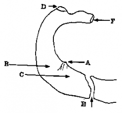
This diagram (Figure 8) shows the upper surface of the first rib.
Does scalenus anterior insert into the section of rib marked "D"? |
False
Spot on! Scalenus anterior inserts into the scalene tubercle ("A") not the tubercle of the rib "D". |
|
This diagram (Figure 8) shows the upper surface of the first rib.
Does the subclavian vein pass over the first rib at "C"? |
True
Yes - the vein passes over the rib in front of scalenus anterior. |
|

This diagram (Figure 8) shows the upper surface of the first rib.
Is the joint between the first costal cartilage and the manubrium "E" a secondary cartilaginous joint? |
False
That's right - this joint is of the immobile, primary cartilaginous variety. Remember a secondary cartilaginous joint has fibrous tissue at the joint as well as cartilage and they occur in the midline of the body; how many can you think of? |
|

This diagram (Figure 8) shows the upper surface of the first rib.
Does the area of the rib marked "F" articulate with the vertebral bodies of C7 and T1? |
False
The first rib articulates only with T1. |
|
|
Is it true that most of the first thoracic ventral ramus passes over the first rib?
|
True
Yes it is. This nerve bundle, together with the subclavian artery, passes over the first rib through the scalene triangle. |
|
|
Do you agree that a newborn child with an atrial septal defect (ASD) is likely to be cyanosed?
|
False
Very well done! This condition results in a left-to-right shunt, with oxygenated blood coming into the left atrium from the lungs entering the right atrium through the defect because the left-sided pressure is higher than the right. Hence there is no cyanosis. However, if the condition progresses to heart failure the opposite shunt may occur, and the child will be cyanosed. |
|
|
Does the condition of the tetralogy of Fallot result in cyanosis?
|
True
Yes and just to remind you, these are the four features of the tetralogy: (1) a ventricular septal defect (2) an enlarged right ventricle (3) a narrowing of the outflow from the right ventricle (pulmonary stenosis) (4) an aorta which overrides both ventricles. |
|
|
Is a barium swallow of help in the diagnosis of an enlarged left atrium in a patient with mitral stenosis?
|
True
The enlarged left atrium would be seen to indent the shadow of the oesophagus on a lateral or oblique chest film. |
|
|
Is it true that the greater, lesser, and least splanchnic nerves enter the cardiac plexus?
|
False
Good, (but you should really know this anyway!) These nerves descend through the posterior thoracic wall, and pierce the crura of the diaphragm to reach the coeliac and aortic plexuses. |
|
|
Does a branch of the brachiocephalic artery supply the thymus gland?
|
False
The thymus is usually supplied by the internal thoracic or inferior thyroid artery. |
|
|
Does the transverse sinus of the pericardium lie between the aorta and pulmonary trunk
|
False
The transverse sinus lies behind both the aorta and the pulmonary trunk. |
|
|
Do you agree that the oblique pericardial sinus is a pericardial recess behind the left atrium?
|
True
The oblique pericardial sinus is surrounded by the inferior vena cava and pulmonary veins as they penetrate the pericardium. |
|
|
Would cutting the right phrenic nerve in the neck cause the right dome of the diaphragm to fall?
|
False
In fact the dome descends when the diaphragm contracts, so cutting or crushing the phrenic nerve would cause the diaghragm to stay raised. |
|
|
Are the phrenic nerves the only motor nerves to the diaghragm?
|
True
Good, yes they are! They also carry proprioceptive fibres from the diaphram, plus sensory fibres from the peritoneum in the upper abdomen and from mediastinal pleura. |
|
|
Does the smooth walled part of the right atrium develop from the embryonic sinus venosus?
|
True
Yes - and for another gold star, remember that the part of the wall covered with musculi pectinati develops from the primitive atrium. |
|
|
Do you agree that the superficial cardiac plexus is located at the root of the superior vena cava?
|
False
That's right in fact, the superficial part of the plexus is related to the concavity of the aortic arch, and the deep part is found in front of the tracheal bifurcation. |
|
|
Is the left recurrent laryngeal nerve related to the ligamentum arteriosum?
|
True
Yes - the left recurrent laryngeal nerve lies on the left side of this structure and passes under the arch of the aorta before going up the groove between the trachea and oesophagus. |
|
|
Is it true that the interior of the right auricular appendage is lined by trabeculae carneae?
|
False
That is the right answer. This type of muscular ridge is found in the ventricles. Note that the auricular appendage on the right is ridged by musculi pectinati, reflecting its origin from the primitive atrium. |
|
|
Is it true that there is a valve at the opening of the inferior cava into the right atrium?
|
True
This is a functionless embryological remnant in adults. |
|
|
Do you agree that the right principal bronchus is both wider and more vertical than the left?
|
True
Yes and it's for that reason that foreign bodies (peanuts, for instance) enter it more frequently than the left bronchus. |
|
|
Is it true that venous blood from the breast can drain to the vertebral venous plexuses?
|
True
The point is that perforating veins from the breast pass through the intercostal spaces to the internal thoracic vein and intercostal veins. From there, blood flows to the vertebral venous plexuses via the intercostal and spinal veins. The clinical importance of this is that cancer cells can also take this path and can seed in a vertebral body. |
|
|
Is the apex beat usually found in the fourth left intercostal space 4.5 inches from the midline?
|
False
Not quite, partly because we are all different sizes! The apex beat is normally found in the 5th space on the left, medial to the mid-clavicular line (we don't use "inches" to locate it). |
|
|
Is it true that communications exist between the bronchial and pulmonary circulations?
|
True
That is right it is true. Some of the bronchial blood is collected in bronchial veins, but the rest drains into pulmonary veins. |
|
|
Does the axillary tail of Spence refer to a string of lymph nodes along the lower border of the pectoralis minor?
|
False
No - in fact the axillary tail of Spence is a prolongation of breast tissue into the axilla. |
|
|
Does the right atrioventricular valve have right, left and posterior cusps?
|
False
Splendid - these are actually the names of the valve cusps of the aorta (anterior, left and right posterior in alternative nomenclature). In contrast, the tricuspid valve has anterior posterior and septal cusps. |
|

This illustration (Figure 9) shows the diaphragm and some of its relations.
Would the line marked "A" be on the same level as the manubriosternal joint which lies anteriorly? |
True
|
|

This illustration (Figure 9) shows the diaphragm and some of its relations.
Is the opening marked "B" found at the level of T12? |
False
Clever - "B" is the oesophageal hiatus which is found at the level of T10. |
|

Is the opening marked 'B" associated with a muscular sling?
|
True
True in fact, a muscular sling from the right crus of the diaphragm is one mechanism for preventing reflux. |
|

This illustration (Figure 9) shows the diaphragm and some of its relations.
Does the thoracic duct pass from the abdomen to the thorax with the structure labelled "C"? |
True
The structure "C" is the aorta, which passes through the aortic opening of the diaphragm. |
|

This illustration (Figure 9) shows the diaphragm and some of its relations.
Does the sympathetic trunk pass behind the lateral arcuate ligament ("D" in this diagram)? |
False
It is the medial ligament that the trunk passes behind - "D" has the subcostal neurovascular bundle behind it. |