Reading...
![]()
Play button
![]()
Play button
![]()
Use LEFT and RIGHT arrow keys to navigate between flashcards;
Use UP and DOWN arrow keys to flip the card;
H to show hint;
A reads text to speech;
206 Cards in this Set
- Front
- Back
|
The mammary gland is classified as what type of gland?
|
Mammary glands are modified sweat glands and therefore have no special capsule or sheath. p. 56
|
|
|
The xiphoid process is a midline marker for what structures?
|
It is a midline marker for the superior level of the liver, the central tendon of the diaphragm, and the inferior border of the heart. p. 53
|
|
|
The left bronchial arteries arise from?
|
The left bronchial arteries (superior & inferior) usually arise directly from the thoracic aorta.-web
|
|
|
The right bronchial arteries arise from?
|
The single right bronchial artery usually arises from one of the following:
-the thoracic aorta at a common trunk with the right 3rd posterior intercostal artery -the superior bronchial artery on the left side -a superior posterior intercostal artery |
|
|
What is the purpose of the fibrous pericardium?
|
It is a dense connective tissue, protecting the heart, anchoring it to the surrounding walls, and preventing it from overfilling with blood. p. 82
|
|
|
What is auscultation?
|
listening to the internal sounds of the body, using a stethoscope. p. 75
|
|
|
What are the superior boundaries of mediastinum?
|
superior mediastinum - extends inferiorly from the superior thoracic aperature to the horizontal plane, passing through the sternal angle and the IV disc of the T4-T5 vertebra. p. 81
|
|
|
What are the inferior boundaries of mediastinum?
|
inferior mediastinum is sub-divided by the pericardium into:
anterior mediastinum - lies btween the body of the sternum and the transverse thoracic muscles anteriorly and the pericardium posteriorly (it contains remnants of the thymus, lymph nodes, fat, and connective tissue) middle mediastinum - containing the pericardium, heart, roots of the great vessels, SVC, pulmonary trunk, arch of azygos vein, and main bronchi posterior mediastinum - containing the espophagus thoracis arota, azygos and hemiazygos veins, thoracics duct , vagus nerves, sympathetic trunks, and splachnic nerves. p. 81 |
|
|
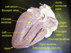
Identify the chambers of the heart.
|
there are 4 chambers:
right and left atria right and left ventricles p. 83 |
|
|
What is the fibrous skeleton of the heart?
|
the structure of dense connective tissue in the heart that separates the atria from the ventricles.
|
|
|
Where is the fibrous skeleton of the heart located?
|
The majority of the skeleton lies within the plane of the base of the ventricles, roughly parallel to the coronary sinus. -web
|
|
|
The wall of each chamber of the heart consists of what three layers?
|
epicardium
myocardium endocardium p. 87 |
|
|
What is epicardium(mesothelium)?
|
The outer layer of the heart and is surrounded by a small amount of fluid enclosed by a fibrous sac called the pericardium
|
|
|
What is myocardium?
|
myocardium, the muscular tissue responsible for the contraction of the heart.
|
|
|
What is endocardium(endothelium)?
|
innermost layer of tissue that lines the chambers of the heart that also covers its valves. p. 87
|
|
|
What are the 5 basic parts of the thorax?
|
- Inlet
- Thoracic Wall - Pleural Cavity - Mediastinum - Outlet |
|
|
What are the contents of the mediastinal thoracic cavity?
|
Central compartment of thorax containing everything except the lungs (heart, thoracic parts of the great vessels, thoracic parts of the trachea, esophagus, thymus and other structures) between the pulmonary cavities
|
|
|
what are the contents of the pericardial thoracic cavity?
|
-walled fribroserous membrane that encloses the heart and roots of its great vessels.
|
|
|
where is the pericardial thoracic cavity located?
|
- lies posterior to body of sternum and 2nd-6th costal cartilages at level T5-T8 vertebrae
- continuous with central tendon |
|
|
What are the bones and cartilages that make up the bony thorax?
|
•Sternum
•24 Ribs •Costal Cartilage •12 Thoracic vertebrae |
|
|
What are the boundaries of the inlet of thorax (superior thoracic aperture)
|
- Posteriorly: T-1
- Laterally: first pair of ribs and their costal cartilage - Anteriorly: superior border of the manubrium of sternum |
|
|
what are the boundaries of the outlet of thorax?
|
Bounded of the outlet of the thorax are:
- T12 - ribs 11 and 12 - costal cartilages 7-10 - xiphisternal junction |
|
|
what are the intrinsic muscles of the thoracic wall?
|
- Internal intercostals: aid in forced expiration
- External intercostals: lift rib cage, aid in normal inspiration: aid the diaphragm - Inner most intercostal - Transverse thoracic: weakly depresses ribs - Subcostal: probably act in same manner as internal intercostal muscles - Levator costarum: elevate ribs - Serratus posterior superior: elevate ribs - Serratus posterior inferior: depress ribs |
|
|
what vessels form the coronary circulation?
|
Coronary circulation is the circulation of blood in the blood vessels of the heart muscle. Although blood fills the chambers of the heart, the muscle tissue of the heart (the myocardium) is so thick that it requires coronary blood vessels to deliver blood deep into it. The vessels that deliver oxygen-rich blood to the myocardium are known as coronary arteries. The vessels that remove the deoxygenated blood from the heart muscle are known as coronary veins.
|
|
|
what are the 3 types of arteries?
|
Systemic arteries can be subdivided into two types - muscular and elastic.
Systemic arteries deliver blood to the arterioles, and then to the capillaries, where nutrients and gasses are exchanged. - large elastic - Large lumen, great elasticity - medium muscular arteries-Active in vasoconstriction (further from heart) Arterioles - the smallest of the true arteries, help regulate blood pressure by the variable contraction of the smooth muscle of their walls, and deliver blood to the capillaries. |
|
|
what are large elastic arteries?
|
an artery with a large number of collagen and elastin filaments in the tunica media, which gives it the ability to stretch in response to each pulse
|
|
|
what are the medium muscular arteries?
|
- walls contain mostly smooth muscle
- able to decrease their diameter (vasoconstrict) to regulate blood flow to different parts of the body as required (femoral artery) |
|
|
what are small arteries and arterioles?
|
relatively narrow lumina and thick muscular walls
|
|
|
what are the two types of hemorrhoids?
|
Internal and External
|
|
|
Why do internal hemorroids occur?
|
Internal hemorrhoids occur due to a prolapse of the rectal mucosa around the internal venous plexus. Because these occur superior to the pectinate line, there is less pain associated with them than with external
|
|
|
why do external hemorrhoids occur?
|
External hemorrhoids are blood clots of the external venus plexus that are covered by skin.
A common cause of hemorrhoids is simply the standing position, in which all the blood above the rectum exerts pressure on the rectal and anal areas. Other conditions which contribute to hemorrhoids are: poor bowel habits, constipation, diarrhea, pregnancy, obesity, and especially frequent straining when having a bowel movement External hemorrhoids are prone to thrombosis: if the vein ruptures and/or a blood clot develops, the hemorrhoid becomes a thrombosed hemorrhoid |
|
|
what is the arterial supply to the perineum?
|
Internal pudendal artery and vein (branch off internal iliac artery)
perineum was supplied by the urogenital and internal pudendal arteries. The urogenital artery originated from the internal iliac artery near the ischiatic spine and after a short course to the lateral surface of the vagina, it divided into cranial and caudal branches. (?) |
|
|
what do capillary beds connect?
|
arteries and veins
|
|
|
When numbering the ribs what is the first landmark to identify before you start counting?
|
from the sternal angle onto the 2nd costal cartilage
the 1st rib cannot be palpated because it lies deep to the clavicle, so count the ribs and intercostal spaces by sliding the fingers laterally from the sternal angle onto the 2nd costal cartilage.p. 67 |
|
|
What is the innervation of the visceral pleura?
|
the Autonomic Nervous system and therefore has NO PAIN RECEPTORS
The parietal pleura is highly sensitive to pain while the visceral pleura is not, due to its lack of sensory innervation |
|
|
what is the innervation of the parietal pleura?
|
the Intercostal Nerves and it DOES have pain receptors
|
|
|
What is the relationship of the heart ventricles to the sternum?
|
- Right Ventricle lies just posterior to the Sternum at the level of the 4th and 5th intercostal spaces
- Left ventricle (mitral valve) is located posterior to the sternum at the level of the 4th costal cartilage |
|
|
What is the direct venous drainage of the thoracic wall?
|
azygous veins
|
|
|
Where does the main thoracic lymphatic duct empty into the venous system?
|
In the Venous system near union of the left internal jugular and subclavian veins, left venous angle of origin of left brachiocephalic vein
|
|
|
What prevents the first rib from being palpated?
|
It lies behind the clavicle
|
|
|
Be able to give, in order, the pathway that sperm would travel through the male duct system into the urethrae.
|
The testes(1) are where sperm(2) are manufactured in the scrotum. The epididymis(3) is a tortuously coiled structure topping the testis, and it receives immature sperm from the testis and stores it several days. When ejaculation occurs, sperm is forcefully expelled from the tail of the epididymis(4) into the deferent duct(5). Sperm then travels through the deferent duct through up the spermatic cord(6) into the pelvic cavity, over the ureter to the prostate behind the bladder. Here, the vas deferens(7) joins with the seminal vesicle(8) to form the ejaculatory duct, which passes through the prostate and empties into the urethra.
When ejaculation occurs, rhythmic muscle movements propel the sperm forward. Seminal vesicles is where fluid contributes to sperm to produce semen |
|
|
What three layers make up the wall of the uterus?
|
Perimetrium -outer serous layer
Myometrium - the thickest layer, which is smooth muscle capable of powerful contractions Endometrium - (lining) inner glandular layer that is shed during menses |
|
|
Whats a true rib?
|
true ribs are 1-7 (vertebrocostal) first 7 pairs of ribs
-attach directly to the thoracic vertebra, and by costal cartilage to the sternum |
|
|
whats a false rib?
|
false ribs are 8-10 (pointed at its end)
-8-10 (vetebrochondral) indirectly attaches to the sternum by way of the 7th costal cartilage (video) |
|
|
whats a floating rib?
|
floating ribs 11-12
11-12 (vetebral) do not have anterior attachment. the 12th is sometimes removed in cosmetic surgery to make a smaller waist -no costal attachment |
|
|
what is the sternal angle?
|
- where manubrium joins the body
- second rib attaches at this angle |
|
|
what are the 3 parts of the sternum?
|
- Manubrium
- Body - xiphoid process |
|
|
What is the significance of the sentinel lymph node in breast cancer?
|
it is the first lymph node and is the lymph node that helps sound the warning that the cancer has spread.
it decreases unnecessary lymph node dissections where this is not necessary, thereby reducing the risk of lymphedema |
|
|
What is the origin of the azygos and the hemiazygos venous system?
|
The azygos system of veins is considered to be the azygos vein, along with its left-sided counterparts, the hemiazygos vein and the accessory hemiazygos vein. Together, they form an anastomosis between the superior vena cava to the inferior vena cava.
|
|
|
Where are azygos vein and the accessory hemiazygos connected?
|
The right bronchial vein drains into the azygos vein and the left bronchial vein drains into the accessory hemiazygos vein or the left superior intercostal vein (not sure)
|
|
|
Arteries receive blood directly from the heart. True or False
|
True
|
|
|
Veins carry blood towards the heart, True or False
|
True
|
|
|
Veins have thick and elastic muscular walls. True or False
|
False - Arteries have thick and elastic muscular walls
|
|
|
veins carry bluish-red de-oxygenated blood (exception: pulmonary veins which carry oxygenated blood from the lungs to the heart) True or False
|
True
|
|
|
While they both carry blood, they do not have much else in common, what are they?
|
Arteries and Veins
|
|
|
Which are true of veins?
a. are flexible b. collapse when not filled with blood c. carry de-oxygenated blood d. all the above e. a and b |
d. all the above
|
|
|
what is gaseous exchange of the lungs?
|
Gaseous exchange is the movement of oxygen into the body, and carbon dioxide out of the body.
|
|
|
Where is the primary location of gaseous exchange in the lungs?
|
The gaseous exchange takes place in the lungs by diffusion through the alveolar surface.
|
|
|
What is coronary circulation?
|
the circulation of blood in the blood vessels of the heart muscle.
|
|
|
Be able to identify the muscles that form the coronory circulation.
|
-the major vessels of the coronary circulation are the left main coronary that divides into left anterior descending
-and circumflex branches, and the right main coronary artery. The left and right coronary arteries originate at the base of the aorta from openings called the coronary ostia located behind the aortic valve leaflets. (still searching) The left and right coronary arteries and their branches lie |
|
|
What are the components of a typical rib?
|
Shaft
Costal Groove Head Neck tubercle Angle |
|
|
The bony thoracic or thoracic cage is made up of what?
|
Sternum
12 pairs of ribs 12 throacic vertebrae |
|
|
the xiphoid process does not articulate with the ribs. True/False
|
True
|
|
|
what are the two openings to the thorax?
|
Inlet - structures enter from the head and neck
Outlet - thru the muscular diaphragm |
|
|
the manubrium articulates with the 2nd rib. True/False
|
False - the manubrium articulates with the clavicle & 1st rib.
|
|
|
what is the clinical signifance of the linea alba (white line)?
|
exploratory surgery is performed on this line because it is a bloddless and nerveless line
|
|
|
what forms by the fascia of the three abdominal muscles?
|
the inguinal ligament (external & internal abdominal oblique & transverse abdominal)
|
|
|
what are the three parts of the small intestine? (in order)
|
Duodenum
Jejunum Illium |
|
|
ribs 3 - 9 are typical ribs? true/false
|
true - each have a head, neck, tubercle, body, angle, and costal groove
|
|
|
Be able to identify the divisions, sphincters, and lining of the stomach.
|
Divisions:
-Cardia of Stomach -Fundus (means lower part of a hollow organ) -Pylorus Lining - Rugi (folds) Sphincters - Two smooth muscle valves, or sphincters, keep the contents of the stomach contained. They are the esophageal sphincter (found in the cardiac region) dividing the tract above, and the Pyloric sphincter dividing the stomach from the small intestine. |
|
|
Name the mesentery, which attaches from the lesser curvature of the stomach to the hilus of the liver?
|
Lesser omentum (Hepatogastric ligament & hepatoduodenal ligaments) connects the lesser curvature of the stomach and the proximal end of the duodenum to the liver.p.139
|
|
|
What three structures run in the free margin of this lesser omentum (messentary)?
|
the portal triad:
Portal vein Hepatic artery Bile duct |
|
|
In relationship to blood vessels what does the term anastomose mean?
|
Arteries often anastomose (communicate) to form networks, which ensures a continuous blood supply pg. 16
anastomose - the connection of two structures |
|
|
. Be able to give a definition of a portal system. (The body has two portal systems – hepatic and hypophyseal(pertaining to the hypophysis (pituitary body)
|
the hepatic portal system is the system of veins comprised of the hepatic portal vein and its tributaries. It is also called the portal venous system
The portal venous system is responsible for directing blood from parts of the gastrointestinal tract to the liver. Substances absorbed in the small intestine travel first to the liver for processing before continuing to the heart The hypophyseal portal system is the system of blood vessels that link the hypothalamus and the anterior pituitary in the brain. It allows endocrine communication between the two structures. |
|
|
What structure attaches to the greater curvature of the stomach?
|
Greater Omentum
|
|
|
Be able to identify the structures, which form the stomach bed.
|
from superior to inferior:
left dome of the diaphragm spleen left kidney suprarenal gland splenic artery pancreas transverse mesocolon colon p. 146 |
|
|
With blockage of the superior mesenteric artery what is the significance of the marginal artery supplying the intestines
|
• The significance of the marginal artery supplying the small intestines is that it connects the Inferior Mesenteric Artery (IMA) with the Superior Mesenteric Artery (SMA) thereby acting as a continuous anastomic channel which may provide important collateral circulation.
p.159 text, wikopedia |
|
|
What is the clinical significance of the Z-line at the esophageal-gastric junction
|
Prevents gastric reflux. p. 142
|
|
|
Define a mesentery & What is its importance?
|
It exists to sustain and support certain portions of the small intestine from the abdomen. The mesentery consists of parts of the peritoneum. The function of the mesentery is to project and shelter nerves and blood vessels from the central system to the organ.
A specialized portion of the dorsal mesentery supports the large intestine and is called the mesocolon, and contains the blood vessels supplying the large intestine. Importance: Having these important structures encased in the strong tissue of the mesentery allows these organs to move more freely within the abdominal cavity. As a result, the brain does not map sensation to these organs very well and often interprets any sort of pain as coming from the midline of the abdomen, causing the occurrence of referred pain. |
|
|
Define a peritoneal ligament
|
A peritoneal ligament consists of a double-layer of peritoneum that connects an organ with another organ or to the abdominal wall. Ex. the liver is connected to the anterior abdominal wall by the falciform ligament. p.138
|
|
|
Define an Omentum
|
A double- layered extension of the peritoneum passing from the stomach and proximal part of the duodenum to adjacent organs.
Two types: -Greater Omentum – Extends superiorly, laterally to the left, and inferiorly from the greater curvature of the stomach and proximal part of the duodenum. -Lesser Omentum (hepatogastric and hepatoduodenal ligaments) – connects the lesser curvature of the stomach and the proximal part of the duodenum to the liver.p.138,9 |
|
|
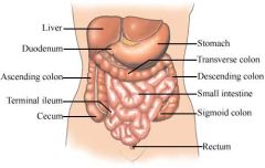
Identify the subdivisions of the small intestine
|
The small intestine consisting of the duodenum, jejunum, and ileum, extends from the pylorus of the stomach to the ileocecal junction, where the ileum joins the cecum, the first part of the large intestine.
-Duodenum – The first and shortest part of the small intestine, is also the widest and most fixed part. Begins at the pylorus on the right side and ends at the duodenojejunal junction on the left side. -Jejunum & Ileum – The Jejunum begins at the duodenojejunal junction and the ileum ends at the ileocecal junction, the union of the terminal ileum and the cecum.p. 152 |
|
|
What is the action of the sympathetic and parasympathetic nervous system on the intestine
|
-Sympathetic(fight or flight) – In general, sympathetic stimulation reduces motility of the intestine, and secretion and acts as a vasoconstrictor, reducing or stopping digestion and making blood (and energy available for “fleeing or fighting.”
-Parasympathetic(rest and digest) – increases motility of the intestine and secretion, restoring digestive activity after a sympathetic reaction.p.. 155 |
|
|
What are the subdivisions of the large intestine?
|
The large intestine consists of :
cecum colon (ascending, transverse, descending, and sigmoid) rectum anal canal |
|
|
What is the Cecum and where does it lie?
|
Cecum – The first part of the large intestine that is continuous with the ascending colon, is a blind intestinal pouch in the right lower quadrant where it lies in the iliac fossa inferior to the junction of the terminal ileum and cecum.p 155
The cecum is usually almost entirely enveloped by peritoneum and can be lifted freely; owever, the cecum has no messentery. |
|
|
What is the Colon and its parts and where do they lie?
|
Colon – The colon is described as having 4 parts – ascending, transverse, descending, and sigmoid –that succeed one another in an arch.
-Ascending colon – passes superiorly on the right side of the abdominal cavity from the cecum to the right lobe of the liver, where it turns to the left as the right colic flexure. -Transverse colon – the largest and most mobile part of the large intestine, crosses the abdomen from the right colic flexure to the colic flexure, where it bends inferiorly to become the descending colon. -Descending colon – passes retroperitoneally from the left colic flexure into the left iliac fossa, where it is continuous with the sigmoid colon. -Sigmoid colon – characterized by its S-shaped loop of variable length, links the descending colon and the rectum. The sigmoid colon extends from the iliac fossa to the third sacral segment. -Rectum & Anal Canal – The rectum is continuous with the sigmoid colon at the level of the S3 vertebra. The rectum is continuous inferiorly with the anal canal.p.155-8 |
|
|
Be able to give the relationship of the spleen to the Costodiaphragmatic recess.
|
Costodiaphragmatic recess – associated posteriorly with the spleen. p159
|
|
|
Be able to give the relationship of the spleen to the gastrosplenic ligament.
|
Gastrosplenic ligament – Spleen contacts the posterior wall of the stomach and is connected to its greater curvature by the gastrosplenic ligament. (sas: attaches spleen to stomach) p.159
|
|
|
Be able to give the relationship of the spleen to the splenorenal ligament.
|
Splenorenal ligament – attaches spleen to left kidneyp. 159
|
|
|
Give the relationship of the spleen to the Hilum.
|
Hilum – Where the splenic branches of the splenic artery and vein enter and leave the spleen. The hilum is often in contact with the tail of the pancreas and constitutes the left boundary of the omental bursa. p159
|
|
|
Give the relationship of the spleen to the Portal Vein.
|
Portal vein – The splenic vein unites with the Superior mesenteric vein posterior to the neck of the pancreas to form the portal vein. p.159
|
|
|
What are the three (text lists 4)identifying characteristics of the large intestine that distinguishes it from the small intestine?
|
1. Teniae coli – three thickened bands of longitudinal muscle fibers.
2. Haustra – sacculations or pouches of the colon between the teniae. 3. Omental appendices – small, fatty appendices (projections) of the colon. 4. Caliber – the internal diameter is much larger. p. 155 |
|
|
What is the importance of intestinal gutters?
|
Dr. S answer: The gutters run along the intestines, clinaclly important in that if their were a rupture of the gall bladder its contents would essentially use the intestinal gutters kind of like a canal system to spread throughout the abdominal cavity.
Druggan answer: Gutters are important for the absorption of gas. |
|
|
What are the divisions of the pelvic bone?
|
-Right and Left hip bones – two large, irregularly shaped bones, each of which forms at puberty by fusion of three bones – ilium, ishium, and pubis.
-Sacrum – Formed by the fusion of 5 originally separate sacral vertebra |
|
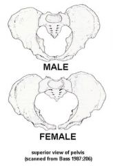
Be able to iddentify the structural difference btwn the male & female pelvic bone.
|
General Structure
M-Thick & Heavy F-Thin & Light (see chart on p.207) |
|
|
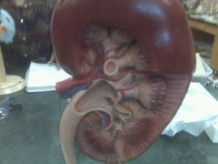
Be able, by description, to identify these internal structures of the kidneys:
Renal pyramid, Major calyx & Minor calyx |
Renal pyramid – cone-shaped tissues of the kidney. Each minor calyx is indented by the apex of the renal pyramid - the renal papilla.
Major calyx - the renal pelvis is formed through the merging of two or three major calices, each of which was formed by the merginng of two or three minor calices. |
|
|
Be able, by description, to identify these internal structures of the kidneys:
renal papilla & renal columns |
-Renal papilla – forms the apex of the renal pyramid.
-Renal columns – The renal column (or Bertin column, or column of Bertin) is a medullary extension of the renal cortex in between the renal pyramids. It allows the cortex to be better anchored. Each column consists of lines of blood vessels and urinary tubes and a fibrous material. |
|
|
Be able, by description, to identify these internal structures of the kidneys:
Renal cortex & Ureter |
Renal cortex – The renal cortex is the outer portion of the kidney between the renal capsule and the renal medulla. In the adult, it forms a continuous smooth outer zone with a number of projections (cortical columns) that extend down between the pyramids. It contains the renal corpuscles and the renal tubules except for parts of the loop of Henle which descend into the renal medulla. It also contains blood vessels and cortical collecting ducts.The renal cortex is the part of the kidney where ultra filtration occurs.
Ureter – muscular ducts with narrow lumina that carry urine from the kidneys to the urinary bladder. |
|
|
Identify the three places where the ureter is constricted.
|
1.At the junction of the ureters and the renal pelvis
2.Where the ureters cross the brim of the pelvic inlet 3.During their passage through the wall of the urinary bladder p.176 |
|
|
What is the relationship of the suprarenal (adrenal) glands to the kidneys and the diaphragm
|
The suprarenal glands are located betwn. the superomedial aspects of the kidneys and the diaphragmatic crura, where they are surrounded by connective tissue containing considerable perinephric fat. The glands are enclosed by renal fascia by which they are attached to the crura of the diaphragm; however, they are separated from the kidneys by fibrous tissue.
|
|
|
Where do the blood vessels to the suprarenal glands originate?
|
The suprarenal arteries arise from three sources.
1.Superior suprarenal arteries ( 6 to 8 ) from the inferior phrenic artery. 2.Middle suprarenal arteries ( 1 or more) from the abdominal aorta near the origin of the superior mesenteric artery (SMA). 3.Inferior suprarenal arteries (1 or more) from the renal artery. p.177 |
|
|
What is the difference btwn. the renal sinus and the renal pelvis?
|
-renal sinus -the main cavity of the kidney that is an expansion behind the hilum and contains the renal pelvis, calyxes, and the major renal vessels
-The renal pelvis is the funnel-like dilated proximal part of the ureter in the kidney. It is the point of convergence of two or three major calyces. Each renal papilla is surrounded by a branch of the renal pelvis called a calyx. The major function of the renal pelvis is to act as a funnel for urine flowing to the ureter. The renal pelvis is the location of several kinds of kidney cancer. |
|
|
What is the relationship of the renal fascia and fat to the kidney?
|
-Perinephric fat (parirenal fat capsule) - surrounds the kidneys and suprarenal glands and is continuous with the fat in the renal sinus.
-Renal fascia – the kidneys, suprarenal glands, and perinephric fat surrounding them are enclosed (except inferiorly) by a membranous layer of renal fascia. Inferiormedially, the renal fascia is prolonged along the ureters as periureteric fascia. -Paranephric fat (pararenal fat body) – External to the renal fascia. The extraperitoneal fat of the lumbar region that is most obvious posterior to the kidney. |
|
|
labeling exercises link (choose a chapter then choose "labeling exercise")
chapter 19 - respiratory chapter 17 - large/small intestine, gall bladder, liver chapter 15 - cardiovascular |
http://highered.mcgraw-hill.com/sites/0072919329/student_view0/chapter19/labeling_exercises.html#
|
|
|
.
|
.
|
|
The diaphragm is innervated by the phrenic nerve. True/False
|
True
|
|
|
What are the internal boundaries of the abdominal cavity?
|
The abdominal wall is composed of four paired muscles:
1.internal obliques 2.external obliques 3.transversus abdominis 4.rectus abdominis ...their fasciae, and their aponeuroses |
|
|
The bile, stored in the gallbladder, primarily breaks down what type of food materials?
|
Carbohydrates, Fats, and/or Proteins
|
|
|
Identify the main muscles of the posterior abdominal wall.
|
-Iliacus lies in the Iliac fossa
-Psoas Major lies along the vertebra from T-12 to L-5 -Quadratus Lumborum lies above iliacus and lateral to the superior portion of the psoas major |
|
|
80. What is the landmark for the division of the true pelvis from the false pelvis?
|
Pelvic Brim
|
|
|
Which of the male reproductive glands makes the largest contribution to semen?
|
Seminal Gland
|
|
|
In the male this gland can obstruct the urethra.
|
Prostate Gland
|
|
|
What are the bones that form the pelvic girdle?
|
Ilium, ischium, pubis, sacrum, coccyx
|
|
|
What is the importance of the subpubic angle?
|
Can determine pelvis of male or female. Greater angle in females
|
|
|
What muscles form the pelvic diaphragm?
|
What muscles form the pelvic diaphragm?
Coccygeus Puborectalis (medial), Pubococcygeus (intermediate) Iliococcygeus (lateral) |
|
|
Where does fertilization normally take place?
|
Ampulla
The ampulla is the second portion of the uterine tube. It is an intermediate dilated portion, which curves over the ovary. It is the most common site of human fertilization. |
|
|
What structure during embryonic development ran in the ligamentum teres of the liver?
|
Left umbilical vein
|
|
|
What is the vesicle trigone and what is its importance?
|
is a smooth triangular region of the internal urinary bladder formed by the two ureteral orifices and the internal urethral orifice
The area is very sensitive to expansion and once stretched to a certain degree, the urinary bladder signals the brain of its need to empty. The signals become stronger as the bladder continues to fill. |
|
|
Be able to identify a structure in the pelvic region by a description of the surrounding anatomical structures
|
|
|
|
What is the clinical significance of the apex of the lung?
|
-When observing the positioning of the lungs in the thoracic cavity you will note the Apex of the lung extends beyond the thoracic cavity lying ABOVE the clavicle and 1st ribs (cupula)
-This is clinically important since injury to the Root of the Neck (knife, bullet etc) could puncture the lung resulting in collapse of that lung |
|
|
What is an ectopic pregnancy? Identify the most common area(s) of the body where they can be located.
|
a complication of pregnancy in which the fertilized ovum is developed in any tissue other than the uterine wall
Most ectopic pregnancies occur in the Fallopian tube |
|
|
Be able to identify the following branches of the internal iliac artery
|
?
|
|
|
Identify the divisions of the uterine tube.
|
-Isthmus-portion connected to the uterus
-Ampulla-Largest portion (middle). -Infundibulum-distal end which opens into the peritoneal cavity. -Fimbriae which are finger-like projections coming from the infundibulum sweep the egg into the tube |
|
|
Which muscle of the pelvic floor can be torn in childbirth?
|
The pubococcygeus
The levator ani is usually considered in three parts: pubococcygeus, puborectalis, and iliococcygeus The pubococcygeus, the main part of the levator, runs backward from the body of the pubis toward the coccyx and may be damaged during childbirth. |
|
|
Be able to identify the layers of the rectus sheath and identify from which abdominal wall muscle layer the sheaths originated
|
-Anterior layer formed by fascia of internal and external obliques
-Posterior layer formed by internal oblique and transverse abdominis fascia |
|
|
Be able to identify all structures of the human heart both outside and inside
|
?
|
|
|
Where does the main thoracic lymphatic duct empty into the venous system?
|
Venous system near union of the left internal jugular and subclavian veins, left venous angle of origin of left brachiocephalic vein
|
|
|
What is (are) the function of the abdominal wall muscles?
|
In addition to forming the abdominal wall, these muscles:
-Are involved with lateral flexion and rotation of the trunk -Help promote urination, defecation, childbirth, vomiting, coughing, and screaming |
|
|
Identify the three largest nerves of the lumbar plexus and in general what structures do they supply?
|
-Obturator Nerve, medial thigh
-Femoral Nerve (largest & longest)-supply the lower limb -Lumbosacral Trunk, pelvic and sacrum |
|
|
Be able to identify the parts of the urinary bladder. Describe its relationship with the uterus and the prostate gland
|
2 ureteral openings and opening of urethra
relationship to the uterus: In the female, the bladder is in relation behind with the uterus and the upper part of the vagina. It is separated from the anterior surface of the body of the uterus by the vesicouterine excavation. A healthy human prostate is slightly larger than a walnut. It surrounds the urethra just below the urinary bladder and can be felt during a rectal exam. |
|
|
Be able to identify the 9 abdominal regions and give an example of a structure or organ that lies in each region
|
Right Hypogastric - liver, gallbladder
Left Hypogastric - small intestine, transverse colon -Epigastric - stomach, esophagus -Right Lumbar - liver, ascending colon -Left Lumbar - small intestine, descending colon Umbilical - stomach, pancreas Right iliac - appendix, cecum & ascending colon Left iliac - sigmoid colon, small intestine Hypogastric (pubic) - small intestine, rectum |
|
|
What is the relationship of the heart ventricles to the sternum?
|
Right Ventricle lies just posterior to the Sternum at the level of the 4th and 5th intercostal spaces
Left ventricle (mitral valve) is located posterior to the sternum at the level of the 4th costal cartilage |
|
|
What is the direct venous drainage of the thoracic wall?
|
azygous veins
|
|
|
What is the origin of the azygos and the hemiazygos venous system? Where are they connected?
|
The azygos vein is a vein running up the right side of the thoracic vertebral column. It can also provide an alternate path for blood to the superior vena cava.
Azygos: It is formed by the union of the ascending lumbar veins with the right subcostal veins at the level of the 12th thoracic vertebra, ascending in the posterior mediastinum, and arching over the right main bronchus posteriorly at the root of the right lung to join the superior vena cava. Hemiazygous: is the main tribuatary; usually arise from “roots” arising from posterior aspect of IVC and/or renal vein which merge with ascending lumbar veins (refer to p.112 |
|
|
Capillary beds connect what?
|
arteries and veins
|
|
|
Identify the borders and surfaces of the lungs
|
Borders of the lungs:
Anterior Posterior Inferior Surfaces of the lungs: Mediastinal Surface (medial), Diaphragmatic Surface (inferior) Costal - Cervical - |
|
|
What are the branches of the internal iliac artery?
|
Nemonic: I Usually Love Going In Places In My Very Own Dirty Underwear Idiot
Iliolumbar Uterine Lateral Sacral (superior & inferior) Gonadal (testicular & ovarian) Internal iliac (common, posterior, anterior) Pudendal, Internal Inferior Vesicle & Superior Vesicle Middle Rectal Vaginal Obturator Ductus Deferens...artery to ductus deferens Umbilical Inferior Gluteal |
|
|
Identify the structures that enter and leave the hilus of the liver. Where do these structures originate?
|
Portal vein, bile duct, and hepatic artery
|
|
|
What is the innervation of the visceral and parietal pleura? Are the pleurae sensitive to pain?
|
-The Visceral Pleura is innervated by the Autonomic Nervous system and therefore has NO PAIN RECEPTORS
-The Parietal Pleura is innervated by Intercostal Nerves and it DOES have pain receptors |
|
|
What is the importance of the anastomosis between the superior and inferior epigastric blood vessels?
|
?
|
|
|
Identify the boundaries of the following thoracic cavities and the contents of each: pleural, pericardial, mediastinal
|
a. Pleural - the body cavity that surrounds the lungs. The lungs are surrounded by the pleura, a serous membrane which folds back upon itself to form a two-layered, membrane structure. The thin space between the two pleural layers is known as the pleural space; it normally contains a small amount of pleural fluid. The outer pleura (parietal pleura) is attached to the chest wall. The inner pleura (visceral pleura) covers the lungs and adjoining structures, i.e. blood vessels, bronchi and nerves.
The parietal pleura is highly sensitive to pain while the visceral pleura is not, due to its lack of sensory innervation b. Pericardial: double-walled fribroserous membrane that encloses the heart and roots of its great vessels. It lies posterior to body of sternum and 2nd-6th costal cartilages at level T5-T8 vertebrae, continuous with central tendon c. Mediastinal: Central compartment of thorax containing everything except the lungs (heart, thoracic parts of the great vessels, thoracic parts of the trachea, esophagus, thymus and other structures) between the pulmonary cavities |
|
|
Be able to identify the boundaries of the following
|
-Inlet of thorax (AKA Superior thoracic aperture) The Inlet (superior thoracic aperture) is bounded posteriorly by T-1, laterally by the first pair of ribs and their costal cartilage, and anteriorly by superior border of the Manubrium of the Sternum
-Outlet of thorax - The thoracic Outlet is bounded by T-12, ribs 11 & 12, costal cartilages 7 – 10, and the xiphisternal junction -Mediastinum ? |
|
|
Be able to identify the structure and list the function of each part of the conduction system of the heart
|
1 Sinoatrial node (SA)
2 Atrioventricular node (AV 3 Left & right Bundle Branches 4 Prukinje fibers Signals arising in the SA node stimulate the atria to contract and travel to the AV node. After a delay, the stimulus is conducted through the bundle of His to the Purkinje fibers and the endocardium at the apex of the heart, then finally to the ventricular epicardium. |
|
|
Be able to trace the blood flow into, through and out of the heart starting at any point. Include both arterial and venous pathways
|
Superior vena cava, inferior vena cava and coronary sinus drain into right atrium
Right atrium tricuspid valve right ventricle Right ventricle pulmonary semilunar valve (exiting the heart) pulmonary arteries lungs Lungs pulmonary veins left atrium Left atrium bicuspid valve left ventricle Left ventricle aortic semilunar valve ( exiting the heart) aorta Aorta systemic circulation ( arteries and capillaries to tissues of the body |
|
|
What are the structural differences between arteries and veins?
|
Lower blood pressure in the venous system make the walls of veins thinner than arteries. There are three types of arteries:
•Large elastic- many elastic layers (aorta and branches from the aortic arch) •Medium muscular arteries- walls contain mostly smooth muscle; able to decrease their diameter (vasoconstrict) to regulate blood flow to different parts of the body as required (femoral artery) •Small arteries and arterioles- relatively narrow lumina and thick muscular walls ? |
|
|
Be able to identify the lobes of the right and left lung and the markings on both the mediastinal and costal surfaces.
|
The Right Lung has 3 lobes-Superior, Middle, & Inferior as well as 2 Fissures (Oblique & Horizontal)
The Left Lung has only 2 lobes (Superior & Inferior) and only 1 fissure (Oblique) Left Middle Lobe has the Cardiac Notch which provides room for the Heart Left lung has lingula at the posterior aspect of the cardiac notch |
|
|
Be able to identify the abdominal openings of the respiratory diaphragm
|
The three large apertures for the IVC, esophagus, and aorta are:
caval opening - aperture in the central tendon primarily for the IVC. Also terminal branches of the right phrenic nerve (located to the right of the median plane). esophageal hiatus - oval aperture for the esophagus in the muscle of the right crus of the diaphragm aortic hiatus - an opening posterior to the diaphragm for the aorta. The aortic hiatus transmits the aorta, azygos vein, and the thoracic duct. |
|
|
Be able to identify the two main lobes of the liver and the subdivisions of the right lobe.
|
Right, Left, Quadrate,& Caudate
Quadrate and Caudate lobes are actually subdivisions of the right lobe Ligamentum Teres (Round Ligament) separates right and left lobes. It is the remains of the fetal Umbilical Vein Falciform Ligament attaches anterior surface of liver to anterior abdominal wall. |
|
|
how does the body of the sternum attach ribs to the sternum?
|
The body has notches on its lateral borders where the cartilage attaches ribs to the sternum
|
|
|
What two categories do the false ribs divide into?
|
1) vertebrochondral: attach to 8-10 and indirectly to sternum by way of 7th through costal cartilage
2) vertebral: attached only to 11-12, and don't have anterior attachment |
|
|
which rib is sometimes removed in order to have a smaller waist?
|
12th
|
|
|
Why does the first rib have several grooves?
|
These are made by blood vessels that pass over it
|
|
|
Where does the intercostal artery, vein and nerve lie?
|
within the costal groove of a typical rib
|
|
|
what are 2 reminders of the segmentation of the body during embryonic development?
|
Vertebrae and ribs
|
|
|
what are the two openings of the thorax?
|
- Inlet: structures enter from head and neck
- Outlet: through muscular diaphragm that divides thorax from the abdomen |
|
|
What structures pass through the outlet/diaphragm?
|
- inferior vena cava
- CN 10 - thoracic aorta - esophagus |
|
|
What is the serratus anterior?
|
Its the large muscle of the thorax that inserts on the lateral thoracic wall and interdigitizes with ribs
The serratus anterior is occasionally called the "boxer's muscle" because it is largely responsible for the protraction of the scapula—that is, the pulling of the scapula forward and around the rib cage that occurs when someone throws a punch. The serratus anterior also helps to stabilize the scapula. In addition, it assists in rotating the scapula (glenoid fossa) upward. |
|
|
where does the external intercostal originate and end?
|
- originates adjacent to vertebral column between 2 ribs
- ends at mid circular line |
|
|
where does the internal intercostal originate and end?
|
- originates at the angle of ribs
- ends adjacent to sternum |
|
|
how can you identify the external and internal intercostal?
|
- External runs in direction of front pocket
- Internal run in direction of back pocket |
|
|
what two cavities does the diaphragm seperate?
|
Thoracic cavity and abdominal-pelvic cavity
|
|
|
What are the 2 lobes of the left lung?
|
- Superior
- Inferior (divided by oblique fissure) |
|
|
What are the 3 lobes of the right lung?
|
- Superior
- Middle - Inferior (divided by oblique and horizontal fissures) |
|
|
what is the significance of the apex of the lung?
|
Apex lies at the root of neck, above clavicle in area called Cubula.
Cubula is extension of pleural cavity and lined by pleural membrane. As a result of this relationship of lung to neck area, injury to neck can cause lungs to collapse. This area is also important for listening to the lungs in the neck region. |
|
|
In what direction does blood flow to/from heart from veins and arteries?
|
- Veins: blood goes towards heart and they are always on the posterior surface of the heart
- Arteries: blood goes away from the heart and they always lie on the anterior surface of the heart |
|
|
what are tendinous intersections?
|
points where segments of rectus abdominus muscle fuse
|
|
|
What are the walls of the abdominal composed of?
|
- Anteriorly, laterally, and posteriorly are primarily composed of muscle
- Posterior midline is composed of lumbar vertebrae |
|
|
what two incisions are made when opening the body?
|
1) Midline cut: from manubrium down to pubic symphysis
2) Rib cage cut: across bottom of rib cage |
|
|
what attaches the liver to the diaphragm?
|
falciform ligament
|
|
|
what is the remanent of the left umbilical vein from development?
|
ligamentum teres
|
|
|
what is the bare area of the liver?
|
Area that is not covered by the peritonium and is surrounded by coronary ligaments
|
|
|
what is the porta hepatis?
|
point where hepatic artery and portal vein enter the liver and where common bile duct leaves liver to empty into 2nd part of the duodenum
|
|
|
what are the remains of the venous shunt through the liver that was present in the fetus?
|
ligamentum venosum
|
|
|
What is the route of the digestive track?
|
Starts in oral cavity -> continues through thorax as esophagus -> passes through diaphragm into stomach -> empties into duodenum (1st part of small intestine) -> continues as jejunum (2nd part) -> becomes part of ileum (3rd part)
|
|
|
where is the pancreas?
|
sits in the curvature of the duodenum
|
|
|
what are the 4 parts of the pancreas?
|
Head, Neck, Body, Tail (sits of hialus of spleen)
|
|
|
what are the divisions of the stomach?
|
- Cardia
- Fundus - Body (largest area) - Pylorus |
|
|
How does the common bile duct empty?
|
By way of the duodenal papillae into the 2nd part of the duodenum
|
|
|
What is the route of the large intestine?
|
Cecum -> travels up right side of body as ascending colon -> turns and travels across abdomen as transverse colon -> turns on left side and travels down as descending colon -> then froms "s" shaped curve called sigmoid colon -> comes to lie in midline of body as rectum
|
|
|
what is the mesentery?
|
- double layered membrane that supports the small intestine
- also attaches the small intestine to posterior abdominal wall |
|
|
what are the 6 layers of the digestive track?
|
1) Epithelium
2)Lamina Propria 3) muscularis mucosa 4)submucosa 5) muscular - circular longitudinal 6) Adventita |
|
|
what is the significance of the 6 layers of the digestive track?
|
changes the thickness and shape from the esophagus to the large intestine
|
|
|
what are the layers of the esophagus?
|
1) lumen
2) stratified epithelium 3)esophageal glands ( in submucosa) 4) circular layer of muscle (inner) 5) loginitudinal layer of muscle (outer) |
|
|
why is the lumen of the esophagus irregular?
|
because the lumen allows the esophagus to enlarge and accomodate passage of food
|
|
|
what is the structure of a lymph node?
|
Its a round/ovale, bean like shaped structure surrounded by a capsule.
Between the capsule and gland is a subcapsular sinus The gland is divided into the cortex, which is the outer ring of the tissue, and the medulla, which is formed by cords. Between these cords are the sinuses. |
|
|
The liver normally occupies most of the upper right quadrant of the abdominal cavity and is partially covered by the ribs on that side. True or False
|
True
|
|
|
In the liver, where does the falciform ligament attach?
|
the subphrenic spaces are separated by the falciform ligament which extends btwn the liver and the anterior abdominal wall, into the left and right recvesses
|
|
|
how does the ligamentum teres attach to the liver?
|
the ligamentum teres is a cord-like ligament located in the free margin of the falciform ligament.
The ligamentum teres is the remnant of the umbilical vein. |
|
|
The gallbladder is protected by the liver. True/False
|
True
The gallbladder is situated on the inferior surface and is thus protected by the liver both superiorly and anteriorly. |
|
|
The cystic duct of the gallbladder joins the common hepatic duct to form the common bile duct. True/False
|
True
|
|
|
Because fat cannot be dissolved in water, a special system has evolved for its digestion and its absorption through the intestinal wall. What is it?
|
the gall bladder
|
|
|
________ is secreted by the liver and stored in the _____________ until needed
|
Bile, gallbladder
|
|
|
When fat is eaten, this stimulates the gallbladder to contract and bile flows down the _________ duct, into the ___________duct, and through the ____________ into the intestine.
|
cystic duct, common bile duct, ampulla
|
|
|
what happens to fat digestion if the gall bladder is removed?
|
if the gallbladder is removed, although bile still flows into the intestine from the liver, fat digestion may be less efficient because the bile is not concentrated.
|
|
|
what is the most common disorder of the biliary tract (gallbladder and bile ducts)?
|
gallstones
|
|
|
-bicupsid valve
-interventricular septum -left atrium -left ventricle -papillary muscles -pectinate muscles -right atrium -right ventricle -tendinous cords -tricupsid valve |
|
|
-cortex
-hilus -major calyx -medulla -minor calyx -renal column -renal papilla -renal pelvis -renal sinus -renal pyramid -ureter |
|
|
The liver receives blood from two sources, what are they?
|
the portal vein (75-80%)
the hepatic artery (20-25%) |
|
|
the hepatic artery is a branch of the celiac trunk. true or false
|
True - the hepatic truck carries well-oxygenated blood from the aorta and is a branch of the celiac trunk
|
|
|
the lesser omentum, enclosing the portal triad (portal vein, hepatic artery, and bile duct) passes from the liver to the lesser curvature of the stomach and the jejunum. true/false
|
False - ...and the doudenum, not the jejunum
|
|
|
what is the largest branch of the celic trunk?
|
splenic artery
|
|
|
the sigmoid colon links the descending colon and the...
|
rectum
|
|
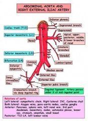
picture of abdominal aorta
|
picture of abdominal aorta
|
|
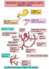
celiac branches pic
|
celiac branches
|
|

celiac branches pic
|
celiac branches
|
|
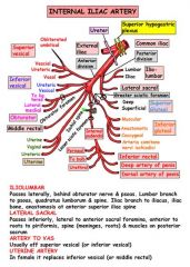
internal iliac branches pic
|
internal iliac branches pic
|