Reading...
![]()
Play button
![]()
Play button
![]()
Use LEFT and RIGHT arrow keys to navigate between flashcards;
Use UP and DOWN arrow keys to flip the card;
H to show hint;
A reads text to speech;
75 Cards in this Set
- Front
- Back
|
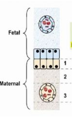
6 layers of placenta that can separate maternal and fetal blood
|
Maternal tissue
-blood vessel endothelium -connective tissue -epithelium of endometrium Fetal tissue -chorion -connective tissue -blood vessel endothelium |
|
|
Placenta types
|
-Epithelial-chorial
-Endothelial-chorial -Hemochorial |
|
|
Epithelial-Chorial placenta
-anatomy -animals |
Anatomy
-maternal epithelium of the endometriumis in contact with the fetal chorion -all 6 tissue layers intact throughout pregnancy Animals -sow -mare -ruminants (modified epithelial-chorial) |
|
|
Why is colostrum so important to animals with epithelial-chorial placentas?
|
-it is difficult for antibodies to travel to the fetus through all 6 tissue layers
|
|
|
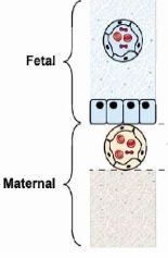
Endothelial-chorial placenta
-anatomy -animals |
Anatomy
-maternal blood vessel endothelium is in contact with fetal chorion -2 layers of tissue (epithelium and connective tissue of endometrium) have been removed at fetal-maternal contact sites Animals -dog -cat -others |
|
|
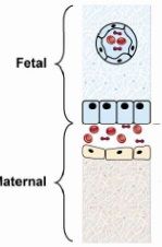
Hemochorial placenta
-anatomy -animals |
Anatomy
-maternal blood is in contact with fetal blood -3 layers of tissue (epithelium and connective tissue of endometrium and blood vessel epithelium) removed at the fetal-maternal contact sites Animals -primates -rodents |
|
Placenta type
|
-epithelial-chorial
|
|
Placenta type
|
-endothelial-chorial
|
|
Placenta type
|
hemochorial
|
|
|
Hemoendothelial placenta
-anatomy -animals |
Anatomy
-5 layers of tissue removed (epithelium and connective tissue of endometrium, maternal blood vessel endothelium, placental chorion, placental connective tissue) Animal -rabbit |
|
|
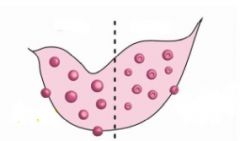
Placenta chorionic villous pattern
-pig |
-diffuse
|
|
|
Placenta chorionic villous pattern
-horse |
-diffuse and micro-cotyledonary
|
|
|
Placenta chorionic villous pattern
-bovine, ovine, caprine |
-cotyledonary
|
|
|
Placentome
-composition |
-caruncle (maternal)
-cotyledon (fetus) |
|
|
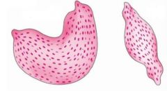
Cotyledonary placenta
-concave caruncles -convex caruncles |
Concave: sheep
Convex: cow |
|
|
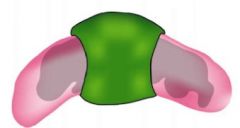
Placenta chorionic villous pattern
-dog & cat |
-zonary
|
|
|
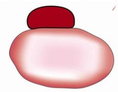
Placenta chorionic villous pattern
-primates & rodents |
-discoid
|
|
Chorionic villous pattern
-animals |
Diffuse placenta
-pig -mare |
|
Choriod villous pattern
-animals |
Cotyledonary
-sheep/goat (concave) -cow (convex) |
|
Choroid villous pattern
-animals |
Zonary placent
-cat -dog |
|
Choroid villous pattern
-animals |
Discoid placenta
-primates -rodents |
|
|
Main components of the male reproductive tract
|
-penis
-secondary sex organs |
|
|
Main things secreted by the testicle
|
-gametes
-steroid hormones |
|
|
Fluid for semen provided by
|
-seminal vesicles
-prostate -bulbourethral gland |
|
|
Pampiniform plexus
-function |
-complex venous network that surround the testicular artery and is a countercurrent heat exchanger to reduce testicular temp.
|
|
|
Testicle
-structure to protect from temp extremes |
-scrotum
-tunic dartos (muscle layer under scrotum) -cremaster m. (drops/sucks up testes) |
|
|
Seminiferous tubules
-composition |
-sertoli cells
-layers of germ cells |
|
|
Time for sperm to form in seminiferous tubule
|
-45-50 days
-mature in epididymis (1 wk) |
|
|
Epididymis function
|
-provide an energy efficient storage of sperm while maintaining fertility
-mix recently formed with older spermatozoa to provide temporal spectrum -facilitate maturation of spermatozoa |
|
|
Vas deferens pass into the body via
|
-inguinal ring
|
|
|
Spermatic cord
-components |
-vas deferens
-blood vessels -nerves |
|
|
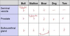
Accessory gland presence/absence in:
-bull -stallion -boar -dog -tom |
|
|
|
Penis
-function |
deposit semen into vagina or cervix/uterus
|
|
|
Ruminants
-penis type |
-fibroelastc (minimal erectile tissue)
|
|
|
Equine
-penis type |
-vascular penis
|
|
|
Canine/Feline
-penis type |
-Os penis
-erection after intromission |
|
|
Sigmoid flexure
-function |
-provides the means by which the penis is held inside the body and sheath of ruminants
-held by retractor muscles |
|
|
Blood pools in which penile structure
|
-Corpus cavernosum
|
|
|
End of penis
-anatomical name |
-glans penis
|
|
|
Bull
-testis orientation -penis type -copulation duration -volume -sperm concentration (per mL) -Total cells in ejaculate (10^9) |
-cauda down
-fibroelastic -1 sec -5 mL -1.2x10^9 -6x10^9 |
|
|
Stallion
-testis orientation -penis type -copulation duration -volume -sperm concentration (per mL) -Total cells in ejaculate (10^9) |
-horizontal
-vascular -20 sec -spring/summer = 100 mL; fall/winter = 50 mL -0.1x10^9 -spring/summer = 20x10^9; fall/winter = 10x10^9 |
|
|
Boar
-testis orientation -penis type -copulation duration -volume -sperm concentration (per mL) -Total cells in ejaculate (10^9) |
-perineal cauda up
-fibroelastic -6 min -200 mL -0.15x10^9 -30-50x10^9 |
|
|
Dog
-testis orientation -penis type -copulation duration -volume -sperm concentration (per mL) -Total cells in ejaculate (10^9) |
-horizontal
-vascular, os -20 min -20 mL -0.025x10^9 -0.5x10^9 |
|
|
Tom
-testis orientation -penis type -copulation duration -volume -sperm concentration (per mL) -Total cells in ejaculate (10^9) |
-perineal cauda up
-vascular, os -seconds -0.04 mL -1.7x10^9 -0.057x10^9 |
|
|
Mullerian ducts become
-female -male |
Female:
-oviducts -uterus -cranial vaginal Male: -regresses |
|
|
Wolffian ducts become:
-female -male |
Female
-regresses Male -vas deferens -epididymis |
|
|
Urogenital sinus becomes:
-females -males |
Females:
-caudal vagina -vestibule Males: -Closes (urethra & prostate) |
|
|
Genital tubercle becomes:
-female -male |
Female:
-clitoris Male: -penis |
|
|
Genital swelling becomes
-female -male |
Female:
-remains open (vulva) Male: -closes (scrotum) |
|
|
Chromosomal abnormalities
-syndromes -effect |
Syndromes
-xxx syndrome -xxxy syndrome -xo syndrome Effect -appear normal but are apparently infertile |
|
|
Intersexes
-define -types -most common in |
-mismatch of chromosomal, gonadal, phenotypic sex
-hermaphrodites -pseudohermaphrodites -freemartins -most common in goats |
|
|
Hermaphrodite
-define |
-both ovarian and testicular tissue are present
-1 testicle + 1 ovary or ovotestis -amt. of testicular tissue determines extent of masculinization |
|
|
Hermaphrodite
-chromosomal females characteristics |
-XX
-normal to abnormal vulva, clitoris, uterus, oviducts -epididymis, vas deferens |
|
|
Hermaphrodite
-chromosomal males characteristics |
-XY
-Testes, Epididymis, Vas deferense -abnormally shaped prepuce -hypoplastic penis -uterus |
|
|
Should hermaphrodite animals be removed from the population?
|
-yes
-genetically linked -remove parents and offspring |
|
|
Hermaphrodites
-presentation |
-present for infertility or abnormal external genitalia
|
|
|
Pseudohermaphrodite
-define |
-agreement of chromosomal and gonadal sex, but internal or external genitalia are ambiguous
|
|
|
Female pseudohermaphrodite
-characteristics |
-XX chromosomes and ovaries
-androgen dependent genitalia is masculinized |
|
|
Female pseudohermaphrodite
-occurs when |
-when the Dam is given androgen or progesterone during gestation
|
|
|
Male pseudohermaphrodite
-characteristics |
-XY chormosomes and testes
-Mullerian ducts are retained -Oviducts, uterus, cervix, cranial vagina present to some extent -normal external genitalia but are cryptorchid |
|
|
Freemartin
-animals |
-bovine
-caprine |
|
|
Freemartin
-define |
-abnormalities in a female co-twin with male fetus
-shared blood fetal vessel allow mullerian inhibiting substance, testosterone and insulin-like peptide 3 to affect tubular tract and external genitalia |
|
|
Freemartin
-abnormalities in female with male co-twin |
-short/absent vagina
-elongated vulva -inc. anal-vulvar distance -rudimentary seminal vesicles -ovotestes |
|
|
Do freemartin relative need to be culled?
|
-no
-not genetically linked |
|
|
Ovarian abnormalities
|
-ovarian agenesis
-ovarian hypoplasia -supernumery ovaries |
|
|
Uterine Tube abnormalities
|
-ductal hypoplasia
-white heifer disease |
|
|
Ductal hypoplasia
-occurs in what animals |
-holsteins
|
|
|
White heifer disease
-describe |
-segmental aplasia of the tubular tract of certain cattle breeds (shorthorn, Belgian blue)
-white coat color is a recessive trait associated with defects in the mullerian system -the animals have enough uterine tissue to cycle normally, but cannot maintain pregnancy |
|
|
Vulva/Vagina abnormalities
|
-persistent hymen
|
|
|
Persistent hymen
-describe |
-segmental hypoplasia
-failure of the mullerian ducts and urogenital sinus to fuse properly |
|
|
Abnormalities from decreased fusion of the mullerian ducts
|
-double cervix
-double vagina |
|
|
Testicular/Spermatic Cord abnormalities
|
-cryptorchidism
-testicular hypoplasia |
|
|
Process of testicular descent
|
1) peri-renal to peritoneal side of inguinal canal
2) through the inguinal canal 3) from scrotal side of inguinal canal to scrotum |
|
|
Cryptorchidism
-heritability |
-heritable
-unethical to surgically correct without rendering the animal incapable of reproduction |
|
|
Testicular hypoplasia
-causes |
-chromosomal abnormalities
-endocrine exposure during gestation |