![]()
![]()
![]()
Use LEFT and RIGHT arrow keys to navigate between flashcards;
Use UP and DOWN arrow keys to flip the card;
H to show hint;
A reads text to speech;
49 Cards in this Set
- Front
- Back
|
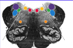
Early in development, the neural tube is separated into a dorsal (posterior) _____ plate and a ventral (anterior) _____ plate by the _____.
Nuclei dorsal to the sulcus limitans are _____ (afferent) and nuclei ventral to the sulcus limitans (in basal plate) are _____ (efferent). This is the key to understanding the anatomical locations of cranial nerve nuclei in the brainstem. |
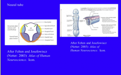
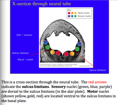
Early in development, the neural tube is separated into a dorsal (posterior) alar plate and a ventral (anterior) basal plate by the sulcus limitans. Nuclei dorsal to the sulcus limitans are sensory (afferent) and nuclei ventral to the sulcus limitans (in basal plate) are motor (efferent). This is the key to understanding the anatomical locations of cranial nerve nuclei in the brainstem. |
|
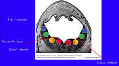
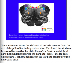
What do the arrows indicate? Identify motor nuclei, sensory nuclei |
|
|
|

|
|
Cross section of adult rostral medulla: Identify Motor nuclei and sensory nuclei. What do the dotted lines indicate? |
|
|
|
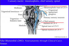
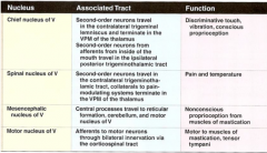
What are the three sensory nuclei for cranial nerve V?
Give general locations of each |
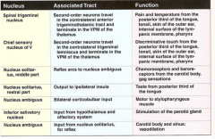
Chief, spinal, mesencephalic
Chief = pons Mesencephalic = mesencephalon Spinal = column from mesencephalon to spinal cord => continuous with substantia gelatinosa (layers I and II). |
|
|
Where are the vestibular nuclei and cochlear nuclei located? What nerve? |
Pons CN VIII |
|
|
What cranial nerves project to the solitary nucleus?
What is the rostral part concerned with? |
Rostral part = CN VII, IX, and X Caudal = cardiorespiratory inputs |
|
Review |

|
|
|
Cranial nerve I is completely _____. It serves olfaction. The sensory receptors reside in the nasal epithelium. These cells send very fine axons (olfactory nerves or filae) through the _____ plate to synapse in the _____ bulb. Axons of projection neurons in the bulb travel as the _____ tract to synapse in olfactory cortex, amygdala, and related forebrain structures. Anosmia is a condition marked by loss of the sense of smell. |
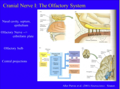
Cranial nerve I is completely sensory. It serves olfaction. The sensory receptors reside in the nasal epithelium. These cells send very fine axons (olfactory nerves or filae) through the cribriform plate to synapse in the olfactory bulb. Axons of projection neurons in the bulb travel as the olfactory tract to synapse in olfactory cortex, amygdala, and related forebrain structures. Anosmia is a condition marked by loss of the sense of smell. |
|
|
Cranial nerve II is the _____ Nerve. It is completely _____ - it transmits visual input to the brain. The receptors are located in the retina, at the rear of the eye. The optic nerve is made up of axons of _____ ganglion cells. |
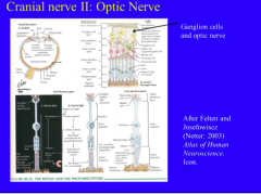
Cranial nerve II is the Optic Nerve. It is completely sensory - it transmits visual input to the brain. The receptors are located in the retina, at the rear of the eye. The optic nerve is made up of axons of retinal ganglion cells. |
|
|
The optic nerves from each eye converge just outside the _____. At this point, about 50% of the fibers from each eye cross to the opposite side of the body at the optic _____ (“cross”). Central to the chiasm, the fibers run as the _____ tract. |
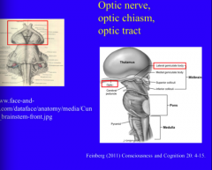
The optic nerves from each eye converge just outside the mesencephalon. At this point, about 50% of the fibers from each eye cross to the opposite side of the body at the optic chiasm (“cross”). Central to the chiasm, the fibers run as the optic tract. |
|
|
The optic tract runs along the outside of the mesencephalon to the _____ geniculate body (the visual thalamus: LGN), which in turn projects to ______ cortex (banks of calcarine fissure). Some optic nerve fibers bypass the LGN and instead travel in the _____ (“arm”) of the superior colliculus to synapse in the _____ (tectum of mesencephalon). This input is important for visual “_____” reflexes. |
The optic tract runs along the outside of the mesencephalon to the lateral geniculate body (the visual thalamus: LGN), which in turn projects to primary visual cortex (banks of calcarine fissure). Some optic nerve fibers bypass the LGN and instead travel in the brachium (“arm”) of the superior colliculus to synapse in the superior colliculus (tectum of mesencephalon). This input is important for visual “startle” reflexes. |
|
|
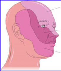
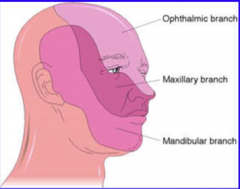
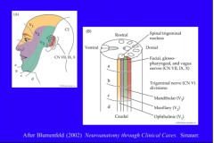
Cranial Nerve V is the Trigeminal nerve. Thissystemistheprimary one for somestheses for the face. Peripherally, there are three main branches to the nerve: V1 = _____, V2 = _____, and V3 = _____. |
Cranial Nerve V is the Trigeminal nerve. Thissystemistheprimary one for somestheses for the face. Peripherally, there are three main branches to the nerve: V1 = Ophthalmic, V2 = Maxillary, and V3 = Mandibular. |
|
|
In addition to motor innervation of muscles of mastication (and a few other muscles), the trigeminal nerves carry fine touch, proprioception, and pain and temperature afferents from the skin of the face region. The locations of the nerve branches and relative position of jaws and muscles are shown on the left. On the right, the areas of the skin sub served by each nerve branch is shown. Note that the back of the head is mostly innervated by spinal nerves from _____. Also note the complicated arrangement for innervation of the _____ ear (pinna) by multiple cranial nerves. |
In addition to motor innervation of muscles of mastication (and a few other muscles), the trigeminal nerves carry fine touch, proprioception, and pain and temperature afferents from the skin of the face region. The locations of the nerve branches and relative position of jaws and muscles are shown on the left. On the right, the areas of the skin sub served by each nerve branch is shown. Note that the back of the head is mostly innervated by spinal nerves from C2. Also note the complicated arrangement for innervation of the external ear (pinna) by multiple cranial nerves. |
|
Identify which branch of trigeminal. |
|
|
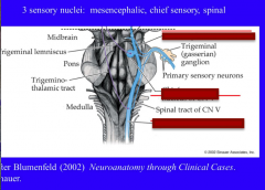
Identify the locations of the three sensory nuclei of trigeminal nerve: mesencephalic, chief sensory, and spinal. |
|
|
|
The chief sensory nucleus is located in the rostral _____ and receives _____ inputs from afferents for fine touch and vibration sense from the face. It is thus analogous to the posterior column nuclei (chief sensory nucleus of V is to fine touch for the face as posterior columns are for fine touch from the body). Epicritic afferents in the trigeminal nerve have their cell bodies in the ______ (or Gasserian) ganglion. This ganglion is located outside of the brainstem and is analogous to the dorsal root ganglia associated with the spinal cord. The central process of these cells synapse in the _____ sensory nucleus. It is important to note that fine touch afferents that run in other cranial nerves (VII, IX, X) have cell bodies in different peripheral ganglia but also synapse in the chief sensory nucleus of V. The chief sensory nucleus is thus the first integration center for all _____ touch and vibration inputs for the face region. |
The chief sensory nucleus is located in the rostral pons and receives epicritic inputs from afferents for fine touch and vibration sense from the face. It is thus analogous to the posterior column nuclei (chief sensory nucleus of V is to fine touch for the face as posterior columns are for fine touch from the body). Epicritic afferents in the trigeminal nerve have their cell bodies in the trigeminal (or Gasserian) ganglion. This ganglion is located outside of the brainstem and is analogous to the dorsal root ganglia associated with the spinal cord. The central process of these cells synapse in the chief sensory nucleus. It is important to note that fine touch afferents that run in other cranial nerves (VII, IX, X) have cell bodies in different peripheral ganglia but also synapse in the chief sensory nucleus of V. The chief sensory nucleus is thus the first integration center for all fine touch and vibration inputs for the face region. |
|
|
The mesencephalic nucleus of V contains the cell bodies of _____ afferents (spindle organs, Golgi tendon organs, etc.) from the muscles of mastication (and probably extraocular muscles and muscles of the tongue). It is essentially a displaced peripheral ganglion in the tegmentum of the _____. The peripheral processes of these cells form the _____ tract of V. |
The mesencephalic nucleus of V contains the cell bodies of proprioceptive afferents (spindle organs, Golgi tendon organs, etc.) from the muscles of mastication (and probably extraocular muscles and muscles of the tongue). It is essentially a displaced peripheral ganglion in the tegmentum of the mesencephalon. The peripheral processes of these cells form the mesencephalic tract of V. |
|
|
The spinal nucleus of V extends from the mesencephalon to the spinal cord, where it becomes continuous with the _____ (layers I and II of dorsal horn). This tract receives inputs from pain and temperature afferents for the face that run in cranial nerve V, as well as cranial nerves ___, ___, and ___. Pain and temperature afferents in branches of V have their cell bodies in the _____ ganglion. The central processes of these cells project into and synapse in the spinal nucleus of V. These axons, as well as second order axons from the spinal nucleus of V, run in the associated _____ tract of V. |
The spinal nucleus of V extends from the mesencephalon to the spinal cord, where it becomes continuous with the substantia gelatinosa (layers I and II of dorsal horn). This tract receives inputs from pain and temperature afferents for the face that run in cranial nerve V, as well as cranial nerves VII, IX, and X. Pain and temperature afferents in branches of V have their cell bodies in the trigeminal ganglion. The central processes of these cells project into and synapse in the spinal nucleus of V. These axons, as well as second order axons from the spinal nucleus of V, run in the associated spinal tract of V. |
|
|
The trigeminal lemniscus runs from the chief sensory nucleus of V to the _____ nucleus (VPM) of thalamus. It is analogous to the medial lemniscus for spinal pathways. |
The trigeminal lemniscus runs from the chief sensory nucleus of V to the ventroposteromedial nucleus (VPM) of thalamus. It is analogous to the medial lemniscus for spinal pathways. |
|
|
The trigeminothalamic tract runs from the spinal nucleus of V to the _____. It is analogous to the spinothalamic tract for spinal pathways. |
The trigeminothalamic tract runs from the spinal nucleus of V to the VPM. It is analogous to the spinothalamic tract for spinal pathways. |
|

Review |
|
|
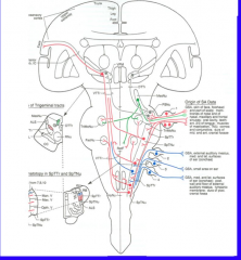
Somatotropic organization:
Identify ophthalmic (V1), CN VII, IX, X, V2, V3, spinal trigeminal nucleus |
|
|
|
The cell bodies of proprioceptive afferents from muscles of mastication (also extraocular muscles and tongue) are located in the _____nucleus of V (shown in green). These cells project bilaterally to the trigeminal motor nucleus (TrMNu) to mediate _____ stretch reflexes. |
The cell bodies of proprioceptive afferents from muscles of mastication (also extraocular muscles and tongue) are located in the mesencephalic nucleus of V (shown in green). These cells project bilaterally to the trigeminal motor nucleus (TrMNu) to mediate monosynaptic stretch reflexes. |
|
|
The cell bodies of afferents for fine touch and vibration sense are located in ganglia _____ of the brainstem (especially the trigeminal ganglion (V), but also geniculate for VII, for inferior and superior glossopharyngeal for IX, inferior and superior vagal for X). The central processes of these cells project to the _____ (shown in red). Second order projections from this nucleus cross the midline and travel in the _____ meniscus to the VPM (thalamus). |
The cell bodies of afferents for fine touch and vibration sense are located in ganglia outside of the brainstem (especially the trigeminal ganglion (V), but also geniculate for VII, for inferior and superior glossopharyngeal for IX, inferior and superior vagal for X). The central processes of these cells project to the chief (principal) sensory nucleus of V (shown in red). Second order projections from this nucleus cross the midline and travel in the trigeminal meniscus to the VPM (thalamus). |
|
|
The cell bodies of pain and temperature afferents (shown in blue) are located in various ganglia (Trigeminal for V, geniculate for VII, for inferior and superior glossopharyngeal for IX, inferior and superior vagal for X). Thus several cranial nerves contribute to this nucleus (V, VII, IX, X). Their central processes project into the _____ of V and synapse in the spinal _____ of V. The second order projections cross the midline and travel in the _____ tract (also shown in red). |
The cell bodies of pain and temperature afferents (shown in blue) are located in various ganglia (Trigeminal for V, geniculate for VII, for inferior and superior glossopharyngeal for IX, inferior and superior vagal for X). Thus several cranial nerves contribute to this nucleus (V, VII, IX, X). Their central processes project into the spinal tract of V and synapse in the spinal nucleus of V. The second order projections cross the midline and travel in the trigeminothalamic tract (also shown in red). |
|

|
|
|
|
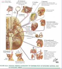
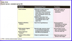
In addition to its motor functions, Cranial nerve VII (facial) has two sensory components: taste sensation for the anterior 2/3 of the tongue and somesthetic afferents (fine touch, vibration, pain and temperature) for a small area around the ______ meatus. Cell bodies for the somesthetic afferents are located in the _____ ganglion. The central processes project to the _____ nucleus of V (epicritic) or _____ nucleus of V (pain and temperature). |
In addition to its motor functions, Cranial nerve VII (facial) has two sensory components: taste sensation for the anterior 2/3 of the tongue and somesthetic afferents (fine touch, vibration, pain and temperature) for a small area around the external auditory meatus. Cell bodies for the somesthetic afferents are located in the geniculate ganglion. The central processes project to the chief sensory nucleus of V (epicritic) or spinal nucleus of V (pain and temperature). |
|

Review |
|
|
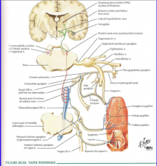
Identify rostral part of nucleus solitarius. |

|
|
|
Nucleus solitarius runs as a column through the _____ and _____. The rostral portions of this nucleus are also called the _____ nucleus and are concerned with _____ sensation. The gustatory nucleus receives inputs from CNs __, __, and __. |
Nucleus solitarius runs as a column through the pons and medulla. The rostral portions of this nucleus are also called the gustatory nucleus and are concerned with taste sensation. The gustatory nucleus receives inputs from CNs VII, IX, and X. |
|
The rostral part of nucleus solitarius is the _____ nucleus. It receives taste input (indicated in red) from CNs __ (anterior 2/3 of tongue), __ (posterior 1/3 of tongue), and X (pharynx and epiglottis). The caudal part of the solitary nucleus is known as the _____ nucleus. It receives _____ (visceral or somatic) sensory input (glands, chemoreceptors, baroreceptors: shown in blue) carried by CNs __, __, and __. |
The rostral part of nucleus solitarius is the gustatory nucleus. It receives taste input (indicated in red) from CNs VII (anterior 2/3 of tongue), IX (posterior 1/3 of tongue), and X (pharynx and epiglottis). The caudal part of the solitary nucleus is known as the cardiorespiratorynucleus. Itreceivesvisceralsensoryinput (glands, chemoreceptors, baroreceptors: shown in blue) carried by CNs VII, IX, and X. |
|
|
What are the two divisions of the one sensory component of the vestibulocochlear nerve? |
1. Vestibular (Balance and acceleration) 2. Auditory (hearing) |
|
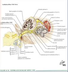
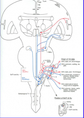
The sensory afferents for audition (hearing) and vestibular sense (body position in space) are located in the _____ ear. Auditory receptors (___ cells) are located in the organ of ____ in the cochlea. These receptors are sensitive to airborne pressure waves,astransducedintofluidwavesinthecochlea. Dependingon location in the cochlea, these receptors are tuned to different frequencies of sound (tonotopic organization). Auditory _____ (afferent or efferent) travel in CN VIII. |
The sensory afferents for audition (hearing) and vestibular sense (body position in space) are located in the inner ear. Auditory receptors (hair cells) are located in the organ of Corti in the cochlea. These receptors are sensitive to airborne pressure waves,astransducedintofluidwavesinthecochlea. Dependingon location in the cochlea, these receptors are tuned to different frequencies of sound (tonotopic organization). Auditory afferents travel in CN VIII. |
|
Review |

|
|
|
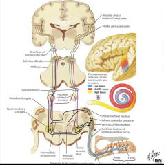
The cell bodies of auditory afferents are located in the _____ ganglion. These cells send axons in CN VIII (cochlear division) to synapse in the dorsal and ventral _____ nuclei. Right: Hair cells and supporting cells comprise the organ of _____, located in the cochlea. Hair cells synapse on cells in the _____ ganglion. Spiral ganglion cells send their axons in the cochlear division of the ______ nerve. Left: The cochlear nerve fibers synapse in the _____ (ipsilateral or contralateral) dorsal and ventral cochlear nuclei (at the pontomedullary junction). From this point, auditory pathways ascend _____ (bilateral or unilateral) to the inferior colliculus. Inferior colliculus neurons project in turn to the medial ______ body(auditorythalamus). Themedialgeniculateprojectsto auditory cortex (Heschel's gyrus: transverse gyri in superior temporal lobe). |
The cell bodies of auditory afferents are located in the spiral ganglion. These cells send axons in CN VIII (cochlear division) to synapse in the dorsal and ventral cochlear nuclei. Right: Hair cells and supporting cells comprise the organ of Corti, located in the cochlea. Hair cells synapse on cells in the spiral ganglion. Spiral ganglion cells send their axons in the cochlear division of the vestibulocochlear nerve. Left: The cochlear nerve fibers synapse in the ipsilateral dorsal and ventral cochlear nuclei (at the pontomedullary junction). From this point, auditory pathways ascend bilaterally to the inferior colliculus. Inferior colliculus neurons project in turn to the medial geniculate body(auditorythalamus). Themedialgeniculateprojectsto auditory cortex (Heschel's gyrus: transverse gyri in superior temporal lobe). |
|
|
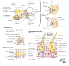
The other division of CN VIII is vestibular. This system is important for adjustment of posture, regulating muscle tone, and for coordination of _____ movements. The hair cells are located in the semicircular canals, saccule and vestibule. Vestibular hair cells synapse on cells in the _____ ganglion (Scarpa’s ganglion). These cells send their axons in the vestibular division of the vestibulocochlear nerve (VIII). |
The other division of CN VIII is vestibular. This system is important for adjustment of posture, regulating muscle tone, and for coordination of eye movements. The hair cells are located in the semicircular canals, saccule and vestibule. Vestibular hair cells synapse on cells in the vestibular ganglion (Scarpa’s ganglion). These cells send their axons in the vestibular division of the vestibulocochlear nerve (VIII). |
|

Review: |
|
|

The axons of cells from the _____ ganglion synapse in the vestibular nuclei (located in the pons and rostral medulla, near floor of fourth ventricle). There are ____ (#) vestibular nuclei (superior, lateral, medial, inferior). The lateral vestibular nucleus gives rise to the lateral _____ tract, a descending motor pathway (balance and extensor tone). The medial vestibulospinal tract arises from the ___ and ____ vestibular nuclei. It only extends to cervical levels of the cord and helps control neck and head position. The medial and superior vestibular nuclei contribute to the ________, which is important for coordinating eye movements. |
The axons of cells from the vestibular ganglion synapse in the vestibular nuclei (located in the pons and rostral medulla, near floor of fourth ventricle). There are four vestibular nuclei (superior, lateral, medial, inferior). The lateral vestibular nucleus gives rise to the lateral vestibulospinal tract, a descending motor pathway (balance and extensor tone). The medial vestibulospinal tract arises from the medial and inferior vestibular nuclei. It only extends to cervical levels of the cord and helps control neck and head position. The medial and superior vestibular nuclei contribute to the medial longitudinal fasciculus (MLF), which is important for coordinating eye movements. |
|
|
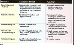
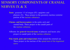
In addition to motor functions, CN IX mediates _____ from the posterior 1/3 of the tongue, _____ sensation from the pharynx and posterior 1/3 of the tongue, chemo- and baroreception from the carotid ____ and carotid ____, and fine touch and pain and temperature from the middle ear and a portion of the external ear. |
In addition to motor functions, CN IX mediates taste from the posterior 1/3 of the tongue, somatic sensation from the pharynx and posterior 1/3 of the tongue, chemo- and baroreception from the carotid sinus and carotid bodies, and fine touch and pain and temperature from the middle ear and a portion of the external ear. |
|
|
CN IX is associated with several nuclei in the brainstem. This nerve sub serves taste from the _____ 1/3 of the tongue. These afferents terminate in the _____ tract (gustatory nucleus). Afferents from Baroreceptors (carotid sinus) and chemoreceptors (carotid body) are carried by IX and project to the _____ part of the solitary nucleus and tract. The Nucleus Ambiguous is a motor nucleus containing neurons that project with CNs IX and X. Pain and temperature afferents from the posterior 1/3 of the tongue, pharynx, tonsils, tympanum, and a small area around the external ear travel travel in CN IX and project into the _____ tract and nucleus of V. Afferents for _____ (type) touch from the posterior 1/3 of the tongue, pharynx, tonsils, tympanum, and a small area around the external ear travel in IX and project to the Chief Sensory Nucleus of V. |
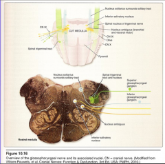
CN IX is associated with several nuclei in the brainstem. This nerve sub serves taste from the posterior 1/3 of the tongue. These afferents terminate in the rostral solitary tract (gustatory nucleus). Afferents from Baroreceptors (carotid sinus) and chemoreceptors (carotid body) are carried by IX and project to the caudal part of the solitary nucleus and tract. The Nucleus Ambiguous is a motor nucleus containing neurons that project with CNs IX and X. Pain and temperature afferents from the posterior 1/3 of the tongue, pharynx, tonsils, tympanum, and a small area around the external ear travel travel in CN IX and project into the spinal tract and nucleus of V. Afferents for Epicritic touch from the posterior 1/3 of the tongue, pharynx, tonsils, tympanum, and a small area around the external ear travel in IX and project to the Chief Sensory Nucleus of V. |
|
Afferents from Baroreceptors (carotid sinus) and chemoreceptors (carotid body) are carried by IX and project to the caudal part of the _____ _____ and tract. |
Afferents from Baroreceptors (carotid sinus) and chemoreceptors (carotid body) are carried by IX and project to the caudal part of the solitary nucleus and tract. |
|
|

|
|
|
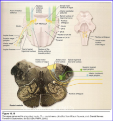
The sensory components of the vagus nerve include taste from the _____ and _____, visceral information from aortic arch baro- and chemoreceptors, somatic information from pharynx and larynx, and touch and _____ and temperature from a small portion of the external ear. |
he sensory components of the vagus nerve include taste from the pharynx and epiglottis, visceral information from aortic arch baro- and chemoreceptors, somatic information from pharynx and larynx, and touch and pain and temperature from a small portion of the external ear. |
|
|

|
|
|
The sensory components of the vagus nerve include taste from the pharynx and epiglottis. These afferents project to the _____ (rostral or caudal) solitary tract and nucleus. Afferents carrying visceral information from aortic arch baro- and chemoreceptors project to the caudal solitary tract and nucleus. Afferents carrying somatic information from pharynx, posterior meninges, and larynx, and touch and pain and temperature from a small portion of the external ear project into the spinal tract and nucleus of V. |
The sensory components of the vagus nerve include taste from the pharynx and epiglottis. These afferents project to the rostral solitary tract and nucleus. Afferents carrying visceral information from aortic arch baro- and chemoreceptors project to the caudal solitary tract and nucleus. Afferents carrying somatic information from pharynx, posterior meninges, and larynx, and touch and pain and temperature from a small portion of the external ear project into the spinal tract and nucleus of V. |
|

|
|
|
|
|
|
|
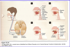
The gag reflex is for protecting and clearing the airway in response to an irritant to the palate, pharynx, and associated areas. The afferent limb of this reflex is carried by afferents of CN ___ projecting to the ___ (rostral or caudal) solitary nucleus. An interneuron is interposed between the afferent and efferent in Nucleus _____. The efferent response is mediated by motoneurons in N. Ambiguous whose axons travel with CN X. |
he gag reflex is for protecting and clearing the airway in response to an irritant to the palate, pharynx, and associated areas. The afferent limb of this reflex is carried by afferents of CN IX projecting to the caudal solitary nucleus. An interneuron is interposed between the afferent and efferent in Nucleus Ambiguous. The efferent response is mediated by motoneurons in N. Ambiguous whose axons travel with CN X. |