![]()
![]()
![]()
Use LEFT and RIGHT arrow keys to navigate between flashcards;
Use UP and DOWN arrow keys to flip the card;
H to show hint;
A reads text to speech;
67 Cards in this Set
- Front
- Back
|
The integumentary system has two major parts: |
* The Cutaneous Membrane (skin) * The accessory structures |
|
|
What are some examples of accessory structures? |
* Hair shaft * Pore of sweat gland duct * Tactile (Meissner's) corpuscle * Sebaceous gland * Arrector pili muscle * Sweat gland duct * Hair follicle * Lamellated corpuscle * Nerve fibers * Sweat Gland |
|
|
What are the two components of the cutaneous membrane? |
* the epidermis * the dermis |
|
|
What are the functions of the integumentary system? |
1) Protection of underlying tissues and organs against impact, abrasion, fluid loss, and chemical attack 2) Excretion of salts, water, and organic wastes by integumentary glands 3) Maintenance of normal body temperature through either insulation or evaporative cooling, as needed 4) Production of melanin, which protects underlying tissue from ultraviolet radiation 5) Production of keratin, which protects against abrasion and serves as a water repellent 6) Synthesis of vitamin D3, a steroid that is concerted to calcitriol, a hormone important to normal calcium metabolism 7) Storage of lipids in adipocytes in the dermis and in adipose tissue in the hypodermis (subcutaneous layer) 8) Detection of touch, pressure, pain, vibration, and temperature stimuli and the relaying of that information to the nervous system. 9) Coordination of immune response to pathogens and cancers in the skin. |
|
|
What does the Epidermis do? |
Protects the dermis, prevents water loss and the entry of pathogens, and synthesizes vitamin D3. Sensory receptors detect touch, pressure, pain, and temperature. |
|
|
What are the two layers of the Dermis? |
1) Papillary Layer 2) Reticular Layer |
|
|
What does the papillary layer do? |
* The papillary layer nourished and supports epidermis |
|
|
What does the reticular layer do? |
The reticular layer has sensory receptors that detect touch, pressure, pain, vibration, and temperature. Blood vessels assist in the thermoregulation. |
|
|
Since the epidermis is avascular, how do the epidermal cells rely gain nutrients? |
Because there are no local blood vessels, epidermal cells rely on the *diffusion* of nutrients and oxygen from capillaries within the dermis. |
|
|
What are keratinocytes? |
Keratinocytes are the body's most abundant epithelial cells that dominate the epidermis. These cells form several layers, or state, and contain large amounts of the protein keratin. |
|
|
Where can thick skin be found? |
Thick skin can be found on the palm of the handstand the soles of the feet. |
|
|
What are the five layers of keratinocytes in a section of the epidermis? |
* Stratum Corneum * Stratum Lucidum * Stratum Granulosum * Stratum Spinosum * Stratum Basale |
|
|
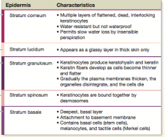
What are the characteristics of the stratum basale layer and stratum germinativum? |
* Deepest layer * Attachment to basement membrane * Contains basal cells (stem cells), melanocytes, and tactile cells (merkel cells) |
|
|
What are the characteristics of the stratum spinosum ? |
* Keratinocytes are bound together by desmosomes. Each time a stem cells divides, one of the daughter cells is pushed superficial to the stratum basale into the stratum spinosum. |
|
|
What are the characteristics of the Stratum Granulosum? |
* Keratinocytes produce keratohyalin and keratin. * Keratin fibers develop as cells become thinner and flatter * Gradually the plasma membranes thicken, the organelles disintegrate, and the cells die. |
|
|
What are the characteristics of Stratum lucidum? |
* Appears as a glassy layer in thick skin only |
|
|
What are the characteristics of the Stratum Corneum? |
* Multiple layers of flattened, dead, interlocking keratinocytes * Water resistant but not waterproof * Permits slow water loss by insensible perspiration |
|
|
What is the difference between insensible perspiration and sensible perspiration? |
* In Insensible perspiration, you can not feel or see the water loss. * In sensible perspiration, you are usually very aware of the perspiration by active sweat glands. |
|
|
What are the two pigments that the epidermis contains? |
* Carotene * Melanin |
|
|
What is carotene? |
Carotene is an organ-yellow pigment that normally accumulates in epidermal cells. It is most apparent in cells of the stratum corneum in light-skinned individuals. |
|
|
What is melanin? |
Melanin is a pigment produced by melanocytes, pigment producing cells. |
|
|
What are the two types of melanin? |
* pheomelanin, a red-yellow form * eumelanin, a brown-black form |
|
|
Where are melanocytes located? |
The melanocytes are located in the stratum basale. |
|
|
What is cyanosis? |
It is the bluish coloration of the skin. It can occur in response to extreme cold or as a result of cardiovascular or respiratory disorders, such as heart failures or severe asthma. |
|
|
How does sunlight cause epidermal cells to convert a steroid into vitamin D3? |
When exposed to ultraviolet radiation, epidermal cells in the stratum spinosum and stratum basale convert a cholesterol-related steroid into *cholecalciferol* (Vitamin D3). The lover then converts this to a product the kidneys can use and the kidneys synthesize the hormone *calcitriol*. Calicitriol is essential for the normal absorption of calcium and phosphorus in the small intestine. |
|
|
Where are the epidermal growth factor? What are the functions of the epidermal growth factor? |
EGF is produced by the salivary glands and glands of the duodenum. * Promoting the division of basal cells in the stratum basale and stratum spinosum * Accelerating the production of keratin in differentiating keratinocytes * Stimulating synthetic activity and secretion by epithelial glands |
|
|
What is dermatitis? |
Dermatitis is an inflammation of the skin that primarily involves the papillary layer. |
|
|
What are cleavage lines? |
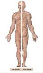
Cleavage lines are also called tension lines and Langer lines. Cleavage lines are clinically significant. A cut parallel to a cleavage line will usually remain closed and heal with little scarring. |
|
|
Arteries supplying the skin lie deep in the hypodermis. They are called ______. |
Plexuses. |
|
|
Where is the cutaneous plexus? |
The deeper network lies along the border of the hypodermic with the reticular layer of the dermis. |
|
|
Where is the sub papillary plexus? |
On the papillary layer. |
|
|
What are collagen fibers? |
Collagen fibers are very strong and resist stretching. |
|
|
What are elastic fibers? |
Elastic fibers permit stretching and the recoil to their original length. |
|
|
True or False. The hypodermic is not a part of the integument. |
True |
|
|
List the two terms for the tissue that connects the dermis to underlying tissues. |
Hypodermis or subcutaneous layer |
|
|
What is a root hair plexus? |
A root hair plexus of sensory nerves surrounds the base of each hair follicle. |
|
|
What does the arrestor pili do? |
The arrestor pili is a bundle of smooth muscle cells. When stimulated, the arrestor pili muscle contracts, pulling on the follicle and forcing the hair to stand erect. Contraction may be the result of emotional states, such as fear or rage. |
|
|
What are sebaceous glands? |
Sebaceous glands or oil glands are holocrine glands that discharge an oily lipid secretion into hair follicles. The lipid is called sebum. |
|
|
What does sebum do? |
Sebum inhibits the growth of bacteria, lubricates and protects the keratin of the hair shaft, and conditions the surrounding skin. |
|
|
What are sebaceous follicles? |
Sebaceous follicles are large sebaceous glands that are not associated with hair follicles. Instead, their ducts discharge sebum directly into the epidermis. |
|
|
What are sudoriferous glands? |
Sudoiferous glands are sweat glands. There are two types: apocrine sweat glands and merocrine sweat glands. |
|
|
Where are apocrine sweat glands found? |
In armpits, around the nipples, and in the pubic region. They actually rely on merocrine secretion. |
|
|
Where are merocrine sweat glands found? |
Merocrine sweat glands are far more numerous and widely distributed than apocrine sweat glands. The plasma and soles have the highest numbers. |
|
|
What are the functions of merocrine sweat glands? |
* Cooling the surface of the skin to reduce body temperature * Excreting protection from environmental hazards * Providing protection from environmental hazards |
|
|
What are ceruminous glands? |
Ceruminous glands are modified sweat glands in the passageway of the external ear. Their secretions combine with those of nearby sebaceous glands, forming a mixture called cerumen, or earwax. |
|
|
What is granulation tissue? |
It is the combination of blood clot, fibroblasts, and an extensive capillary network at the base of a wound. |
|
|
The portion of the hair follicle where cell divisions occur is the ______.
|
Matrix |
|
|
The stratum corneum of the nail root, which extends over the exposed nail, is called _______. |
Eponychium . |
|
|
The fibrous protein that is responsible for the strength and water resistance of the skin surface is called ________. |
Keratin |
|
|
In order for bacteria on the skin to cause an infection in the skin, they must accomplish all of the following except? |
Penetrate to the level of the capillaries. |
|
|
Which of the following layers is composed of adipose and areolar tissues and is the site of subcutaneous injections? |
Hypodermis |
|
|
When a scar tissue formation beyond the requirements of tissue repair, ____ is formed. |
A keloid |
|
|
Which of the following is NOT an effect of aging on the integumentary system? |
Melanocyte activity increases, darkening the skin. |
|
|
What is the epidermis beneath the nail? |
A pale present shape of base of nail |
|
|
What is the pale crescent shape base of nail? |
Lunula |
|
|
What is the order of layers of hair shaft? |
* Medulla * Cortex * Cuticle * Internal Root Sheath * External Root Sheath * Glassy Membrane |
|
|
What is the subcutaneous layer that separates the integument from the deep fascia around other organs? |
Hypodermis |
|
|
Which epidermal layer is found only in thick skin? |
Stratum Lucidum |
|
|
What are the two basic factors interacting to produce skin color? |
Circulatory supply and pigment concentration |
|
|
What are the primary tissues composing the hypodermis? |
areolar and adipose |
|
|
An important function of the hypodermic is to ________. |
Stabilize the position of the skin in relation to underlying tissues. |
|
|
What is the natural factor responsible for varying shades of hair color? |
Type of Melanin present |
|
|
Where can you find an apocrine sweat gland? |
* Armpits * Nipples * Pubic region |
|
|
The body of the nail _______. |
Consists of dead, tightly compacted cells |
|
|
A decrease in the number of _______ leads to increased damage and infection associated with age. |
Langerhans cells |
|
|
The peptide that is secreted by the pituitary gland and increases the rate of melanin production is _______. |
MSH |
|
|
A hair at the end of the growth cycle is called? |
* Club Hair |