![]()
![]()
![]()
Use LEFT and RIGHT arrow keys to navigate between flashcards;
Use UP and DOWN arrow keys to flip the card;
H to show hint;
A reads text to speech;
43 Cards in this Set
- Front
- Back
|
pericardium |
|
|
The serous membrane that surrounds the heart (except cranially where the great vessels are located) |
pericardium |
|
|
the two layers of pericardium |
visceral(epicardium) pericardium and perietal pericardium |
|
|
The layer which is continuous with the parietal paricardium |
mediastinal pleura |
|
|
the potential space between the visceral and parietal pericardia |
pericardial cavity |
|
|
the layer of fibrous connective tissue between the parietal pericardium and mediastinal pleura |
pericardial sack |
|
|
base |
|
|
apex |
|
|
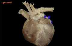
left auricle |
|
|
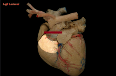
paraconal interventricular groove |
|
|
the fat filled groove around the circumfrence of the heart that marks the external boundary between the atria and ventricles |
coronary groove |
|
|
the external landmarks of the internal septum between right and left ventricles |
interventricular groove |
|
|
subsinuosal interventricular groove |
|
|
right coronary artery |
|

|
left coronary artery |
|
|
The main part of the right atrium |
sinus venarum |
|
|
the ridges on the interior surface of the auricle |
pectinate muscles |
|
|
the wall or septum between the right and left atria |
interatrial septum |
|
|
intervenous tubercle |
|
|
the opening between the right atrium and right ventricle |
right atrioventricular orifice |
|
|
fossa ovalis |
|
|
the fetal remnant of the fetal foramen ovalle |
fossa ovalis |
|
|
opening of the coronary sinus |
|
|
great cardiac vein |
|
|
the termination of the great cardiac vein that drains deoxygenated blood from the heart muscle |
coronary sinus |
|
|
pulmonary trunk |
|
|
the pulmonary trunk divides into what 2 vessels |
left pulmonary artery and right pulmonary artery |
|
|
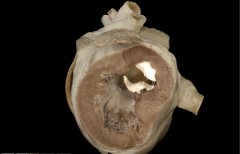
right atrioventricular (AV) valve |
|
|
chordae tendinaea |
|
|
papillary muscles |
|
|
trabeculae carneae |
|
|
the muscular ridges on the interior of the ventricles |
trabeculae carneae |
|
|
conus arteriousus |
|
|
the area where the right ventricle narrows down to enter the pulmonary trunk |
conus arteriousus |
|
|
pulmonary valve |
|
|
pulmonary veins |
|
|
left atroventricular(AV) valve |
|
|
the opening between the left atrium and left ventricle |
left atrioventricular orifice |
|
|
the left tricuspid valve analogous to the pulmonary valve |
aortic valve |
|
|
circumflex branch of the left coranary artery |
|
|
paraconal interventricular branch of the left coronary artery |
|
|
ligamentum arteriosum |
|
|
the fetal remnant of the fetal ductus arteriosus |
ligamentum arteriosum |