![]()
![]()
![]()
Use LEFT and RIGHT arrow keys to navigate between flashcards;
Use UP and DOWN arrow keys to flip the card;
H to show hint;
A reads text to speech;
38 Cards in this Set
- Front
- Back
|
A band |
DARK bands; contain overlapping myosin and actin filaments |
|
|
Acetylcholine (ACh) |
Neurotransmitter released from the axon terminals of the motor end plate of a motor neuron. - stimulates a skeletal muscle cell to contract - docks with ACh receptor in the motor end plate and is finally decomposed with the help of acetylcholinesterase |
|
|
Actin |
"Thin filaments" actin filament is a double stranded chain of actin molecules. Each actin molecule is a spherical protein that is the repeated unit in an actin filament. - Has two other proteins associated with it: 1) TM (tropomyosin) 2) TP (troponin) |
|
|
Aponeurosis |
a flat sheet of pearly-white fibrous connective tissue that takes the place of a tendon in sheet-like muscles having a wide area of attachment. Ex) epicranial aponeurosis |
|
|
Axon terminal |
The end of a neuron that resembles a "knob" like structure. |
|
|
Cardiac muscle |
Muscle ONLY found in the Heart - tubular structure - single nucleus per cell - striations - intercalated discs: permit one cell to stimulate another to contract, called "cell to cell" communication |
|
|
Disuse atrophy |
Decrease in muscle mass due to disuse.
- Caused from being bedridden. Proteases (enzymes digest proteins) are activated and actin/myosin are digested, decreasing the size of the muscle cell. - NOT permanent bc actin/myosin can be remade - MAIN cause of muscle atrophy |
|
|
Endomysium |

Connective tissue that surrounds the skeletal muscle cells. Binds one muscle cell to another and insulates the cell for electrical activity |
|
|
Epimysium |

Tough, fibrous C.T. that surrounds the whole muscle |
|
|
Fascia |
Fibrous connective tissue that surrounds the outside of the whole muscle |
|
|
Fascicle |
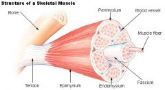
cluster of skeletal muscle cells |
|
|
Hemoglobin |
protein pigment: gives cells bright red color in oxygenated form and dark red color in deoxygenated form - located in red blood cells Function: transport oxygen to tissues throughout the body. |
|
|
Hypertrophy |
Increase in muscle mass
Ex) Body builder lifting weights - Weighted stress causes muscle cells to produce more actin and myosin therapy increasing the size of skeletal muscle cells. |
|
|
I band |
LIGHT bands; contains ONLY actin filaments |
|
|
Intercalated disc |
permit one cell tostimulate another to contract. Thisis “cell to cell” communication. |
|
|
Isometric contraction |
The muscle does NOT change in length and the tension never exceeds the load Ex) muscles for maintaining posture use isometric contractions |
|
|
Isotonic contraction |
As the tension increases, the muscle changes in length. Ex) muscles involved in walking and running involve isotonic contractions |
|
|
Lactate |
When muscles don't get replenished with oxygen, anaerobic respiration occurs and lactate accummulates.glucose >> pyruvate >> lactateLeads to muscle fatigue from a buildup of lactate, decrease in Ca in sarcoplasm, depleted creative phosphate, oxygen, glycogen and other nutrients.
|
|
|
Ligament |
Fibrous connective tissue that anchors bone to bone |
|
|
Motor end plate |
The pocket or depression in the sarcolemma that receives the axon terminal of the motor neuron. The sarcolemma contains the ACh receptors for the neurotransmitter Acetylcholine and the enzyme acetylcholinesterase. |
|
|
Muscular dystrophy |
Genetic disease where there is a deterioration of muscles from having a lack of dystrophin (sarcolemma stabilizer) - The sarcolemma then tears during contraction leading to muscle atrophy and connective tissue build up which destroys the muscle cell. Treatment: gene and stem cell therapy Duchenne muscular dystrophy (DMD) = most common. - death is often associated with muscle weakness in respiratory muscles (intercostals and diaphragm) - Women are usually carriers and men contract the disease. |
|
|
Myasthenia gravis |
Autoimmune disease that targets the neuromuscular junction.
- Antibodies block ACh receptor sites, so ACh cannot bind. - Leads to paralysis; drooping eyelids, difficulty swallowing and walking >>gen. muscle weakness. Treatment: Acetylcholinesterase inhibitors and immunosuppressants. |
|
|
Myofibril |
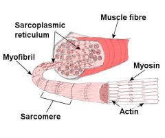
cluster of myosin and actin protein filaments |
|
|
Myoglobin |
protein pigment: gives skeletal muscle its reddish color along with the hemoglobin in the blood - located in skeletal muscle cells Function: temporarily stores oxygen as a reserve for cellular respiration |
|
|
Myosin |
"Thick filaments" - myosin filament cluster of about 200 myosin molecules - myosin head consists of 2 heads and one long tail. Each head has 2 binding sites 1) Actin binding site 2) ATP binding site |
|
|
Neuromuscular junction |
a junction between nerve and muscle; it is a chemical synapse formed by the contact between the presynaptic terminal of a motor neuron and the postsynaptic membrane of a muscle fiber.
|
|
|
Perimysium |
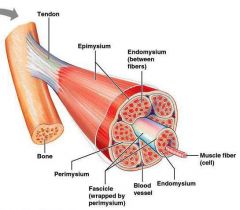
Connective tissue that surrounds each fascicle. |
|
|
Rigor mortis |
Stiffening of muscles that begin 3-4 hrs after death. Hits peak rigidity at 12hrs. Then later dissipates, leading to flaccid muscle tone.
How: dying cells have influx of Ca; leading myosin heads to attach when breathing stops. ATP synthesis stops. Myosin heads therefor cannot detach. Flaccidness comes as proteases digest actin/myosin. |
|
|
Sarcolemma |
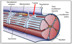
Sheath that surrounds the muscle. a.k.a. Plasma membrane of skeletal muscle cell |
|
|
Sarcomere |

The regional area between two Z-lines (discs); also, the functional unit for muscle contraction |
|
|
Sarcoplasm |
cytoplasm of skeletal muscle cell |
|
|
Sarcoplasmic reticulum (S.R.) |
muscle tissue's version of the E.R. - an elaborate network thats more extensive compared to other cells f(x): stores Ca++ ions - contains specialized regions called triads ( 3 structures): 2 outer T-tubules and middle terminal cisternae 1) T (transverse) tubules: function is to conduct nervous impulses to every single region of the muscle cell. 2) Terminal cisternae: site where calcium is released from the SR to the surrounding sarcoplasm |
|
|
Skeletal muscle |
all major muscles throughout the body - striated - voluntary |
|
|
Smooth muscle |
Involuntary - found in digestive tract, blood vessels, respiratory system, etc. - smaller, flatter cells - single nucleus per cell - no striations - cells often stack on top of each other to form a sheet |
|
|
Synapse |
a junction between two nerve cells, consisting of a minute gap across which impulses pass by diffusion of a neurotransmitter. |
|
|
Tendon |
"cable-like" structure Fibrous connective tissue that anchors muscle to bone. |
|
|
Tropomyosin (TM) |
Regulates binding of myosin head - bound with troponin, TM slides back and forth over the actin filaments. - It can either be OPEN (exposing myosin binding sites) or CLOSED (covering binding sites). - Movement to the OPEN state requires Calcium to bind to Troponin. |
|
|
Troponin (TP) |
Bound to Tropomyosin to form TM/TP complex. - When Calcium binds to troponin, it allows the TM/TP complex to slide OPEN, exposing the myosin binding sites. |