![]()
![]()
![]()
Use LEFT and RIGHT arrow keys to navigate between flashcards;
Use UP and DOWN arrow keys to flip the card;
H to show hint;
A reads text to speech;
96 Cards in this Set
- Front
- Back
|
Bacterial skin disease can be divided into what 5 broad categories? |
1. superficial pyoderma 2. deep pyoderma 3. bacterial granulomatous dermatitis 4. systemic infections w/ toxin-producing bacteria 5. pododermatitis |
|
|
Pyoderma is dermatitis caused by pyogenic bacteria. What are two common species of bacteria that cause this disease? |
staphylococci streptococci cornybacterium actinomyces spp. dermatophilus congolensis |
|
|
Superficial pyoderma: What is the typical duration? Is scaring common? Does it affect lymph nodes? Is prognosis good or bad? |
Short duration heals without scaring rarely effects lymph nodes good prognosis |
|
|
What are two examples of superficial pyoderma? What is the typical presentation? What is the causative agent? |
1. "puppy pyoderma" in dogs impetigo on muzzle on paws staphylococci 2. exudative dermatitis in pigs= greasy pigs deep fissures, greasy crusts, parakeratosis staphylococci hyicus *often fatal due to dehydration and toxicity |
|
|
There are two different types of deep pyoderma. What are they? What is the causative agent? |
1. Staphylococcal folliculitis and furunculosis 2. Abscesses: caused by a variety of bacteria (staphylococcus, corynebacterium, acrobacterium...) |
|
|
Deep pyoderma: What is the typical duration? Is scaring common? Does it affect lymph nodes? Is prognosis good or bad? |
chronic/longterm duration (often secondary) may result in scaring usually involves lymph nodes prognosis usually worse than superficial |
|
|
Bacterial granulomatous dermatitis can be caused by three different types of bacteria. What are they? |
1. mycobacteria 2. filamentous bacteria 3. non-filamentous bacteria |
|
|
mycobacterial granulomatous dermatitis is most common is which species? This disease is typically caused by what kind of mycobacteria (very general)? |
cats
opportunistic atypical mycobacteriosis (atypical because these bacteria are usually slow growing, but in these infections they grow quickly) |
|
|
What is an example of granulomas caused by nonfilamentous bacteria? What are some bacteria that cause this type of infection? |
Botryomycosis staphylococcus, streptococcus, actinobacillus lignieressi |
|
|
Erysipelas is categorized as what type of bacterial skin disease? What is it commonly called in pigs? Species effected? clinical signs (acute vs. chronic)? |
systemic, toxin producing bacteria diamond skin disease pigs, sheep, birds, humans (zoonotic) subcutaneous hemorrhage (acute) dermatitis, endocarditis, arthritis (chronic) |
|
|
What kind of bacteria typically result in toxic shock syndrome? What is the involvement of the skin in this condition? |
Staphylococcus aureus or group A streptococcus erythema (hypotension, v+/d+) open wounds predispose animals to these infections |
|
|
Name one example of a bacterial digital infection of ruminants |
Necrobacillosis: bovine interdigital dermatitis caused by Fusobacterium necrophorum also: interdigital dermatitis of sheep caused by Dichelobacter nodosus |
|
|
Fungal skin infection can be divided into what three categories based on location on the host? |
cutaneous mycoses subcutaneous mycoses systemic mycoses |
|
|
What is one example of a zoonotic cutaneous mycoses? What is the pathology and clinical presentation? |
Dermatophytosis (ringworm) Caused by Microsporum spp. and Trichophyton spp. Pathology: invade and destroy keratin, leading to folliculitis and furunculosis Clinical presentation: alopecia, scales, crusts. |
|
|
What is Malassezia pachyderatis? What is the clinical presentation? |
cutaneous mycoses caused by yeast presentation: alopecia, hyperpigmentation, lichenification mostly secondary (to atopic dermatitis) |
|
|
What is an example of subcutaneous mycoses? What species does it typically infect? |
Sporothrix schenkii granulomatous dermatitis and panniculitis cats (and humans! but only contagious by implantation) |
|
|
how are systemic mycoses usually infectious (what method of infection)? What is the typical presentation in the skin? |
infection by inhalation pyogranulomatous dermatitis |
|
|
Viral skin diseases can be classified either as epitheliotropic or systemic. What are examples of each type? |
epitheliotropic: pox viruses, vesicular diseases systemic: distemper, hog cholera |
|
|
What is the typical presentation characteristic of viral skin diseases? |
proliferative dermatitis with eosinophilic cytoplasmic inclusion bodies |
|
|
Avian pox has what two forms? What is the difference? |
"dry" pox: proliferative dermatitis on legs and head "wet" pox: oral cavity |
|
What is this? |
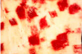
Ecthyma contagiosum caused by parapoxvirus ovis infects sheep and goats affects lips, eyelids, oral mucosa, feet mortality only if it interferes with feeding |
|
|
What is myxomytosis? |
viral skin disease of rabbits (especially in California) characterized by myxedema of eyelids and external genitalia proliferative dermatitis systemic infection (commonly with secondary bacterial infections) |
|
|
Canine distemper is characterized by what type of skin lesions? What is the pathogenic organism? |
Impetigo, less commonly with hyperkeratosis of foot pads and nasal planum paramyxoviridae, or canine distemper virus |
|
|
Hog cholera is also known as ________. What is the clinical presentation of this disease? |
classical swine fever, caused by flaviviridae, porcine pestivirus clinical presentation is severe cutaneous hemorrhage. |
|
|
habronema larvae cause what kind of clinical presentation in horses? |
eosinophilicgranulomas with intralesional larvae. cutaneous habronemiasis |
|
|
This mite lives in the hair follicle of many species, and causes primary alopecia. What is it, and what is the name of the disease it causes? |
Demodex demodicosis |
|
|
There are several different types of mange, caused by mites other than demodex. What are two different types and what are some clinical signs? |
Chorioptic mange Psoroptic mange Sarcoptic mange alopecia (secondary), hyperplasia, scales, crusts |
|
|
Which infections skin conditions are potentially zoonotic? |
mycobacteria: forming bacterial granulomatous dermatitis erysipelas (diamond skin disease, systemic bacterial infection) dermatophytosis: cutaneous mycoses Sporothrix schenkii: subcutaneous mycoses Sarcoptic mange: caused by sarcoptes species |
|
|
What are three examples of Type I hypersensitivity skin diseases? |
urticaria atopic dermatitis food hypersensitivity parasite hypersensitivity anaphylactic shock |
|
|
What is Type I hypersensitivity caused by? What is the clinical presentation? |
Anaphylaxis IgE binding and mast cell degranulation (release of histamine, heparin, serotonin) clinical presentation: pruritis, wheals, pustules, papules |
|
|
What is the important differentiating presentation between Type I and Type IV hypersensitivity? |
Type I: pruritis Type IV: no pruritis |
|
|
What is the histopathology/cytology typical of Type I hypersensitivity? |
acute: eosinophilic dermatitis chronic: epidermal hyperplasia, lymphoplasmocytic dermatitis |
|
|
Atopic dermatitis is a multi-factorial disease. What kinds of factors contribute to the disease process? |
genetic factors allergens environmental factors bacterial infections food psycologic factors (stress) defective lipids in the stratum corneum |
|
|
What is the pathogenesis of Type IV hypersensitivity? Clinical presentation? |
Cell-mediated (delayed) APCs (DCs, macrophages, keratinocytes, T cells) acute: degeneration of keratinocytes chronic: epidermal hyperplasia, lymphoplamacytic dermatitis clinical presentation: little or no pruritis, pustules, papules |
|
|
What are some examples of Type IV hypersensitivity? |
Tuberculin reaction insect bite hypersensitivity contact dermatitis |
|
|
What types of hypersensitivity can be caused by drug-mediated responses? |
I, II, III, and IV |
|
|
Flea bit hypersensitivity is what type? |
Type I or Type IV |
|
|
Type III hypersensitivity: pathogenesis? patterns? example? |
pathogenesis: immune complex-mediated, complement activation, neutrophils and reactive oxygen species patterns: localized (arthus type) or systemic (serum sickness) example: lupus erythematosus |
|
|
Type II hypersensitivity: pathogenesis? clinical presentation? example? |
pathogenesis: cellular cytotoxicity, antibody-dependent (IgG and IgM), complement activation clinical presentation: vesicles, bullae, acantholysis example: pemphigus |
|
|
How are autoimmune diseases categorized? |
1. vesicles or bullae (acantholysis) 2. depigmentation and/or ulceration (degeneration of keratinocytes with interface dermatitis) |
|
|
Autoimmune diseases characterized by vesicles or bullae can be further broken down into two types. What are these? What are examples? Which is more clinically severe? |
intraepidermal: pemphigus foliaceous along basement membrane: pemphigus vulgaris (more severe) |
|
|
What is the difference between pemphigus foliaceous, pemphigus vulgaris, and bullous pemphigoid? |
pemphigus foliaceous: autoantibodies target desmosomes between keratinocytes, suprabasilar vesicles pemphigus vulgaris: autoantibodies target desmosomes between keratinocytes, subcorneal vesicles form bullous pemphigoid: autoantibodies target hemidesmosomes (connections btwn keratinocytes and BM) |
|
|
depigmentation/ulceration autoimmune diseases can be further broken down into two categories. What are these, and what are examples? |
1. skin only: discoid lupus erythematosus 2. systemic (including skin) systemic lupus erythematosus |
|
|
discoid lupus erythematosus is often called ______, and is exacerbated by _________ |
"collie nose" UV light |
|
|
systemic lupus erythematosus is characterized by autoantibodies against what cellular component? |
antinuclear autoantibodies: against DNA, RNA, nuclear proteins, histon antigens |
|
|
If you see an animal with bilaterally symmetrical alopecia, what should be at the top of your differential list? |
endocrinopathy |
|
|
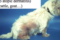
Endocrinopathies are typically characterized by what kind of clinical skin presentation? |
bilateral alopecia non-pruritic (may have pruritis from secondary infections/conditions) systemic signs often see recurrent pyoderma histopathology: hyperkeratosis, epidermal hyperpigmentation, follicular dilation, lack of hair shafts |
|
|
What are 5 examples of endocrinopathies that cause skin pathology? |
1. hypothyroidism 2. hyperadrenocorticism 3. hyperestrogenism 4. hypo and hypersomatotropism 5. castration/growth hormone responsive dermatitis |
|
|
calcinosis cutis is a systemic symptom of what endocrinopathy that also presents with skin pathology? |
hyperadrenocorticism |
|
|
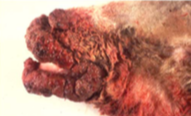
what are the clinical signs and microscopic lesions of zinc deficiency? In pigs, what bacterial skin infection has a similar presentation? |
clinical signs: papules, scaly plaques, crusts, fissures lesions: parakeratosis, acanthosis, secondary pustular dermatitis, epidermal hyperplasia In pigs: similar to superficial pyoderma, staphylococcal exudative dermatitis (greasy pigs) |
|
|
what nutritional deficiency leads to poor hair quality as well as hypomyelinogenesis and anemia? |
copper deficiency |
|
|
exposure to UV radiation can have a variety of presentations depending on how chronic the condition is. What is typically seen with acute damage? |
erythema edema sunburn cells (apoptotic keratinocytes) hyperkeratotsis (ortho and para) comedones |
|
|
exposure to UV radiation can have a variety of presentations depending on how chronic the condition is. What is typically seen with chronic damage? |
acanthosis solar elastosis fibrosis pseudocarcinomatous hyperplasia |
|
|
exposure to UV radiation can have a variety of presentations depending on how chronic the condition is. What kinds of neoplasms can be seen with chronic exposure? |
squamous cell carcinoma (white haired areas, cat, cattle) hemangioma, hemangiosarcoma (glaborous skin, dog) melanoma (human) |
|
|
Photosensitization can be categorized into three different subdivisions. What are these? |
1. primary: exogenous (type I) 2. primary: endogenous (type II) 3. secondary: hepatogenous (type III) |
|
|
What are examples of dermatophathologies caused by physical injury? |
chronic trauma causing calluses lack of attrition (overgrown hooves) acral lick dermatitis (hyperkeratosis, fibrosis, dermatitis) psychogenic alopecia (caused by licking) |
|
|
How does ergot poisoning cause damage to the skin? |
fungus on grain or grass seed heads produces toxin: Ergotamine vasoconstriction and thrombosis infarction dermatitis, necrosis/gangrene |
|
|
What types of toxins cause skin necrosis? |
ergot poisoning spider and snake bites |
|
|
What kind of clinical presentation and histophathology is seen with injection site reactions in dogs? |
lesions: focal alopecia, hyperpigmentation histopath: lymphoplasmacytic panniculitis, vasculitis leading to follicular atrophy |
|
|
In serious cases, vaccine site reactions in cats can result in ________ and eventually develop into __________ |
granulomatous dermatitis sarcomas |
|
|
What is seborrhea? |
hyperkeratosis of epidermis and follicles secondarily, see change of flora (staph) resulting in characteristic smell |
|
|
What causes seborrhea?
|
primary: idiopathic disorder of cornification secondary: due to unrelated disorders (allergy, endocrinopathies, dietary deficiencies, internal dx, etc) |
|
|
Seborrhea is clinically divided into two categories based on presentation. What are these? |
seborrhea oleosa (greasy) seborrhea sicca (dry, scaly) |
|
|
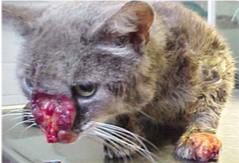
eosinophilic granulomas can be easily confused with what neoplastic disorder? why? |
mast cell tumor both produce nodules/papules aspirate of both will show eosinophils and multinucleate giant cells |
|
|
In dogs, the accumulation of calcified material over pressure points is known as ___________ |
calcinosis circumscripta |
|
|
What are some causes of hyperpigmentation? |
primary: acanthosis nigricans (genetic) secondary: inflammation, irritation, metabolic disorders proliferative lesions: lentigines (lentigo, non-neoplastic, macular to raised) |
|
|
hypopigmentation of the skin is called _______ hypopigmentation of the hair is called _______ |
leukoderma leukotrichia |
|
|
What are some causes of hypopigmentation? |
inherited hypomelanosis (most extreme: albinism) acquired (from injury): mild (pigment can rebound) or severe (death of melanocytes, no recovery) |
|
|
What are some causes of laminitis? |
alimentary carbohydrate overload toxemia sepsis all cause edema of the coronary band |
|
|
ichthyosis is also known as _________ and is most common in which two species? |
fish scale disease cattle and dogs |
|
|
What is the difference between atrichia and hypotrichosis? |
atrichia: congenital aplasia, absence of hair hypotrichosis: less than normal amount of hair. typically hereditary. |
|
|
collagen dysplasia results in what clinical presentation? |
hyperextensibility decreased tensile strength, so skin tears easily |
|
|
What are characteristics of benign vs. malignant neoplasms? |
Benign: slow, expansive, local Malignant: fast, invasive, disseminates/metastasizes |
|
|
Is it possible to differentiate clinically between a nodule (inflammation) and a neoplasm? |
often NOT (unless cytology is done) |
|
|
Is it possible to differentiate clinically between a benign or malignant neoplasm? |
often NOT (sometimes not even with cytology, often requires biopsy) |
|
|
Define these terms: hyperplasia metaplasia dysplasia neoplasia anaplasia |
hyperplasia: increased number of normally differentiated cells. controlled metaplasia: one type of cell is transformed into another dysplasia: cells are disorganized (pre-neoplastic) neoplasia: disorganized, uncontrolled, persists without stimulus anaplasia: cells that are undifferentiated, don't really look like anything (bad news) |
|
|
How is a neoplasm clinically staged? |
based on clinical examination T: size and location N: region lymph nodes involved M: distant sites (e.g. lung) involved |
|
|
How is a neoplasm graded? |
based on hitopathologic characteristics Number of mitotic figures nuclear to cytoplasmic ratio degree of cell differentiation necrosis |
|
|
What are 3 non-neoplastic mass lesions? |
follicular cyst skin tag sebaceous hyperplasia |
|
|
Why do follicular cysts sometimes seem to grow rapidly overnight? |
follicular cyst may rupture, resulting in severe pyogranulomatous dermatitis |
|
|
Sebaceous hyperplasia/adenomas can easily be confused with what type of neoplasm? why? |
melanoma often heavily pigmented |
|
|
What neoplastic masses are common in young dogs? |
cutaneous histiocytoma papilloma (viral neoplasm) |
|
|
What neoplastic mass lesions are epithelial? mesenchymal? |
epithelial: squamous cell carcinoma, trichoblastoma, papilloma mesenchymal: mast cell tumor, cutaneous histiocytoma, fibrosarcoma, hemangioma, hemagiosarcoma, sarcoid |
|
|
What kind skin neoplasm cannot be categorized as epithelial or mesenchymal? What germ layer do these cells arise from? |
melanoma melanocytes arise from neuroectoderm |
|
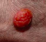
this "raspberry" appearance is typical of what type of neoplasm? what is the prognosis? |
cutaneous histiocytoma excellent prognosis (may actually spontaneously regress) |
|
|
What could a cutaneous histiocytoma by confused with? |
granulomatous inflammation |
|
|
What is the prognosis for mast cell tumors? What should be done to ensure the best approach? |
behavior varies, best to assume the worst take wide margins grading by histopathology is essential |
|
|
vaccine protocols in cats are increasingly recommending injecting in the tail or extremities. Why is this? How does this disease progress? |
vaccine-associate sarcoma granulomatous dermatitis -> sarcomas invasive, poorly demarcated, infiltrative, may require amputation |
|
|
hemangiomas are common dermal to subcutaneous neoplasms. What is their clinical presentation? prognosis? |
dark red to black (don't confuse with melanoma) well demarcated excellent prognosis upon complete excision |
|
|
hemangiosarcomas can be cutaneous or visceral. How do these types differ in origin, presentation, and prognosis? |
cutaneous: from skin, fair prognosis with complete excision visceral: metastasizes early and quickly, poor prognosis, death from blood loss or organ compression (from blood in pericardium or peritoneum). |
|
|
In dogs, melanomas are often _______ In cats, melanomas are often _______ In horses, melanomas are often________ |
In dogs, melanomas are often _benign__ In cats, melanomas are often _malignant_ In horses, melanomas are often_benign or malignant__ |
|
|
In what location are melanomas typically the most malignant? |
oral cavity |
|
|
most papillomas are caused by: |
infections with papillomaviruses |
|
|
what is the most common tumor in the horse? what causes it? |
sarcoid associated with bovine papillomavirus |
|
|
in horses, this type of neoplam is associated with severe tissue destruction with necrosis and ulceration |
squamous cell carcinoma |
|
|
what are the two most common types of neoplasms seen in the nail bed (toe)? |
squamous cell carcinoma melanoma |