![]()
![]()
![]()
Use LEFT and RIGHT arrow keys to navigate between flashcards;
Use UP and DOWN arrow keys to flip the card;
H to show hint;
A reads text to speech;
102 Cards in this Set
- Front
- Back
|
What are the three functions of lymphatic tissue? |
1. immunity- fluid filtered, immune cells stored 2. lipid absorption (lacteals in small intestine) 3. Fluid recovery- absorb plasma proteins and fluid |
|
|
If a pt has some kind of interference with lymphatic drainage, what is the end result? |
severe edema |
|
|
Large lymphatic vessels are composed of three layers. What are they and what are they made of? |
1. tunic interna: endothelium and valves 2. tunica media: elastic fibers and smooth mucsle 3. tunica externa: thin outer layer |
|
|
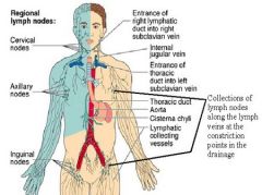
Which structure drains major portions of the body? |
lymphatic trunks |
|
|
Which structure of the lymphatic system courses through many lymph nodes? |
collecting vessels |
|
|
The vein that collects the lymph fluid? |
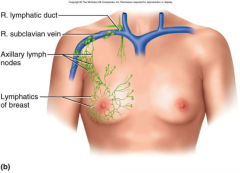
Subclavian Vein |
|
|
Describe the route of lymphatic drainage from each sides of the body. |
|
|
|
Lymph flows at _____________ pressure and speed and is moved along by rhythmic contractions of the ____________. |
LOW Lymphatic vessels |
|
|
The two types of pumps that are involved in lymph flow. If you wanted to rapidly increase lymph flow, what would you tell a pt? |
1. thoracic pump 2. skeletal pump start exercising |
|
|
Lymph tissues also has ____ which prevent back flow. |
valves |
|
|
How does the structure of the vessels leading into the lymph node slow the flow of the lymph fluid? |
one efferent vessel several afferent vessels to slow the flow of lymph out of the node |
|
|
The outer membrane of the lymph node is called a capsule which gives off trabeculae. The function is to |
compartamentalize the nodes |
|
|
The compartments of the lymph nodes contain parenchyma, stroma. What is each composed of? |
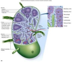
Parenchyma: lymphocytes and APC Stroma: reticular CT |
|
|
The parenchyma of the lymph nodes are subdivided into cortex and medulla. What is found within these structures? Function? |
1. reticular cells and macrophages 2. fx: phagocytes foreign matter lymphoctyes respond to antigens lymphatic nodules location of B cell activation |
|
|
The collective term for all lymph node disease |
Lymphadenopathy |
|
|
A swollen, painful node responding to a foreign antigen is called. |
lymphadenitis |
|
|
A swollen, firm, and usually painless lymph node is likely |
metastatic cancer |
|
|
What type of cells are tonsils covered in? Describe where a pathogen gets in and what it encounters. |
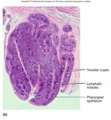
epithelial cells pathogen get into tonsillar crypts and encounter lymphocytes |
|
|
Locate the 6 tonsils. |
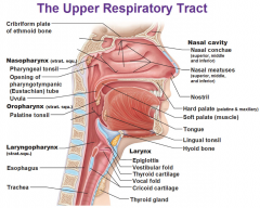
Palantine- pair linqual-pair pharyngeal aka adenoids (single) |
|
|
What type of cells form the blood thymus barrier in cortex? What is the function of the barrier? |
reticular epithelium isolate forming T cells from foreign antigens |
|
|
What is secreted by the cortex of the thymus? |
hormones that promote development and activation of T lymphocytes thymopoeiten, thymulin, thymosis |
|
|
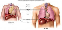
What happens to the thymus as a person matures? |
involution. Mostly gone by the age of 14. mostly just fatty tissue in the elderly |
|
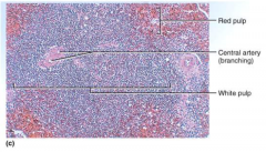
A fresh specimen of the spleen shoes the red and whit pulp. What is in each? |
Red: sinuses filled with erythrocytes White: lymphocytes, macrophages, surround small branches of splenic artery |
|
|
The four major functions of the spleen. |
1. blood production in fetus 2. blood resevoir 3. RBC disposal 4. immune response; filters blood and is quick to respond to antigens |
|
|
An enlarged spleen that is likely due to leukemia, lymphomas, portal hypertension and liver disease. |
Splenomegally |
|
|
In the event of a traumatic event when the spleen is ruptured. What is the treatment? |
Splenetcomy |
|
|
What are the 5 types of leukocytes? |
neutrophils basophils eosinophils monocytes lympocytes |
|
|
Circulating lymphocytes in the blood |
80% T cells 15% B cells 5% NK cells- natural killer cells macrophages |
|
|
Molecules that induce production of antibodies are called |
Antigens |
|
|
Cell surface markers are called |
Major Histocompatability complexes encoded on chromosome 6 example: HLA that is recognized by leukocytes |
|
|
Three proteins that are used to recognize and attack antimicrobial proteins. Where are they produced? What type of immune response are they responsible for? |
Interferons Complement system Cytokines produced by leukocytes and other CT non-specific immune response |
|
|
Proteins that are secreted by certain cells invaded by viruses |
Interferons |
|
|
Describe how an interferon works. |
1. provides general protection against virus 2. diffuses to neighboring cells and stimulates them to produce antiviral proteins. 3. activates natural killer cells and macrophages that destroy infected host cells and stimulate destruction of cancer cells. |
|
|
To activate the complement protein (C) in blood what must be present? What happens to C3 protein. |
pathogen C3 protein splits into C3a and C3b |
|
|
For each of the following pathways, the type of immune response activated. Classical alternative lectin |
Classical: requires antibody: specific Alternative: nonspecific Lectin:nonspecific |
|
|
The defensive response to tissue injury is |
inflammation |
|
|
Name three functions of inflammation |
1. limits spread of pathogens, then destroys them 2. removes debris 3. initiates tissue repair |
|
|
Small proteins that regulate inflammation and immunity that include interferons, TNF, chemotactic factors |
Cytokines |
|
|
The cardinal signs of inflammation are |
1. redness (caused by increased hyperemia) 2. swelling (edema caused by increased capillary permeability and filtration) 3. heat (caused by hyperemia) 4. pain caused by inflammatory chemicals (bradykinin, prostaglandins) that are secreted by damaged cells, pressure on nerves |
|
|
Kinins, histamine, and leukotrienes are secreted by mast cells, basophils, and damaged cells. The release of these chemicals stimulate what two physiologic responses? |
1. vasodilation--> hyperemia --> redness and heat -increases cell metabolic rate for cell duplication and healing. -dilutes toxins, provides O2, nutrients and waste removal 2.increase capillary permeability -allows blood cell and plasma proteins to get into tissue (proteins are AB, Complement proteins, fibrinogen) -clotting sequesters bacteria AND forms scaffold for tissue repair |
|
|
Fibrinogen in the tissue forms the ___ which traps pathogens. |
clot |
|
|
Which leukocyte is the quickest to respond to cellular damage and inflammation? This neutrophils then lead to which 4 physiologic responses: |
Neutrophils 1. phagocytosis 2.respiratory burst 3.secrete cytokines for recruitment of macrophages and neutrophils 4.macrophages and T cells secrete colony-stimulating factor to stimulate leukopoiesis |
|
|
Which cell type is the primary agent of cleanup and arrives 8-12 hours after damage? |
monocytes that become macrophages once they leave the blood stream |
|
|
What does edema at the sight of damage or foreign invader do to the venous flow and lymphatic flow? Why? |
decreases venous flow increases lymphatic flow Favors the removal of bacteria and debris |
|
|
What are the components of pus? |
tissue fluid, cellular debris, dying neutrophils, and microbes |
|
|
a wound that closes before it is healed. Perfect anaerobic condition for bacteria. May lead to sepsis |
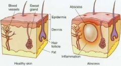
abcess |
|
|
The three types of necrosis |
1.coagulative: denaturation through acidosis ex. MI 2. Liquefactive: infectious agents with immune attack: ex. pus 3. Caseous: chronic infections & inflammation ex. cheesy granulomas TB |
|
|
The cytokine that is released by blood platelets and endothelial cells that stimulates fibrinogenesis and angiogenesis during tissue repair. |
Platelet derived growth factor |
|
|
Hyperemia facilitates tissue repair in which two ways? |
1. provides needed materials 2. increases metabolic activity via heat |
|
|
The scaffold for tissue repair is provided by |
fibrin |
|
|
How does pain aid in tissue repair? |
limits use of body part allowing for repair |
|
|
Cellular immunity is mediated by what cells? |
T cells |
|
|
Humoral immunity is mediated by? |
B cells Antibodies |
|
|
The four components of specific immunity include. |
1. specificity and memory 2. cellular immunity 3. humoral immunity 4. Lymphocytes |
|
|
Where are T cells made? Where do they mature? Where are they deployed? |
made: red bone marrow mature: thymus deployed: naive T cells colonize lymphatic tissue and organs |
|
|
Which protein stimulates maturing T cells in the thymus to produce antigen receptors? |
thymosins *immunocompetent T cell has antigen receptors in place* |
|
|
What type of cells become memory cells? |
T cells. *long lived, higher in numbers than naive T cells" |
|
|
What is the T cell recall response? |
upon re-exposure to same pathogens, MEMORY cells launch a quick attack. |
|
|
Plasma cells are also known as |
B lymphocytes or B cells |
|
|
Describe lifecycle of a B cell. 2 |
B cell --> B cell clones --> produce specific AB B cell --> B memory cell |
|
|
B memory cells have a 30 year half life, and provide which type of immunity? |
long term * presence of specific AB means exposure* *vaccinations work through the B cell mediated pathway* |
|
|
Describe the role of macrophages when encountering a foreign organism, cellular debris, or cancer cells. |
1. engulf and inject particles 2. amoeboid movement 3. stimulate the immune system 4. recruit T-cells: identify the pathogen 5. Wound healing- eat up pathogen and damaged tissue |
|
|
Which type of cells attack foreign cells and diseased cells mediated by memory? |
T cells |
|
|
What are the three classes of T cells? What does each type do? |
1. Cytotoxic T cells -- direct attack 2. helper T cells- promote Tc cell and B cell action and nonspecific defense mechanisms 3. memory T cells-provide immunity to future exposing to antigen |
|
|
Describe the four steps that NK cells does to take action. |
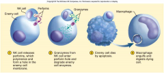
1. |
|
|
The three steps in humoral immunity. |
1. recognition 2. attack 3. memory |
|
|
called the antibody-mediated beta cellularis immune system, is the aspect of immunity that is mediated by macromolecules (as opposed to cell-mediated immunity) found in extracellular fluids such as secreted antibodies, complement proteins and certain antimicrobial peptides. |
Humoral Immunity |
|
|
an immune response that does not involve antibodies, but rather involves the activation of phagocytes, antigen-specific cytotoxic T-lymphocytes, and the release of various cytokines in response to an antigen. |
cell mediated immunity |
|
|
The five stages in the humoral mediated immune response. KNOW THE CHART |
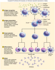
1. antigen recognition 2. antigen presentation 3. clonal selection 4. differentiation 5. attack |
|
|
What type of cells produce antibodies (immunoglobulins) |
B cells |
|
|
Concentrated antibodies derived from donor blood used to boost immune response for certain infections ex. Hep A |
gamma globulins |
|
|
The process whereby antibodies are removed from plasma. |
Plasmophoresis (used to treat MG, SLE, GB) |
|
|
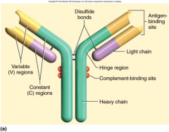
Describe the structure of an AB. |
|
|
|
How are antibodies classified? |
by the amino acid sequence of C region of antibody |
|
|
Type of AB that is a monomer in plasma, dimer in mucus, saliva, tears, milk, intestinal secretions prevents adherence to epithelia |
IgA |
|
|
type of AB that is a monomer B cell membrane antigen receptor |
IgD |
|
|
type of AB that is a monomer found on mast cells stimulates the release of histamine attracts eosinophils immediate hypersensitivity reactions |
IgE |
|
|
type of AB that is a monomer 80% circulating crosses the placenta to fetus secondary immune response complement fixation |
IgG |
|
|
type of AB that is a pentamer 10% in plasma, primary immune response agglutination complement fixation |
IgM |
|
|
Syndrome classified as polyneuropathy of the CNS causing ascending paralysis. Weakness in the extremities progressing to the trunk which can eventually paralyze diaphragm. |
Guillen Barre Syndrome Due to an autoimmune response to infectious organism (viral or bacteria) |
|
|
The treatment for Guillen Barre |
plasmapheresis IV immunoglobulins |
|
|
The four mechanisms in the attack of a pathogen via humoral immunity. |
1. neutralization- AB mask pathogenic region of AB 2. complement fixation: antigen bind to IgM or IgG--> shape change--> complement binding 3. agglutination: AB can bind to 2-10 enemies at a time, immobilizing them 4. precipitation: AB binds to antigen molecules--> AB/AN complex precipitates -->phagocytized by eosinophil |
|
|
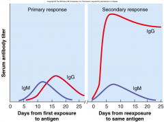
Describe how the humoral immunity response changes between the primary and secondary response. |
|
|
|
Excessive immune reaction against antigens that most people can tolerate |
hypersensitivity (allergens) |
|
|
The four types of hypersensitivity/allergy responses.
What is the type? What is involved? What is the response? |
1.Type I: IgE -- acute reaction 2. Type II: IgG & IgM-- subacute 3. Type III: IgG & IgM- subacute 4. Type IV: cell mediated: delayed (lymphocytes) |
|
|
Anaphylaxis, asthma, and anaphylactic shock are due to what type of hypersensitivity? |
Type I acute |
|
|
Most common chronic illness in children. Inhalation of allergen, releases histamines, which bronchiole constricts |
Asthma Type I: acute |
|
|
Occurs in sensitized people allergen binds IgE, mast cells, basophils release inflammatory chemicals |
Anaphylaxis Type I acute |
|
|
Bronchiole constriction, dyspnea, vasodilation, shock, death txt epinephrine |
Anaphylactic Shock Type I actue |
|
|
Antibody dependent cytotoxic hypersensitivity IgG or IgM mediated -binds to antigens on cells: complement activation and lysis -may bind to cell surface receptors and either interfere with function or over-stimulate the cell |
Type II hypersensitivity subacute |
|
|
Immune Complex Hypersensitivity IgG or IgM form widespread AB/AN complex complex precipitate and trigger intense inflammation |
Type III: subacute **involved in acute glomerulonephritis and in systemic lupus erythematosus SLE*** |
|
|
Delayed hypersensitivity 12-72hr APC in lymph nodes display antigens to helper T cells, which secrete interferon and cytokines that activate cytotoxic T cells and macrophages |
Type IV hypersensitivity cosmetic and poison ivy allergy TB skin test |
|
|
All of the following are examples of what? - Myasthenia Graves -Systemic Lupus Erythematosis -MS (CNS myleniation) -RA - synovial fluid -Guillen Barre |
Autoimmune diseases |
|
|
An autoimmune disease develops due to |
Failure of self tolerance -cross reactivity -abnormal exposure of self-antigens -changes in structure of self-antigens production of autoantibodies |
|
|
How are autoimmune diseases treated? |
immunosuppressants tissue transplantation steroids cyclosporin-- fungal derivative that inhibits T-cells and B-cells |
|
|
An immunodeficiency disorder which is caused by a hereditary lack of T and B cells. The person becomes vulnerable to opportunistic infection |
SCID Sever Combine Immunodeficiency Disorder |
|
|
An immunodeficiency disorder where by helper T cells, macrophages, and dendritic cells are invaded and "tricked" to internalize a the virus by receptor mediated endocytosis |
HIV ***uses enzyme reverse transcriptase*** RNA --> RT --> DNA |
|
|
Treatment for HIV |
antiretroviral "reverse transcriptase inhibitors" |
|
|
increase in cells (mitosis) |
hyperplasia |
|
|
increase in cell size |
hypertrophy |
|
|
cancer cells |
neoplastic |
|
|
decrease in number of cells |
hypoplasia |