Reading...
![]()
Play button
![]()
Play button
![]()
Use LEFT and RIGHT arrow keys to navigate between flashcards;
Use UP and DOWN arrow keys to flip the card;
H to show hint;
A reads text to speech;
52 Cards in this Set
- Front
- Back

#15
|
Sclera- white of the eyes-protects eye |
|

#3
|
Cornea- anterior bulging from the sclera-lets light enter the eyes
|
|
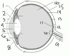
#13
|
Choroid- the highly vascular, dark brown later of the vascular tunic (uvea)-nutrition to the eye
|
|

#1
|
Ciliary body-thickened ring of tissue surrounding the lens-control lens shape
|
|

#6
|
Iris- colored part of the eye- controls dilation of pupil
|
|

#5
|
Pupil- behind the iris- allows light to enter the eye
|
|

#14
|
Retina- Inner layer of the eyeball- collect light, phagocytize, store vitamin A
|
|

#11
|
Fovea Cenrtalis- in the center of the macular lutea-only cones, high acuity color vision
|
|

#12
|
Macula Lutea-in the dead center of the posterior of the eye- mostly cones
|
|

#9
|
Optic Nerve
|
|

#10
|
Optic Disk- where the optic nerve leaves the eye
|
|

#16
|
Vitreous Humor-jelly- transmits light, supports superior structures, keeps intraocular pressure
|
|

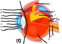
#2
|
Aqueous Humor- in anterior chamber, supplies oxygen and nutrients to the lens and cornea
|
|

#7
|
Canal of Sclemm-venous channel that surrounds the eye in the angle of the sclera-cornea junction- drains aqueous humor
|
|

#4
|
Lens-in anterior chamber, changes shape to allow focus
|
|

#8
|
Conjunctiva- mucous membrane that lines the eye lid and eye
|
|

Area before line @ #16
|
Anterior Segment- 2 parts Cornea to Iris- Anterior Chamber Iris to Lens- Posterior Chamber |
|
Area after line @ #16
|
Posterior Segment
|
|

#1
|
Lacrimal gland
|
|

#2
|
Nasolacrimal duct
|
|

#3
|
Lacrimal sac
|
|
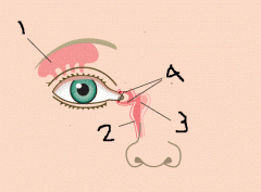
#4
|
Canaliculi- tear duct
|
|

#1
|
Superior Rectus
|
|

#2
|
Lateral Rectus
|
|

#3
|
Inferior Oblique
|
|

#4
|
Inferior Rectus
|
|

#5
|
Medial Rectus
|
|
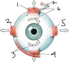
#6
|
Superior Oblique
|
|

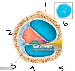
#1
|
Ganglion cell layer
|
|

#2
|
Bipolar cells
|
|
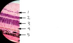
#3
|
Nuclei of rods and cons
|
|

#4
|
Rods and cones
|
|

#5
|
Choroid layer
|
|

#1
|
Scala Vertibuli
|
|

#2
|
Cochlear Duct
|
|

#3
|
Basilar membrane
|
|
#4
|
Organ of corti
|
|

#5
|
Scala tympani
|
|

#6
|
Tectorial membrane
|
|

#1
|
Eustachian tube
|
|

#2
|
round window
|
|

#3
|
Vestibule
|
|
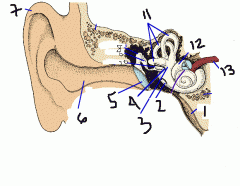
#4
|
Oval window
|
|

#5
|
Tympanic membrane
|
|

#6
|
External auditory meatus
|
|

#7
|
Pinna
|
|

# 8
|
Malleus
|
|

#9
|
Incus
|
|

#10
|
Stapes
|
|

#11
|
Semicircular canals
|
|

#12
|
Cochlea
|
|

#13
|
Cochlear Nerve
|