![]()
![]()
![]()
Use LEFT and RIGHT arrow keys to navigate between flashcards;
Use UP and DOWN arrow keys to flip the card;
H to show hint;
A reads text to speech;
24 Cards in this Set
- Front
- Back
|
URINARY SYSTEM Horse Kidney, anatomy |
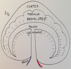
- Heart-shaped, fused papillae ("unilobar"). - Renal crest which feeds to pelvis. - Renal pyramides with base and apex. - Renal glands (foam in eq urine) |
|
|
URINARY SYSTEM Bovine Kidney, anatomy |
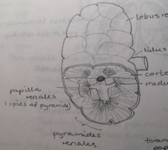
- Multilobar with external lobation. - Multipapilar (internal). - Renal pyramids with base and apex. - Pyramids feed into calices. - No renal pelvis. - Left kidney pushed onto right side of abd. cavity (bo., ov., cap.) |
|
|
URINARY SYSTEM Canine+feline kidney, anatomy |
- Fused papillae ("Unilobar") - Renal crest which feeds to pelvis. - Renal pyramids. - Cats have 1 renal papilla, cortex is yellow due to high lipid content. |
|
|
URINARY SYSTEM Pig kidney, anatomy |
- Multilobar (no external division). - Multipapillar. - + Pelvis renalis. - Minor calix (apex of papilla), major calix (leading to pelvis). |
|
|
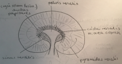
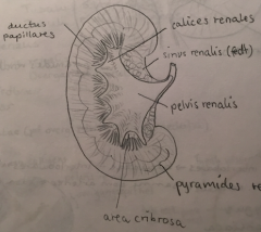
URINARY SYSTEM Kidney, general anatomy |
- Cortex and medulla. - Fused papillae with renal crest (ca., eq., ov.), single renal papilla (fe.), multiple papillae (bo., su.). - Only bo. have external lobular division. - Cortex red in most species, yellow in cats (high lipid content). - Medulla divided into renal pyramids with a base/apex (towards pelvis). - Papillary ducts->minor calices->major calices->pelvis->ureter. - Renal sinus surrounds pelvis and consists of adipose tissue. |
|
|
URINARY SYSTEM Kidney, vasculature |
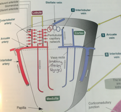
- Interlobar artery along boundary of each lobe. - Branches at right angle to form arcuate a., runs along corticomedullary junction. - Intolobular a. branch from arcuate a. and extend into cortex. These are end-arteries (susceptible to ischemic necrosis). - Interlibular vein->arcuate vein->interlobar vein. - Efferent arterioles give rise to vasa recta: vessels around the Loop of Henle. |
|
|
URINARY SYSTEM The Nephron |
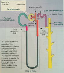
- Functional unit of the kidney. - Glomerulus, Capsule of Bowman, prox. conv. tubule, thick desc. limb, Loop of Henle, thick asc. limb, dist. conv. tubule, collecting tubule/duct. - Epithelium of the dist. conv. tubule forms the Macula Densa, where in contact with Capsule of Bowman. |
|
|
URINARY SYSTEM The Capsule of Bowman |
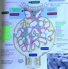
- Tuft of capillaries, lined by fenestrated epithelium and supported by podocytes (foot processes). - Mesangial cells are present in a glycoproteinmatrix (the mesangium) that they produce. They also phagocytize, produce inflammatory mediators and contract capillaries (controls glomerular blood flow). - Inside-out: capillary-basement membrane-fenestrated endothelial cell-podocytes (structure)-urinary space-squamous epithelial cell (capsule)-basement membrane. |
|
|
URINARY SYSTEM The Juxtaglomerular Apparatus |
- Four components: 1) Afferent arteriole with modified myoepithelial cells producing renin. 2) Efferent arteriole. 3) Macula densa 4) Extraglomerular mesangium - Main role: register blood pressure and release renin if decreased (Renin-Angiotensin-Aldosterone system) |
|
|
URINARY SYSTEM Renin-Angiotensin-Aldosterone System |
- BP down (registered by juxtaglomerular apparatus). - Renin released from Juxtaglomerular cells. - Converts circulating angiotensinogen til angiotensin I. - ACE (angiotensin-converting enzyme) in the lung endothelial cell converts angiotensin I to angiotensin II: - Constricts afferent renal arterioles, maintains renal BP, stimulates aldosterone secr. from adrenal gland (Na reabsorbtion), stimulate ADH release from pituitary gland (H2O reabsorbtion). -> increased BP. |
|
|
URINARY SYSTEM Types of glomerulonephritis |
1) Embolic nephritis/Suppurative glomerulitis
- Bacteremia, microabscesses in cortex (Actinobacillus equuli foals). 2) Viral glomerulitis - Viremia, pinpoint red lesions cortex (canine hepatitis) 3) Immune-mediated glomerulonephritis - Persistant infection, deposition in glomeruli, pinpoint red lesions cortex, dx immunoflourenscense 4) Glomerular amyloidosis AA amyloidosis (acute phase protein), deposition of fragments, tan and waxy kidneys 5) Glomerulosclerosis - "End-stage kidney", fibrous connective tissue, diffuse or multifocal, shrunken kidney |
|
|
URINARY SYSTEM Azotemia and uremia |
- Azotemia:
Intravascular increase of nitrogenous waste products. Can lead to renal failure and uremia. - Uremia: The clinical syndrome caused by azotemia toxicosis. |
|
|
URINARY SYSTEM Uremia, non-renal lesions |
- Pulmonary edema (increased vascular permeability). - Fibrinous pericarditis (incr. vasc. perm.). - GI ulcers (ammonia secretion and vascular necrosis). - Ulcerative and necrotic stomatitis (ammonia secretion in saliva). - Hypoplastic anemia (lack of erythropoietin production in kidney). - Soft-tissue mineralization (altered Ca-P metabolism). - Fibrous osteodystrophia (altered Ca-P metabolism). - Parathyroid hyperplasia (altered Ca-P metabolism). |
|
|
URINARY SYSTEM Chronic renal failure, Explain altered Calcium-Phosphorus metabolism (Uremia) |
- Kidney function below 25% of normal. - Phosphorus not adequately secreted. - -> hyperphosphatemia. - Ionised calcium in serum reduced as a result of precipitation of Ca and P. - Parathryoid hormone is released to stimulate Ca-release from bone resorption. - Worsened by kidneys' reduced ability to convert Vit D to calcitriol, resulting in decreased intestinal absorption of Ca. -> Parathyroid chief cell hyperplasia (renal secondary parathyroidism), osteodystrophia, soft tissue calcification (pleura, stomach, lungs, kidney). |
|
|
URINARY SYSTEM Acute renal failure |
- When >75% function lost. - Prerenal (decreased perfusion), renal (compromised kidney function) or postrenal (obstruction). - Results in azotemia and uremia. - Can be caused by hypoperfusion, intoxication, systemic disease. - Sx: sudden onset, uremia signs, proteinuria, elevated creatinine, phosphorus, decreased Ca, bicarbonate. |
|
|
URINARY SYSTEM Acute Tubular Necrosis |
- Most important cause of acute renal failure - Result of nephrotoxic damage or ischemia to tubular epithelial cells. - Nephrotoxins because 25% of blood flow comes through kidneys and is metabolized within renal tubules to active metabolites. - Ischemia numerous causes. - Characteristic histologic find: "tubulorrhexis" = disruption of tubular basement membrane. - Gross lesions: pale, swollen, bulges, excessively moist, striations, medulla pale or congested. - Micro: glomeruli normal (resistant to ischemia), necrosis signs (swell, then disintegrate). - Sx: Oliguria/Anuria |
|
|
URINARY SYSTEM Name some toxic reasons for acute tubular necrosis |
- Ethylene glycol (antifreeze) - Hemoglobin/Myoglobin in large conc. (nyreslag) - Antibiotics (aminoglycosides, tetracycline) - NSAIDs (aspirin, carprofen, flunixin) - Plants (Pigweed, Red Maple) - Vit D toxicity (calcification mitochondria, necrosis) - Cisplatin (antineoplastic) |
|
|
URINARY SYSTEM Pyelonephritis |
- Suppurative inflammation of the renal pelvis with tubulointestinal extension. - Bacterial in origin, always ascending from lower urinary system (in vet.), therefore more common in females. |
|
|
URINARY SYSTEM Tubulointerstitial nephritis |
- Bacteremia or viremia, spread to kidney tubules and interstitial - Leptospira, canine hepatitis, malignant catarrhal fever |
|
|
URINARY SYSTEM Granulomatous nephritis |
- = Tubulointerstitial nephritis with granuloma formation (chronic). - Feline coronavirus, Aspergillus, toxocare etc. |
|
|
URINARY SYSTEM Renal hyperemia and kongestion, definition |
- Hyperemia = increased flow. - Kongestion = decreased drainage. Can be caused by inflammation, hypovolemic shock, cardiac insuff. and more |
|
|
URINARY SYSTEM "Renal cortical necrosis" (DIC) |
- When DIC causes widespread thrombosis in glomerular capillaries -> widespread cortical infarction and necrosis. - Partial or complete. - Bilateral. |
|
|
URINARY SYSTEM Infarction, kidneys |
- Due to occlusion of renal capillary from: - Thrombembolism from valvular endocarditis, - Or parasitic infection and inflammation, neoplastic emboli, mural thrombus (thrombus formed in heart or large vessel). - Most commonly wedge-shaped inflammation or fibrosis in cortex, less commonly larger areas or whole kidney. |
|
|
URINARY SYSTEM Cystitis |
- E. coli (all species) - Corynebacterium renale (cattle) - Eubacterium suis (pigs) - Klebsiella sp. (horses) - Also Proteus, Strep. and Staph in several. |