![]()
![]()
![]()
Use LEFT and RIGHT arrow keys to navigate between flashcards;
Use UP and DOWN arrow keys to flip the card;
H to show hint;
A reads text to speech;
27 Cards in this Set
- Front
- Back
|
HEART Basic Anatomy |
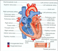
- "Try Pulling My Aorta" - Trabeculae, papillary muscles, chordae tendinae - Foramen ovale, lig. arteriosum. - Superior/Inferior vena cava into right atrium. - Pulmonary veins into left atrium. - Asc. aorta->brachiocephalic trunk, left common carotid artery, left subclavian a.->desc. aorta - pulm. trunk->right pulm. arteries, left pulm. arteries. |
|
|
HEART Myocyte |
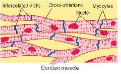
- Cylindrical, branching, elongated nuclei. - intercalated discs - Conducting tissue, incl. SA node, AV node, AV bundle (Bundle of His), parking fibers |
|
|
HEART Concentric vs. eccentric hypertrophy |
- Concentric hypertrophy, caused by addition of sarcomeres to myocytes in parallel, results in an increase in cardiac wall thickness and reduced chamber volume. - Eccentric hypertrophy, caused by addition of sarcomeres in series, leads to a large, dilated ventricle with relative wall thinning (eccentric = store armbevægelser) |
|
|
HEART Cardiac Tamponade |
also known as pericardial tamponade, is when fluid in the pericardium (the sac around the heart) builds up and results in compression of the heart. Onset may be rapid or more gradual |
|
|
HEART Portals of entry |
- Hematogenous
- Local extension - Parasitic migration - Embolism - Trauma - Idiopathic (catheters etc.) |
|
|
HEART Jet lesions |
- Localised areas of subendocardial fibrosis, usually in the atria, thought to be due to abnormal jets of blood caused by valvular lesions. |
|
|
HEART Types of inflammation |
- Pericarditis - Fibrinous/Purulent - Endocarditis - Valvular endocarditis (bacterial infection, valves) - Mural endocarditis (bacterial infection, heart wall) - Ulcerative endocarditis (uremia) - Myocarditis - Hematogenous - Suppurative, necrotizing, hemorrhagic, lymphocytic, eosinophilic - Ex. parvovirus, distemper, clostridial diseases, toxoplasmosis, neospora |
|
|
HEART Valvular diseases |
- Hematocysts - Lymphocysts - Calcification (Vit D/Tox) - Endocardiosis (myxomatous) - Endocarditis - Ruptured chordae tendinae |
|
|
HEART Degeneration/Accummulations |
- Fatty degeneration - Abundant lipid droplet in sarcoplasm of myocytes - Hydrophic degeneration - Vacuolar accummulation in myocytes damaged by toxic injury. - Lipofuscinosis/"Brown atrophy" - Aged/cachexia, brown granules in myocytes = amorphous debris ("residual bodies"). - Myofibrillar degeneration/"Myocytolysis" - Distinctive sublethal injury to cardiac cells, eosinophilic sarcoplasm, myofibrillar lysis. |
|
|
HEART Causes of necrosis |
Nutritional deficiencies - VitE/Selenium, K, Cu, thiamine, Mg Toxicities - catecholamines, ionophores, carcinogenic plants and many more Physical injury - CNS lesions ("Heart-brain syndrome"), gastric volvulus, electrical defibrillation Shock - Hemorrhagic shock |
|
|
HEART Cardiomyopathies |
- Primary/Idiopathic - Hypertrophic cardiomyopathy - Dilated Cardiomyopathy - Restrictive cardiomyopathy - Secondary - Hereditary, nutritional (taurine), toxic, trauma, shock, endocrine, infectious, neoplastic. - Diseases that have extra cardiac cause but directly affect cardiac muscle |
|
|
HEART Grading heart murmurs |
Grade I: Only heard in quiet surroundings after prolonged listening Grade II: Soft murmur, easily heard Grade III: Moderate intensity murmur Grade IV: Loud murmur, no precordial thrill Grade V: Loud murmur with palatable precordial thrill Grade VI: Very loud murmur, precordial thrill, can be heard with stethoscope lifted from chest wall |
|
|
HEART AV-Block |
- 1st degree - Delayed conduction btw atria and ventricles, no clinical signs other than ECG: constant, prolonged PR-interval. - 2nd degree - Type I: Gradually incrasing delay in conduction, increasing PR-interval. Type II: Some P-waves are not let through AV-node, ECG: some standalone P waves without QRS. - 3rd degree - Complete block of AV-node. Atria and ventricles contract separately and totally random. ECG random distribution of P and QRS complexes. |
|
|
HEART Neoplasms |
- Rhabdomyoma, rhabdomyosarcoma: rare, grey nodules. - Schwannoma: cardiac nerve tumor in cattle, incidental finding. - Hemangiosarcoma: dogs, primary or secondary |
|
|
HEART Pulsus paradoxus |
abnormally large decrease in systolic blood pressure during inspiration. The normal fall in pressure is less than 10 mm Hg. When the drop is more than 10 mm Hg, it is referred to as pulsus paradoxus. |
|
|
CARDIOVASCULAR SYSTEM Vasa Vasorum |
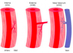
The vasa vasorum (Latin, "the vessels of the vessels") is a network of small blood vessels that supply the walls of large blood vessels, such as elastic arteries (aorta) and large veins (venae cavae). |
|
|
CARDIOVASCULAR SYSTEM Postmortem clotting vs. thrombus |
- Postmortem: red (jelly) or white ("chicken fat") clots can be readily removed or flushed at necropsy. - Thrombus: adherent to underlying tissue |
|
|
CARDIOVASCULAR SYSTEM Aneurysm |
- = localised dilation or outpouching of a thinned or weakened portion of an artery. - Idiopathic, Infection (strongly vulgaris eq.), nutritional (copper def. su.) - Usually rupture -> death. (varicosity/phlebectasia if local/generalised veins) |
|
|
CARDIOVASCULAR SYSTEM Arteriosclerosis, arterial medial calcification |
- Arteriosclerosis Age-related, intimal fibrosis of large arteries secondary to chronic degeneration of arterial wall ->, loss of elasticity and luminal narrowing, abdominal aorta commonly affected. - Arterial medial calcification Mineralization of arterial walls, often also endocardial mineralization, carcinogenic toxicosis, Vit D toxicosis, renal insufficiency. |
|
|
CARDIOVASCULAR SYSTEM Thrombosis |
- = Intravascular coagulation during life - Predisposing factors: 1) Endothelial damage 2) Turbulence or stasis 3) Hypercoagulative state - Adheres to endothelium (post-mortem clots can be readily removed) |
|
|
CARDIOVASCULAR SYSTEM Disseminated Intravascular Coagulation/DIC |
- Formation of micro thrombi throughout cardiovascular system. - Caused by endothelial damage with exposure of sub endothelial collagen and initiation of the coagulation cascade - Bacterial endotoxemias, generalised viral infections, dirofilaria, neoplasia, shock, hemolysis, extensive tissue necrosis (animals with burns) - Depletion of clotting factors lead to widespread haemorrhage |
|
|
CARDIOVASCULAR SYSTEM Infarction |
- Thombosis and embolism lead to occlusion of an end-arteriole, causing anoxia and ischemic necrosis. - Myocardia (coronary artery), kidney and more |
|
|
CARDIOVASCULAR SYSTEM Embolism |
- = occlusion of arteries by lodgment og foreign materials - Thrombemboli, neoplastic cells, bacteria, fat, parasites, foreign body, air bubbles, fracture segments |
|
|
CARDIOVASCULAR SYSTEM Varicosity/Phlebectasia definition |
- = venous dilation from weakened vascular wall in a vein. - Varicosity = localised - Phlebectasia = generalised - Uncommon in animals |
|
|
CARDIOVASCULAR SYSTEM Phlebitis, omphalophlebitis definition |
- phlebitis = Inflammation of a vein - Common, often complicated by thrombosis - Omphalophlebitis = inflammation of umbilical vein/'Navel ill' |
|
|
CARDIOVASCULAR SYSTEM Lymphangieectasis, definition |
- = dilation of lymph vessel - congenital or acquired (blockage) |
|
|
CARDIOVASCULAR SYSTEM Lymphangitis |
- Inflammation of lymph vessel - a feature of many disease, often distal limbs, cordlike structures |