Reading...
![]()
Play button
![]()
Play button
![]()
Use LEFT and RIGHT arrow keys to navigate between flashcards;
Use UP and DOWN arrow keys to flip the card;
H to show hint;
A reads text to speech;
49 Cards in this Set
- Front
- Back
|
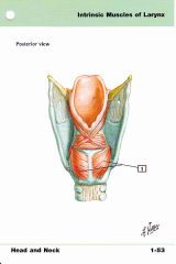
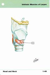
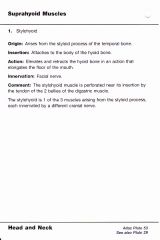
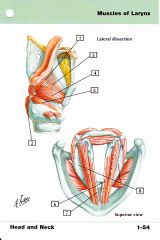
Sites of the Supraglottis
|
Suprahyoid epiglottis
Infrahyoid epiglottis False vocal cords Aryepiglottic folds |
|
|
Sites of the larynx
|
Supaglottis
Subglottis Glottis |
|
|
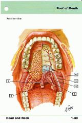
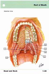
Sites of the oral cavity
|
mucosal lip
buccal mucosa lower alveolar ridge upper alveolar ridge retromolar trigone Floor or mouth Hard palate Anterior two thirds of tongue |
|
|
Sites of the oropharynx
|
Base of tongue
Vallecula Soft palate uvula anterior tonsillar pillar posterior tonsillar pillar pharyngeal tonsils glossotonsillar sulci Lateral and posterior pharyngeal walls |
|
|
Sites of the Hypopharynx
|
Post-cricoid region
Pyriform sinuses Lateral and Posterior hypopharyngeal wall |
|
|
Borders of the Oropharynx
|
Superior: superior surface of the soft palate
Inferior: Superior surface of the hyoid bone(vallecula) Anterior: Circuvellate papillae |
|
|
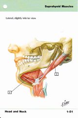
Major salivary gland sites
|
Parotid glands
Submandibular glands Sublingual glands |
|
|
Hypopharynx Borders
|
Superior: An imaginary plane connecting: anteriorly, the base of the vallecula (inferior border of hyoid) and laterally to pharyngoepiglottic folds.
Posterior/Lateral: Posterior and lateral hypopharyngeal walls. Inferior: Lower border of the cricoid Medially: Lateral wall of epiglottis, aryepiglottic folds, lateral laryngeal walls |
|
|
Cribiform plate
|
CN I: Olfactory Nerve
Anterior ethmoidal artery |
|
|
Optic Canal
|
CN II: optic nerve
Opthalmic Artery |
|
|
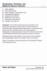
Superior orbital fissure
|
CN III: oculomotor nerve
CN IV: trocheal nerve Opthalmic division of CN V Opthalmic Vein Orbital branch of the middle meningeal artery Recurrent branch of the lacrimal arteries Sympathetic plexus |
|
|
Foramen spinosum
|
Recurrent branch of the mandibular nerve
Middle meningeal artery and vein |
|
|
Internal acoustic meatus
|
CN VII: facial nerve
CN VIII: vestibularcochlear nerve Internal auditory artery from basilar artery |
|
|
Foramen ovale
|
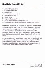
CN V V3: mandibular dvision of trigeminal nerve
|
|
|
Hypoglossal canal
|
CN XII: hypoglossal nerve
Meningeal branch of ascending pharyngeal artery |
|
|
Foramen magnum
|
Spinal cord/medulla oblongata
Spinal accessory nerve Vertebral arteries Anterior and posterior spinal vessels |
|
|
Foramen rotundum
|
CN V V2: maxillary division of trigeminal nerve
|
|
|
Jugular foramen
|
Anterior: Inferior petrosal sinus
Intermediate portion: CN IX (glossopharyngeal), X (vagus), XI (spinal accessory nerves) Posterior portion: Transverse sinus, meningeal branches of the occipital and ascending pharyngeal arteries |
|
|
CN IX: glossopharyngeal nerve
|
Swallowing
Enervates the carotid body Taste to posterior 2/3 of tongue |
|
|
CN I: olfactory nerve
|
Smell
|
|
|
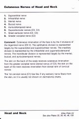
CNV V V1: Opthalmic nerve function
|
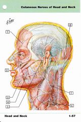
Sensory innervation of the scalp, forehead, nose and globe
|
|
|
CNV V V1: Opthalmic nerve course
|
Courses in the lateral cavernous sinus wall below CN4, exiting the skill through the superior orbital fissue and entering the orbit where it divides into the lacrimal, frontal, and nasociliary nerves
|
|
|
Pyriform Sinus Definition
|
A subsite of the hypopharynx. It is a cone shaped structured covering the posterior 3rd of the larynx. The wide end is at the level of the hyoid and the narrow apex is near the middle of the cricoid. There is no barrier to tumor spread here. The apex level on CT scan is the cricoarytenoid joint.
|
|
|
Cervical LN Level V Borders
|
Superior: Cranial edge of body of hyoid
Inferior: Tranverse cervical vessels Anterior: Posterior edge of SCM Posterior: Anterolateral border of the trapezius Lateral: Platysma Medial: Paraspinal muscles |
|
|
Cervical LN Level VI Borders
|
Superior: Inferior edge of thyroid cartilage
Inferior: Sternal manubrium Anterior: Skin, platysma Posterior: Separation between trachea and esophagus Lateral: Medial edges of thyroid gland, skin, and anteromedial edge of SCM |
|
|
Retropharyngeal Nodes
|
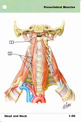
Superior: Base of skull
Inferior: cranial edge of the body of the hyoid Anterior: Fascia under the pharyngeal mucosa Posterior:Prevertebral muscles Lateral: Medial edge of the internal carotid Medial: Midline |
|
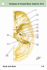
Foramen of the base of skull
|
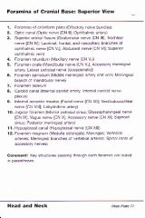
Contents and names of foramen
|
|
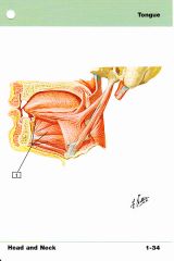
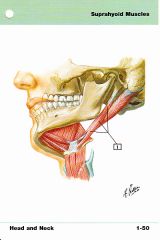
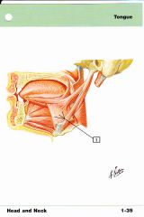
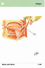
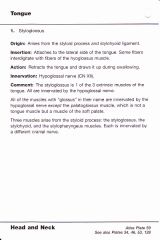
Extrinsic muscles of the tongue: Saggital View
|
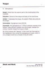
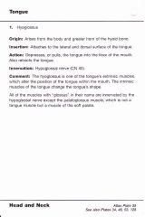
Extrinsic Muscles of the tongue: Names, Origins and Insertions
|
|
|
|
|
|
|
|
|
|
|
|
|
|
|
|
|
|
|
|
|
|
|
|
|
|
|
|
|
|

|
|
|
|
|
|
|
|
|
|
|
|
|
|
|
|
|
|
|
|
|
|
|
|
|
|
|
|
|
|
|
|
|
|