Reading...
![]()
Play button
![]()
Play button
![]()
Use LEFT and RIGHT arrow keys to navigate between flashcards;
Use UP and DOWN arrow keys to flip the card;
H to show hint;
A reads text to speech;
152 Cards in this Set
- Front
- Back
|
What layer are the photoreceptor nuclei located in?
|
Outer nuclear layer
|
|
|
In between what two layers of the retina is Henle's fiber layer and what is it?
|
Henle's fiber layer is between the outer nuclear layer and the outer plexiform layer. Henle's fiber layer consists of photoreceptor cell axons branching out radially from the fovea
|
|
|
Ganglion cells are divided into small midget cells and large magno (M) cells? What is another name for the midget cells?
|

Midget or parvo cells
|
|
|
What is another name for magno (M) cells?
|
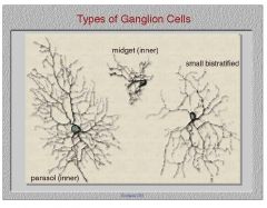
Parasol cells
|
|
|
What percent of retinal ganglion cells can be classified as parvo cells? What percent are parasol cells?
|
80% are midget cells a.k.a. parvo (P) cells, 10% are parasol (M) cells.
|
|
|
How big is the foveola in mm?
|

0.3mm
|
|
|
How big is the fovea in mm?
|

1.8mm
|
|
|
How big is the macula in mm?
|
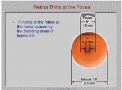
5.8mm
|
|
|
Do amacrine cells synapse in the outer plexiform layer of the retina or the inner plexiform layer of retina?
|
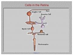
the inner plexiform layer
|
|
|
1mm retina=___ degrees visual angle
|
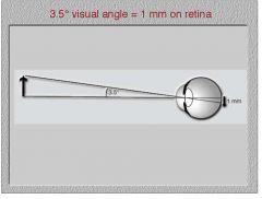
3.5 degrees visual angle
|
|
|
Does the what pathway or the where pathway proceeds from the dLGN to the Inferotemporal cortex?
|
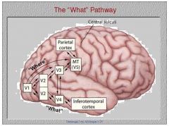
The what pathway
|
|
|
Does the what pathway or the where pathway proceed form the dLGN to the parietal cortex
|

|
|
|
Which layers of the LGN are part of the parvo pathway? Which are part of the magno pathway?
|

Layers 1 and 2, the ventral layers are part of the magno pathway. Layers 3,4,5,6, the dorsal layers are the parvo pathway
|
|
|
Which layers of the LGN originate in the ipsilateral eye? Which layers originate in the contralateral eye?
|
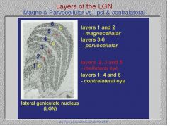
U-235 Layers 1,4, and 6 are crossed. Layers 2,3, and 5 are uncrossed.
|
|
|
What wavelength of light is most likely to be absorbed by rhodopsin (rod photopigment)?
|
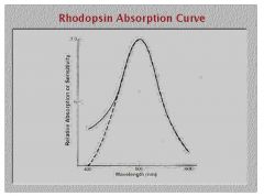
507nm
|
|
|
Do rod or cone photoreceptors exhibit a synaptic ending flat pedicle shape?
|
cones
|
|
|
How long does it take for 50% of cone pigments to recover from bleaching?
|
1.5 min
|
|
|
How long does it take rod pigments to recover from bleaching?
|
5 min
|
|
|
What is the difference in sensitivity between scotopic and photopic of a different wavelength termed?
|
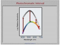
photochromatic interval
|
|
|
What wavelength of light absorbs the most light under photopic conditions?
|
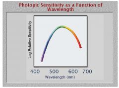
555nm
|
|
|
What is the term for the relative increase in brightness of longer wavelength stimuli as lighting conditions change from scotopic to photopic?
|
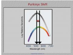
Purkinje Shift
|
|
|
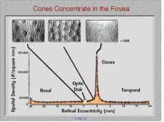
At what number of degrees from the fovea are rods most densely packed?
|
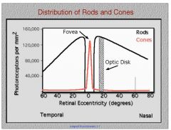
20 degrees from the fovea
|
|
|
At 20 degrees from the fovea, the rods reach a peak density of how much?
|

|
|
|
How many rods are in the fovea?
|

none, so you can't see small dim object such as a star when it's foveally fixated
|
|
|
What is the density of cones at the fovea?
|

150,000, same as rods
|
|
|
Are extrafoveal or foveal cones larger?
|

extrafoveal cones tend to be larger.
|
|
|
Are cones present in the peripheral retina?
|
yes, and they're larger though less concentrated than at the fovea
|
|
|
How many quanta of light are needed to produce a response in a rod?
|
1
|
|
|
How many quanta of light are required to be visible?
|

7 quanta
|
|
|
Under the minimally distinct border method, do smaller differences in luminance create fuzzier or clearer borders?
|
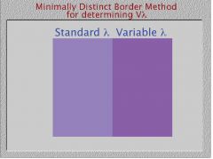
fuzzier
|
|
|
Are rods or cones more sensitive at short wavelengths?
|
rods are more sesnsitive, they have a lower threshold
|
|
|
At what wavelength do rods and cones become nearly equally sensitive?
|
at the longest wavelengths in the visible spectrum
|
|
|
What is the name of the fact that light entering the center of the pupil is more likely to be absorbed than lighet entering the periphery of the pupil?
|
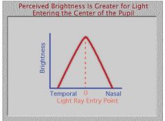
Stiles-Crawford effect
|
|
|
Is the Stiles-Crawford effect more predominant in infants or adults?
|
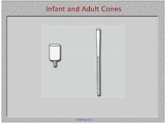
Adults have longer and narrower cones
|
|
|
What law states that Time times Intensity equals a Constant, so long as the presentation time is within the Critical Duration
|
Bloch's law
|
|
|
What is the critical duration for rods?
|
100ms
|
|
|
What is the critical duration for cones?
|
10ms
|
|
|
Which photoreceptors exhibit higher resolution of temporal frequency?
|
cones
|
|
|
Which photoreceptors exhibit a higher resolution of spatial frequency?
|
cones
|
|
|
Which law states that area times Intensity equals a constant, so long as the area is within the critical area
|
Ricco's law
|
|
|
What part of the pupil does Ricco's law hold true?
|
central pupil
|
|
|
What law explains retinal summation in the periphery?
|
Piper's law
|
|
|
What property of light determines hue?
|
difference in wavelength, ROYGBIV
|
|
|
What do you call the lamp that mimics luminant C or looking north in mid November (cloudy)?
|
MacBeth lamp
|
|
|
Which wavelengths can people distinguish the smallest difference in size?
|
the middle of the visual spectrum
|
|
|
Which wavelength appears most desaturated?
|
yellow
|
|
|
What do you call the phenomena that shows a change in hue correlated to a change in brightness?
|
Benzold-Brucke phenomena
|
|
|
Will the resultant color be brighter or darker using subtractive color mixing, e.g. paints and pigments?
|
darker, add them all together and you get black
|
|
|
When using additive color mixing what color results from adding all colors together?
|
white light
|
|
|
If a neutral density filter transmits 10% of the light through it what is it's optical density?
|
look up
|
|
|
Is optical density addtive color mixing or subtractive color mixing?
|
additive color mixing
|
|
|
What do you call two different colors that have different wavelength composition that appear to be the same hue?
|
metamers
|
|
|
Under the CIE color mixing system what size wavelength do X, Y, and Z correcsponde to?
|
X-long (red)
Y-medium (gree) Z short (blue) |
|
|
What are the three properties of color?
|
1. hue
2. saturation 3. brightness |
|
|
At what value of X, Y, and Z do you get white light?
|
X=0.33, Y=0.33, Z=0.33, all equal
|
|
|
Where on the CIE diagram are colors most saturated?
|
towards the edges, away from the middle
|
|
|
Using the CIE diagram determine the hue
|
1. Connect the two wavelengths to determine the mixture.
2. Draw a line through White (0.33) and mixture to find the hue. |
|
|
How do you find the complement color on CIE diagram?
|
Connect the wavelength to W and see where the line passes through on the other side.
|
|
|
How do you define nonspectral purples?
|
Just draw a line through W to define the complement color of purple
|
|
|
What is the Young-Helmholz theory of color vision?
|
trichromatic theory
|
|
|
What is the opponent process theory of color vision?
|
look up
|
|
|
Where is the Trichromatic theory correct?
|
photoreceptor level
|
|
|
Where is the opponent process theory correct?
|
ganglion cell level
|
|
|
Are acquired or congenital color defects more often manifest symmetrically between the two eyes?
|
congenital
|
|
|
Do acquired or congenital color vision deficits usually present with normal visual function elsewhere?
|
congenital
|
|
|
Are patients with congenital or acquired color vision problems more likely to name colors correctly?
|
congenital
|
|
|
Are acquired or congenital color vision problems more likely to get progressively worse?
|
acquired
|
|
|
What is the suffix of color vision anomaly classification terms for a patient with only two photopigments working?
|
anopes
|
|
|
What is the suffix of color vision anomaly classification terms for a patient with three photopigments but one is not normal?
|
anomaly
|
|
|
Where do normal people turn people normally adjust the hue on the Nagel anomaloscope?
|
the middle 43
|
|
|
What type of dichromat would turn the brightness down on the red side of the spectrum?
|
protanope
|
|
|
What type of dichromat would see all colors as equal brightness on the anomaloscope?
|
deuteranope
|
|
|
What common condition effects diabetes?
|
S cones
|
|
|
In which anomalos trichromat is the M wavelength shifted toward the L?
|
Deuteranomalous
|
|
|
The yellow macula lutea absorbs short blue wavelengths creating a dark spot when looking at a blue field. This dark spot is also used to determine where a patient is fixating. What do you call this spot?
|

Maxwell's spot
|
|
|
What causes Haidinger's brush to appear to rotate?
|
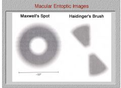
Henle's layer (the inner retinal layers pushed aside at the fovea) has pigments that polarize light that cause the brush to appear to rotate
|
|

1. Can action potentials be at different amplitudes?
2. Once you get an action potential, will it continue without additional help? |
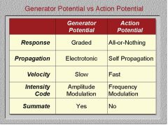
1. Graded potentials can be at different amplitudes.
2. Yes action potentials are self propagating. |
|
|
|
|
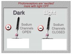
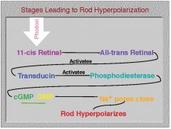
Is glutamate released more when the lights are off or when they are on at the outerplexiform layer?
|
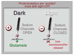
More glutamate is released in the dark in the outer plexiform layer.
|
|

1. What type of bipolar cells receive input from many photoreceptors?
|

1. diffuse bipolar cells
|
|
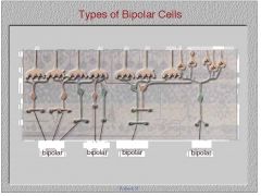
1. What bipolar cells receive input from often only one photoreceptor?
2. Do rod photoreceptors and cone photoreceptors pass through the same kind of bipolar cells? |
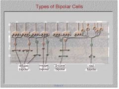
1. midget bioplar cells
2. Rods pass through rod bipolar cells, a different path than cones. |
|
|
1. What do you call a synapse in which whatever response you have at the photoreceptor is reversed at the bipolar cell?
2. Does glutamate inhibit or excite an on bipolar cell? |

1. sign inverting synapse
2. glutamate inhibits an on bipolar cell so it will be shut off in the dark |
|
|
Which bipolar cells are more active or hyperpolarizing when the light is on?
|
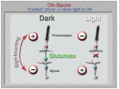
on bipolar cells, that's how they get their name
|
|
|
Are off bipolar cells more or less active when the light is off?
|
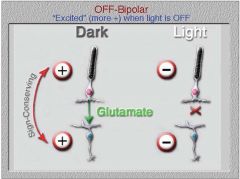
Off bipolar cells possess a sign conserving synapse meaning that whatever happens at the photoreceptor happens at the bipolar cell. So if the photoreceptor is shut down in the dark, so is the bipolar cell. Remember light slows down the photoreceptors release of inhibitory glutamate
|
|
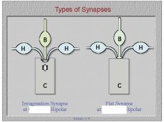
Do on or off bipolar cells have an invaginating synapse?
|
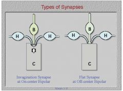
On of course
|
|
|
1. What effect does lateral inhibition have on contrast?
2. Is this effect most important for temporal, color, or spatial vision? |
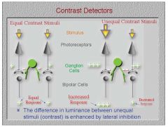
1. Inhibitory retinal cells amplify the difference in luminance.
2. This is important in spatial vision because it allows us to see where objects begin and end. |
|
|
Do ganglion cells or bipolar cells possess the on/off type of cell? that is center surround type cell
|
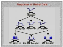
Ganglion cells possess the on/off type in addition to the on type and off type. On off type cells respond to a change in light intensity.
|
|
|
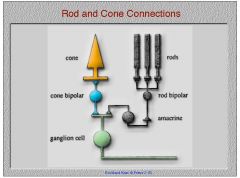
1. Do rods connect directly to ganglion cells?
2. Do cones connect directly to ganglion cells? |
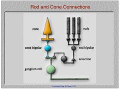
1. The rod system never connects directly to a ganglion cell. It only connects indirectly through a special rod bipolar cell and indirectly through an amacrine cell, kind of piggbacking onto the cone system. The amacrine cell in the rod pathway is called the A2 amacrine cell.
|
|
|
Is the NFL thicker at the fovea or in the periphery? Why?
|
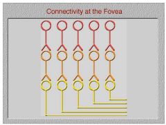
1. The NFL is thicker at the fovea because of the lack of convergence from the outer retinal cells requiring more ganglion cells
|
|
|
1. What property of light must be increased so that it is detected centrally as well as peripherally?
|
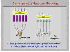
1. Light intensity must be increased for foveal detection.
|
|
|
What is a receptive field by definition
|
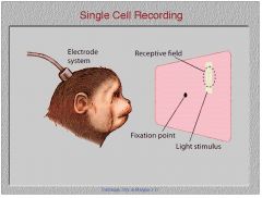
A receptive field is that place out in space that a stimulus can be placed and effect a particular neuron that we are recording from.
|
|
|
Is the surround field of an on center bipolar cell center surround receptive field due to excitatory inputs from bipolar cells or inhibitory inputs from bipolar cells
|
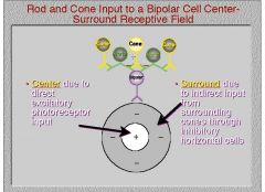
The surround portion of the bipolar cell receptive field is due to inhibitory inputs from surrounding bipolar cells
|
|
|
Will an on center bipolar cell hyperpolarize or depolarize when you present an annulus that stimulates the surround portion of the receptive field?
|
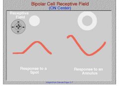
The surround for an on center bipolar cell turns it off or in other words, hyperpolarizes it making the potential more negative.
|
|
|
When the annulus of an off center receptive field is stimulated, will the cell hyperpolarize or depolarize?
|
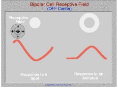
When the annulus of an off center bipolar cells receptive field is stimulated, the cell will depolarize, voltage potential gets more positive.
|
|
|
Is an on response characterized by a few spikes when the light goes on a property of ganglion cells, bipolar cells, or amacrine cells?
|
In amacrine cells, an on response means a couple of spikes when the light goes on.
|
|
|
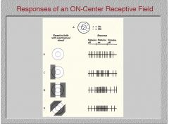
What does a spot of light falling on the excitatory center of a ganglion cell receptive field do to the number of spikes produced?
|
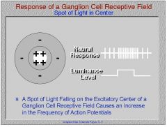
Ganglion cells exhibit action potentials rather than graded potentials, hence a spot of light on the excitatory center of the receptive field will cause an increase in the number of spikes.
|
|
|
What neural response will result from diffusely illuminating a ganglion cell?
|
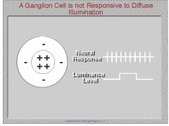
baseline activity since the excitatory center and inhibitory surround are being stimulated equally
|
|
|
What does the Westheimer function plot tell us about ganglion cell sensitivity to different size lights?
|
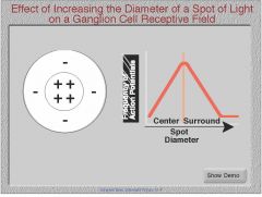
The frequency of action potentials is close to zero for a very small stimulus, then increases with stimulus size until you reach the size of the ganglion cell receptive field. The frequency of action potentials then decreases until your spot is big enough to cover the surround as well as center of the receptive field.
|
|
|
Does the orientation of a stimulus increase or decrease the frequency of ganglion cell action potential responses?
|
Retinal receptive fields are round hence a line stimulus at any orientation will still hit the same amount of center and surround
|
|
|
Is the dendritic field diameter of a retinal ganglion cell greater in the periphery or less?
|
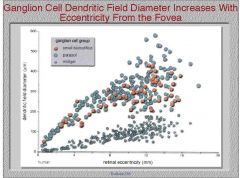
greater dendritic cell diameter as eccentricity increases
|
|
|
Does the where or what pathway travel from V1 to V2 to V4 to inferotemporal cortex?
|

the what pathway
|
|
|
Does the where or what pathway travel from V1 to V2 to V4 to MT cortex to parietal cortex?
|

the where pathway
|
|
|
Between what two visual areas do we see exchange in information between the where and what pathways?
|
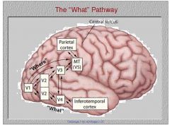
V3 from the where pathway and V4 from the what pathway exchange information
|
|
|
Are retinal on/off center ganglion cells considered part of the M or P pathway?
|
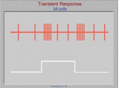
On off center ganglion cells produce a transient response and are only sensitive to changes in light
|
|
|
1. Are on center receptive field ganglion cells and off center receptive field ganglion cells considered a part of the M or P visual pathways?
2. Do they produce a transient or a sustained response? |
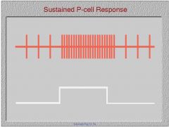
1. They are part of the P pathway.
2. They produce a sustained response to light. |
|
|
Will an on center/off surround retinal M cell decrease its response level if both the on center and off surround are stimulated instead of just the on-center part of the receptive field?
|
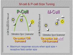
No, retinal M cells response does not decrease when the off surround part of the receptive field is stimulated.
|
|
|
Will a small difference in luminance produce a big change in the response of an M cell at low contrast level? Will this change at a high contrast level?
|
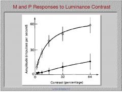
M cells respond a lot to luminance change at low contrast but no so much at higher contrast.
|
|
|
How much do P cells respond to change in luminance at a low percentage of contrast? What about a higher percentage of contrast.
|

P cells respond very little to not at all to luminance changes at a low percentage contrast, but don't taper off in response at higher percentage contrast.
|
|
|
Does hue change in an isoluminant grating? Does an isoluminant grating isolate P cells or M cells?
|
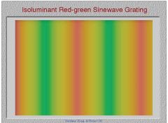
Luminance is held constant. Hue still changes. P cells are isolated.
|
|
|
What wavelength are M cells most sensitive to in the surround portion of their receptive field? center?
|
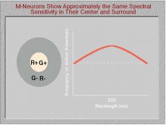
M cells are equally sensitive to 550nm wavelength light in the center and surround portion of their receptive fields
|
|
|
What is the term for the property of P cells due to the fact that their action potentials respond more or less frequently to different colors?
|
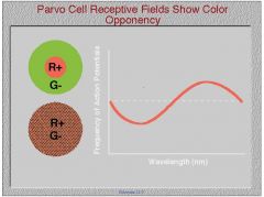
color opponency
|
|
1. Do P cells respond in a spatially linear or nonlinear fashion?
2. What about M cells? |
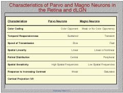
1. P cells respond in a spatially linear fashion.
2. M cells respond in a spatially linear or nonlinear fashion. |
|
|
Where are corresponding points on the LGN?
|
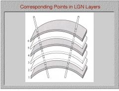
in the same vertical line one on top of the other.
|
|
|
Is the stria of Genera present in both striate and extrastriate cortex?
|
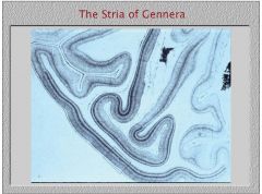
No, the stria of Genera is what differentiates striate cortex from the rest of cortex?
|
|
|
1. Is striate cortex mostly exposed on the surface of the brain?
|
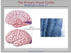
1. No striate cortex is mainly exposed on the calcarine fissure.
|
|
|
Is layer 1 of striate cortex located further inward or outward in the brain?
|
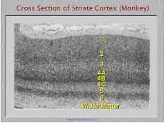
most outward is 1, most inward is layer 6
|
|
|
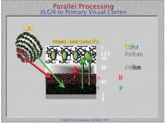
What layer of striate cortex do M cells project to?
|
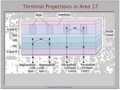
M cells project to layer 4B and 4C alpha
|
|
|
What layer of striate cortex do P cells project to?
|

layer 4C beta
|
|
|
1. Do P cells project to the blob or interblob region of striate cortex?
2. What layers of striate form blob and interblob regions when stained with cytochrome oxidase? |

1. P cells project to the blob region
2. Blob and interblob regions are formed when striate cortex layers II and III are stained. |
|
|
At what layer of striate cortex do the receptive fields become more elongated rather than just circular as in the dLGN?
|

Once you get outward from layer 4C (which still has circular receptive fields)
|
|
|
What do you call a target that is in the same orientation as the receptive field of a simple cell in layers outer to 4C of striate cortex?
|
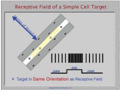
the preferred orientation
|
|
|
What do you call the orientation of a target at which a simple cell in striate cortex does not respond at all?
|
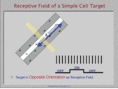
the null position
|
|
|
Is the forming of a simple cell's receptive field from the receptive field of dLGN neurons due to linear or nonlinear summation?
|
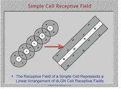
dLGN neuron receptive fields add up linearly to form simple cell receptive fields.
|
|
|
What cells in the visual pathway are the first to respond binocularly or due to input from both eyes?
|

simple cells at striate cortex layers outer to 4C
|
|
|
1. Are complex cell receptive fields formed by linear or nonlinear summation?
|
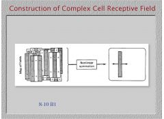
nonelinear summation
|
|
|
Are complex cells more responsive to the particular position of a stimulus or to a particular direction of motion of a stimulus?
|
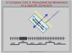
motion
|
|
|
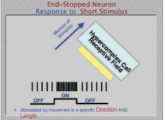
What do you call the length specificity exhibited by a hypercomplex cell?
|

end stopping
|
|
|
Is a hypercomplex cell a different type of cell than a complex or simple cell?
|
No, it's just a simple or complex cell that exhibits the property of endstopping. Hypercomplex is a misnomer.
|
|
|
What do you call cells that are stimulated by movement in a specific direction and of a specific length?
|
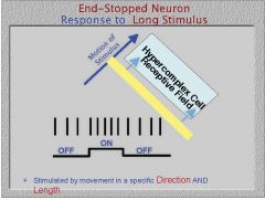
hypercomplex cells
|
|
|
Are the ocular dominance columns from the good eye of an amblyope wider than a binocular patients?
|
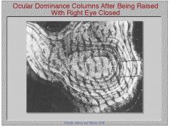
Yes, the ocular dominance columns are wider than normal for an ambloyopes good eye and smaller than normal for his bad eye.
|
|

If you vertically penetrate the striate cortex, will this give rise to an ocular dominance column or an orientation column?
|
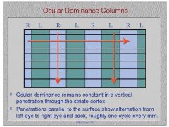
an ocular dominance column
|
|
|
If you insert an electrode perpendicular to the cortical surface, will this give rise to an ocular dominance column or an orientation column?
|
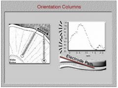
orientation column, same orientation of receptive field but different eyes
|
|
|
What receptive fields are represented in a hypercolumn?
|
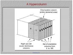
1. all orientations
2. both eyes 3. all spatial frequencies (not shown) |
|
|
1. Do M or P cells input into the blobs?
2. What about the interblobs? |

1. P cells input into the blobs
2. P cells also input into the interblobs. |
|
|
Are blobs or interblobs color specific?
|

blobs
|
|
|
Are the interblobs made up of complex or simple cells?
|

The interblobs are simple cells that respond mostly to orientation and to shape.
|
|
|
1. From layer 4C alpha in striate cortex, what layer of striate cortex do M cells input into?
2. Do they input into simple or complex cells there? |
layer 4B, which contains complex cells responsive to motion
|
|
|
When stained with cytochrome oxidase, what regions of V2 do we see?
|
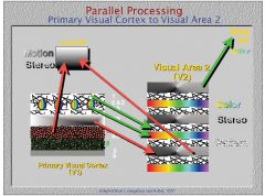
thin stripes, thick stripes, and interstripes
|
|
|
What do the thin stripes in V2 receive input from?
|

blobs
|
|
|
What striate cortex region does the thick stripes in V2 receive input from?
|

blobs
|
|
|
What region of striate cortex do the interstripe regions of V2 receive input from?
|

interblob regions
|
|
|
What are cells in the thick stripe region of V2 selective for?
|

color
|
|
|
What are cells in the thick stripe region of V2 responsible for?
|
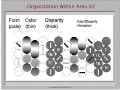
motion and binocular disparity
|
|
|
What are cells in the interstripe region of V2 selective for?
|

form
|
|
|
Where do the thick stripe region cells of V2 project?
|

MT cortex for cells selective for motion and stereovision
|
|
|
Where do the thin stripe region cells of V2 project to?
|

area V4 to cells selective for color
|
|
|
What do the cells of inferotemporal cortex respond best to?
|
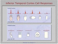
biological features like hands and faces
|
|
|
What are the columns in MT cortex selective for?
|
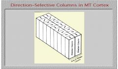
motion unlike striate cortex which is selective for eyes and orieintations
|
|
|
What is the term for when the neuron's center and surround detect differences
in motion between objects and their backgrounds? Where does this happen? |
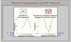
motion segregation, occurs in MT cortex
|
|
|
Where do you first see cells in the visual cortex that can respond to optic flow? What is optic flow?
|
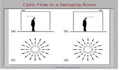
MST (Middle superior temporal cortex), optic flow is the expansion or contraction of objects as you move toward or away from them and is important your view of locomotion through space.
|
|
|
A lesion to what area of the brain would cause a loss of the ability to see color? What that loss called?
|
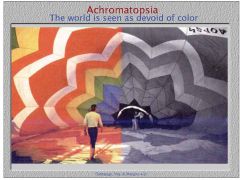
V4, acrhomotopsia
|
|
|
A lesion to what area of the brain would cause an inability to see motion? What do you call this inability?
|
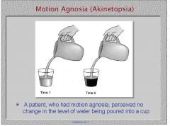
1. MT cortex
2. akinetopsia |