Reading...
![]()
Play button
![]()
Play button
![]()
Use LEFT and RIGHT arrow keys to navigate between flashcards;
Use UP and DOWN arrow keys to flip the card;
H to show hint;
A reads text to speech;
39 Cards in this Set
- Front
- Back
|
|
|
|
|
|
|

|
|

|
|
|
|
|
|
|
|
|
|
|
|
|

|
|
|
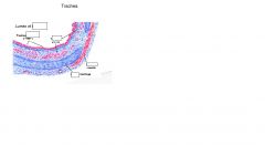
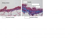
epithelium
|
lines luminal surfaces
|
|
|
lamina propria
|
underlying tissues
|
|
|
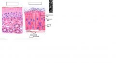
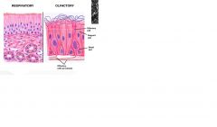
nasal respiratory mucosa
|
compromises :
pseudostratified columnar ciliated epthlelial cells goblet cells sensory brush cells stem cell like basal cells |
|
|
lamina propria of nasal respiratory mucosa contains
|
serous and mucous glands
blood vessels nerves loose connective tissue |
|
|
function of nasal respiratory mucosa
|
warm and humidify incoming air before it reaches the lungs
|
|
|
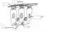
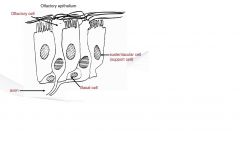
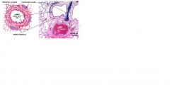
olfactory mucosa is situated in
|
situated in caudodorsal region of nasal cavity including some surfaces of the ethmoid conchae.
|
|
|
olfactory epithelium contrains
|
olfactory cells (cell bodies of olfactory nerve, cranial nerve I, which travel through the cribiform plate of the ethmiod bone directly to the olfactory bulb of the brain for sensing smell)
support cells called sustenacular cells basal cells |
|
|
olfactory lamina propria contains
|
numerous sensory axons
cavenous veins bowmans glands |
|
|
trachea epithelium
|
pseudostratified columnar ciliated epithlial cells lining airway lumen
mucus producing goblet cells |
|
|
trachea lamina propria
|
vascularised supporting tissue containing glands and connective tissue.
elastic fibers in both lamina propria and submucosa to allow for extension during inspiration and elastic recoil during expiration. |
|
|
trachea submucosa
|
elastic fibers
seromucinous glands |
|
|
main support of trachea
|
c shaped hyaline cartilage rings (incomplete) connected to trachealis smooth muscle.
|
|
|
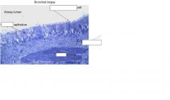
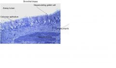
bronchi epithilium
|
pesudostratified columnar ciliated epitheilial cells lining airway lumen.
supporting cartilaginous plates to keep airways from collapse. |
|
|
spirally arranged smooth muscle bundles are located
|
below the lamina propria of the bronchi. contraction and relaxation of these smooth muscle bundles control the degree of airway opening and closing.
|
|
|
bronchial lamina propria
|
is compromised of
connective tissues elastic tissues blood vessles |
|
|
blood vessels in lamina propria of the bronchi stem from
|
the bronchial circulation and provide nutrients to airway wall tissues. in disease states they are also the main exit site for inflammatory cells from the systemic circulation.
|
|
|
bronchioles epithelium
|
pesudostratified columnar ciliated epithlial cells, but smaller ones have cuboidal epithilium.
non ciliated clara cells interspersed no mucus glands no cartilage plates |
|
|
opening and closing of bronchioles is due to
|
bronchiole smooth muscle interaction with elastic fibers surrounding alveolar tissue
|
|
|
respiratory bronchioles lined eith
|
non ciliated cuboidal epitheilium and a small amount of smooth muscle
|
|
|
mucociliary apparatus/clearance system
|
part of respiratory tracts innate defense mechanism which provides protection against deleterious effects of inhaled pollutants, allergens, fungal spores, pollen, dust, bacteria, etc.
it is the first line of defense against inhaled foreign materials. |
|
|
mucociliary aparatus consists of three functional compartments
|
epithelial cilia
airway surface liquid (Sol) layer protective mucus layer |
|
|
in healthy animals rate of mucus secretion and clearance are
|
balanced
|
|
|
mucociliary clearance velocity is faster in
|
larger airways compared to small airways
|
|
|
mucus thick enough to.... but thin enough ...
|
trap bacteria and other inhaled particles
move easily by cilia of epithilium |
|
|
clara cells
|
play an important part in defense mechanisms of smaller bronchiolar airways.
non ciliated cells secrete a variety of products including clara cell secretory protein and a molecule with lung surfactant like properties. responsible for detoxifying harmful substances inhaled into the lungs using cytochrome p450 enzymes found in their smooth ER. also act like stem cell when there is damage to bronchiolar epithelium they can prliferate and differentiate into ciliated cells to regenerate epithilium. |
|
|
sneezing relfex
|
evolved to clear irritants from nose.
|
|
|
sneeze reflex initiated by
|
chemoreceptors in the nose which send afferent neural impulses via the trigeminal nerve to the sneezing center in the medulla region of the brain.
|
|
|
steps of sneezing
|
1. deep inspiration
2. brief clossure of glottis 3. rapid contraction of the respiratory muscles to build up intralung pressue 4. opening og glottis which results in rapid expulsion of air mainly through nasal passage. |
|
|
cough reflex
|
clears irrtants from the pharynx, larynx, trachea and bronchi.
|
|
|
cough reflex intiated by
|
irritants in the larynx and trachea which stimulates the vagal afferent nerve which travels to the medulla in the brain.
|
|
|
steps in coughing mechanism
|
1. deep inspiration
2. brief closure of glottis 3. rapid contraction of respiratory muscles to build up intralung pressue 4. opening of glottis to expel air at a rapid pace through mouth. |