Reading...
![]()
Play button
![]()
Play button
![]()
Use LEFT and RIGHT arrow keys to navigate between flashcards;
Use UP and DOWN arrow keys to flip the card;
H to show hint;
A reads text to speech;
60 Cards in this Set
- Front
- Back
|
C
|
conc. of gas in blood
|
|
|
F
|
Fracional conc. in dry gas
|
|
|
P
|
Partial pressure
|
|
|
Q
|
volume of blood
|
|
|
Q dot
|
volume of blood per unit time
|
|
|
R
|
Respiratory exchange ratio
|
|
|
S
|
Saturation of Hemoglobin
|
|
|
V
|
volume of gas
|
|
|
V dot
|
Volume of gas per unit time
|
|
|
A
|
Alveolar
|
|
|
B
|
Barometric
|
|
|
D
|
Dead Space
|
|
|
E
|
Expired
|
|
|
I
|
inspired
|
|
|
L
|
lung
|
|
|
T
|
Tidal
|
|
|
a
|
arterial
|
|
|
c
|
capillary
|
|
|
c'
|
end-capillary
|
|
|
i
|
ideal
|
|
|
v
|
venous
|
|
|
v with a - on it
|
mixed venous
|
|
|
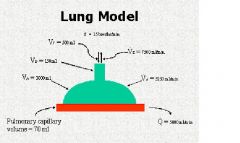
Normal tidal volume
|
500 ml
|
|
|
normal total ventalation
|
7,500 ml/min
|
|
|
normal aveolar gas
|
3,000 ml
|
|
|
normal pulmonary capillary blood
|
70ml
|
|
|
normal pulamanary blood flow
|
5,000 ml/min
|
|
|
normal alveolar ventilation
|
5,250 ml/min
|
|
|
normal frequency of respirations
|
15 per minute
|
|
|
Normal amount of anatomical dead space in respiratory system
|
150 ml
|
|
fill in the normal values
|
values above
|
|
|
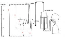
two conditions under which volumes are measured
|
Static- no flow
Dynamic- flow |
|
|
Name the four standard lung volumes
|
1. Tidal volume (VT)
2. Inspriatory reserve volume(IRV) 3. Expiratory reserve volume (ERV) 4. Residual volume (RV) |
|
|
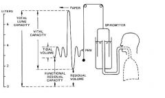
Residual volume
|
volume of air left in lungs after full expiration,, in males normal is approx. 1.5L
|
|
|
Tidal volume
|
volume of air taken in and exhaled, normal approx. 500ml per breath
|
|
|
Inspiratory reserve volume
|
volume air that can be inspired after a normal breath has already been taken
|
|
|
expiratory reserve volume
|
volume of air that can be exhaled after normal expiration
|
|
|
Inspiratory capacity
|
IC=VT+IRV
Max air able to inhale during normal resting expiration males approx.3.5L |
|
|
Functional residual capacity
|
FRC= RV+ERV
vol of air left in lungs after normal expiration |
|
|
Vital capacity
|
VC= ERV+VT+IRV
volume of air that can be expired from the lungs after maximal inspiration |
|
|
Total lung capacity
|
TLC= RV+ERV+VT+IRV
volume of air in the lungsat the end of maximal inspiration |
|
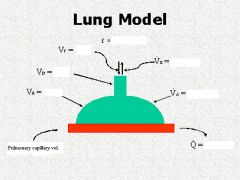
what values are being measured in these areas?
|
above
|
|
|
Name two ways that the residual volume can be measured?`
|
1. Gas dilution
eg. helium- has to mix to read accuratly 2. Plethysomgraphy- thick of a phone booth- accounts all gases by measurment of pressure. |
|
|
Graham's law
|
the rate that any two gases diffuse is inversly proportional the square root of their molecular weight.
|
|
|
Ideal gas law
|
PV=nRT
where p=pressure v=volume n=number of moles of gas T=temp in Kelvin R=ideal gas constant |
|
|
BTPS
|
body temperature, pressure, and saturated...Actual lung volumes are always expressed at BTPS
|
|
|
STPD
|
standard temp, pressure, dry..... used when measuring gas exchange in metabolism,,, such as O2 consumption to CO2 elimination.
|
|
|
ATPS
|
Ambient temperature, pressure, saturated....
|
|
|
What is Graham's equation
|
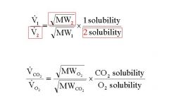
notice the inversion of the values
|
|
|
Henry's law
|
this law describes how much gas is dissolved in a liquid, relative to the pressure of the gas around the liquid.
C = P x S C= content of gas in liquid phase P= Partial pressure of gas in air phase S= solubility of gas in liquid phase |
|
|
Vapor pressure
|
Will exert a pressure in a gas mixture that is proportional to the temperature. Normal is 47 mmHg. Water vapor is important b/c it influences the partial pressure of oxygen as air moves into the lungs from the environment.
|
|
|
Dalton's law of Partial Pressure
|
The total pressure of a mixture of gases is the sum of all the partial pressures.
The pressure exerted by an individual gas is directly proportional to its fractional concentration in the mixture. DRY CONDITIONS |
|
|
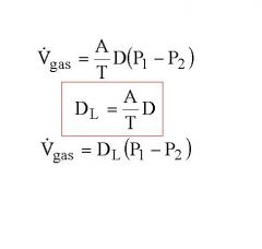
What is the diffusion constant?
|
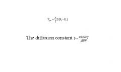
It is used to determine the diffusion of a gas across the aveolar wall.
|
|
|
Explain the two resistences O2 has trying to enter an RBC.
|
1. the resistence of diffusion across the aveolar and plasma membrane
2. the resistence of the O2 to bind with the hemoglobin. |
|
|
Notice it says partial pressure... this demonstrates that the nitrogen is perfussion limited b/c the partial pressure of nitrogen in the air of the aveoli is high, and its diffusion across the aveolar membrane is fast and unimpeded. In contrast, the partial pressure of CO in the lungs is low, and its perfusion into the blood is slower and lower.
|
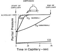
What is this diagram explaining?
|
|
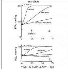
What is this figure showing?
|
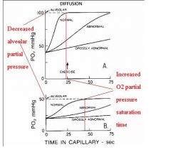
Notice the increased time nessesary to obtain saturation (partial pressure saturation)in the capillary
|
|
|
What is diffusion capacity?
|
Diffusion capacity is a measument of the ability of respiratory memebranes to permit transport of gases. Think - thickness and permiability
|
|
|
Name the 5 muscles that work during inspiration?
|
1.diaphram
2. SCM 3. Scalines 4. External intercostals 5. Anterior Serrati |
|
|
What muscles are used for expiration?
|
1. Abdominal rectus
2. Internal intercostals under resting potential however, no muscle energy is used, it is the built up potential energy that allows for the release (from inspiration) |
|
|
What is hysteresis?
|
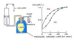
Hysteresis is the cause for the insp/exp curves to be different above. Think of how a new balloon and compare it to that of a balloon after it has been inflate numerous times. Hysteresis is simply the non-equal pressure release caused by viscosity steal by the friction of aveolar inflation on one another. Slows down the release of air. Term Aveolar recruitment
|