![]()
![]()
![]()
Use LEFT and RIGHT arrow keys to navigate between flashcards;
Use UP and DOWN arrow keys to flip the card;
H to show hint;
A reads text to speech;
42 Cards in this Set
- Front
- Back
|
What are the risk factors for DVT? |
Family history Thrombophilia Previous history of thromboembolism Drugs e.g. OCP, tamoxifen, HRT Malignancy Age >40 Varicose veins Chronic illness esp. CCF Recent surgery Immobility Long flights Pregnancy, puerperium Obesity Dehydration |
|
|
What examination findings in DVT might you find? |
Swelling Asymmetry Erythema May have low grade fever Warmth Tenderness Pitting oedema *Don't test Homan's sign (pain on sharp dorsiflexion of foot) as it may dislodge a thrombus |
|
|
What investigations for DVT should you consider? |
Duplex USS - accurate for above-knee, improving for distal calf Repeat in 1 week if normal Contrast venography: use if USS doubtful Can use D-dimer to exclude DT where probability is low Bloods - APTT, INR, platelets, EUCs (renal function) |
|
|
What drugs should be used for treatment of DVT? |
LMW Heparin e.g. Enoxaparin (Clexane) OR Fondaparinux OR Can use unfractionated heparin infusions in hospital PLUS Warfarin - Aim for INR 2-3 (Must be used in combination) *NOACs e.g. rivaroxaban not on etg for rx however can be used under PBS |
|
|
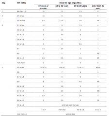
What is the dosing of enoxaparin for treatment of DVT? |
enoxaparin 1.5 mg/kg SC, daily OR enoxaparin 1 mg/kg SC, twice daily (preferable in patients at risk of bleeding e.g. elderly, obese or malignancy) |
|
|
What are the contraindications for enoxaparin (Clexane)? |
Heparin Heparin derivative hypersensitivity Acute bacterial endocarditis Uncontrolled haemorrhage risk incl major bleeding disorder, focal lesion, active ulcerative condition, haemorrhagic stroke Thrombocytopenia assoc with +ve in vitro test for anti platelet Ab * Precaution in: renal (esp ClCr < 30 mL/min), hepatic impairment |
|
|
What are the adverse effects of enoxaparin? |
Thrombocytopenia Thrombocytosis Haemorrhage Anaemia Eosinophilia GI upset Headache Alopecia Oedema Fever Confusion Inj site reaction incl skin necrosis (discontinue), haematoma, pain Elevated LFTs, platelets Hyperkalaemia Hepatocellular, cholestatic hepatic injury Osteopenia Osteoporosis (use > 3 mths) Cutaneous, systemic allergic reaction, spinal, neuraxial haematoma (rare) Hyperlipidaemia (very rare) |
|
|
What is the dosing of rivaroxaban (xarelto) for treatment of DVT/ PE? |
15 mg twice daily for 1st 3 wks, followed by 20 mg once daily for continued treatment |
|
|
What is the basic mechanism of action of rivaroxaban? |
Highly selective direct factor Xa inhibitor |
|
|
What are the contraindications for using rivaroxaban? |
Bleeding: Clinically significant active bleeding (eg intracranial, GI bleeding) Lesions at incr clinically significant bleeding risk Spontaneous haemostasis impairment Liver: Significant hepatic disease assoc with coagulopathy leading to clinically relevant bleeding risk (incl impairment (Child-Pugh B, C)) Kidneys: Dialysis - renal impairment (ClCr < 15 mL/min for 10 mg tab, ClCr < 30 mL/min for 15 mg, 20 mg tabs) Drugs: Concomitant strong CYP3A4 + P-gp inhibitors eg HIV protease inhibitors (eg ritonavir), systemic azole antimycotics (eg ketoconazole) Other: Pregnancy and lactation |
|
|
What are the adverse effects of rivaroxaban? |
GI upset Haemorrhage Blood disorder eg thrombocytopenia, anaemia; incr menstrual bleeding Compartment syndrome Hypotension/ syncope Hypoperfusion induced renal failure; incr muscle tone, cramping Wound healing complications Headache Oedema incl peripheral, allergic, angioedema; Raised LFTs; cholestasis, hepatitis/ jaundice, hypersensitivity (rare) |
|
|
How long should a VTE provoked by a transient major risk factor be treated for? |
3 months |
|
|
How long should an unprovoked distal DVT be treated for? |
3 months |
|
|
How long should a first unprovoked proximal DVT or PE be treated for? |
6 months |
|
|
How long should a person with first unprovoked VTE plus: - active cancer - multiple thrombophilias - antiphospholipid antibody syndrome be treated for with anticoagulation? |
Indefinitely |
|
|
How long should a recurrent unprovoked VTE be treated for? |
Indefinitely |
|
|
What is the basic mechanism of action of enoxaparin? |
The antithrombotic activity is related to inhibition of thrombin generation and inhibition of two main coagulation factors: factor Xa and thrombin. |
|
|
What is a non-pharmacological treatment of DVT? |
Graduated compression stockings |
|
|
What is the basic mechanism of action for warfarin (Coumaden or Marvan)? |
Inhibiting the synthesis of vitamin K dependent coagulation factors |
|
|
What is the dosing of Warfarin when initiating treatment? |
*Murtagh's suggests starting at 5mg for 2 days then adjust base on INR |
|
|
What are the contraindications to Warfarin? |
Bleeding: Blood dyscrasia Where haemorrhage poses greater hazard than anticoagulation eg haemorrhagic tendency assoc with active ulceration, overt bleeding of GI, genitourinary, respiratory tract, cerebrovascular haemorrhage, cerebral aneurysm, dissecting aorta Heart: Pericarditis Pericardial effusion Bacterial endocarditis Other: Recent, contemplated eye, CNS, traumatic surgery Threatened abortion; eclampsia, pre-eclampsia; pregnancy Unsupervised senility, dementia, alcoholism, psychosis Spinal puncture; major regional, lumbar block anaesthesia Malignant hypertension |
|
|
What are the adverse effects of Warfarin? |
Haemorrhage Skin, tissue necrosis Systemic cholesterol microembolism complications incl purple toes syndrome GI upset Fever Hepatic effects Priapism Tracheobronchial calcification (rare) |
|
|
What are the symptoms of PE? |
SOB Retrosternal chest pain Syncope Diaphoresis Vomiting Cyanosis Agitation Haemoptysis Massive PE - hypotension, acute right heart failure or cardiac arrest |
|
|
What sore of pain is associated with PE? |
Dully heavy retrosternal pain that radiates to the lateral chest (pleuritic) |
|
|
What signs are found on examination in PE? |
Tachycardia Decreased pulmonary S2, S3 or S4 +/- adventitious sounds |
|
|
What might you find on a CXR with PE? |
Nothing +/- localised oligaemia or infarction |
|
|
What are the signs of PE on an ECG? |
Normal or R heart strain S1, Q3, T3 |
|
|
What is the first line investigation for PE? |
Radionucleide imaging - the ventilation/ perfusion (V/Q) study |
|
|
What is the gold standard investigation for diagnosing PE? |
Digital subtraction angiography *However CTPA is the mainstay of investigation for PE due to the invasive nature of digital subtraction angiography (requires femoral catheritisation) |
|
|
What non-pharmacological treatments are available for PE? |
Supportive therapy - Oxygen and analgesia |
|
|
What medication is used for the treatment of PE? |
As for DVT except if haemodynamically compromised use - unfractionated heparin 80 units/kg loading dose IV, followed by 18 units/kg/hour IV infusion, adjusted according to APTT and consider - fibrinolytic therapy: alteplase (patients 65 kg or more) 10 mg IV bolus, followed by 90 mg IV infusion over 2 hours. |
|
|
What is important to monitor if someone is on a heparin type of anticoagulant and why? |
Platelet count for heparin induced thrombocytopenia |
|
|
Malaise + weight loss + cough is a diagnostic triad for what? |
Lung cancer |
|
|
What is the most common cause of lung cancer in both sexes? |
Cigarette smoking |
|
|
What are the risk factors for lung cancer? |
Lifestyle factors: • Tobacco smoking Environmental factors: • Passive smoking • Radon exposure • Occupational exposure e.g. asbestos, diesel exhaust • Air pollution Personal factors: • Age • Family history of lung cancer • Previous lung diseases |
|
|
What type of cancer accounts for over 95 % of primary lung malignancies? |
Bronchial carcinoma |
|
|
What features on history might you expect in someone presenting with lung cancer? |
- 50-70 years old - 10-25% asymptomatic at time of diagnosis - If symptomatic usually advanced and not resectable - Cough 42% - Chest pain 22% - Wheezing 15% - Dyspnoea 5% - Weight loos |
|
|
What investigations might you consider in someone presenting with symptoms of lung cancer? |
CXR CT scanning Fibre-optic bronchoscopy PET scanning Flourescence bronchoscopy (helps early detection) Tissue diagnosis where possible |
|
|
What other differentials (other than bronchial carcinoma) might you consider for a solitary pulmonary nodule on CXR? |
Common: Solitary metasis Grauloma e.g. TB Hamartoma Less common: Bronchial adenoma AVM Hydatid |
|
|
What are the different classifications of bronichial carcinoma? |
Small cell lung poorly differentiated (15-20%) AND Non-small cell (approx 20-30% each) - Squamous cell - Adenocarcinomoa - Large cell carcinoma |
|
|
What is the main aim of management in non-small cell lung carcinoma? |
Specialist referral Curative resection |
|
|
What are the treatment options for small cell lung carcinoma? |
Specialist referral Surgery not option as it metastasises rapidly - usually 80% metastasised by time of diagnosis Chemotherapy - extends life from 3-20mnths Radiotherapy - palliative |