![]()
![]()
![]()
Use LEFT and RIGHT arrow keys to navigate between flashcards;
Use UP and DOWN arrow keys to flip the card;
H to show hint;
A reads text to speech;
24 Cards in this Set
- Front
- Back
What structures are outlined (general)? What type of epithelial cells are these composed of? What does the lumen contain? |
Renal tubules - simple cuboidal epithelium Lumen contains provisional urine |
|
|
Tubules found in the pars convoluta |
Proximal convoluted tubule Distal convoluted tubule (Connecting ducts) |
|
|
Tubules found in the pars radiata + outer cortex |
Descending thick limb Ascending thick limb Collecting duct |
|
|
Which nephron structures look the same based on histology? |
Proximal convoluted tubule + descending thick limb Distal convoluted tubule + ascending thick limb Connecting ducts + collecting ducts |
|
|
Characteristics of PCTs and descending thick limbs on EM |
Large cells Numerous microvilli Numerous mitochondria Numerous basolateral infoldings |
|
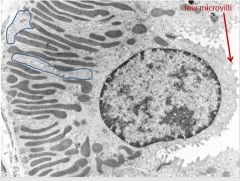
What potential nephron structures could this EM represent? |
PCT or thick descending limb
|
|
What is represented here? |
Basolateral infoldings of PCTs/descending thick limbs of loop of Henle Dark structures = mitochondria |
|
|
LM characteristics of PCT and descending thick limbs |
Nuclei spread out (due to large cells) Fuzzy apical surface of obstructing lumen (microvilli) Eosinophilic cytoplasm (due to numerous cytoplasm) Borders between cells not apparent (due to basolateral infoldings) |
|

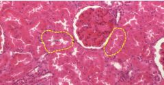
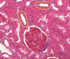
What are the two possibilities for the outlined structures? |
PCT or descending thick limbs (depends on location) - in this case, we are in the pars convoluta so these are proximal convoluted tubules |
|
Outlined structures if we are in the pars radiata? |
Descending thick limbs |
|
|
EM characteristics of distal convoluted tubules + ascending thick limbs |
-Small cells -Few microvilli -Numerous mitochondria -Numerous basolateral infoldings |
|
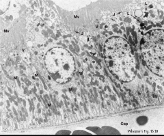
What are some structures visible on this EM? Possibilities for nephron structures? |
-Small cells -Few microvilli -numerous mitochondria (elongated dark structures in basal aspect of cells) -Numerous basolateral infoldings Distal convoluted tubules or thick ascending limbs |
|
|
LM characteristics of distal convoluted tubules and ascending thick limbs of loop of Henle |
-Nuclei are close together (small cells) -Sharp apical surface w/ wide lumen (few microvilli) -Eosinophilic cytoplasm (numerous mitochondria) -Borders between cells not apparent (basolateral infoldings) |
|
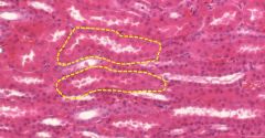
If this micrograph was taken in the pars convoluta, what structures are outlined here? |
Distal convoluted tubules - nuclei close together, sharp apical surface + wide lumen, eosinophilic cytoplasm, borders between cells not apparent |
|
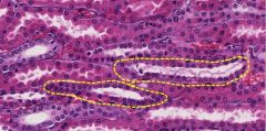
If this LM was taken from the pars radiata, what structures are outlined? |
Thick ascending limbs - nuclei close together, sharp apical surface + wide lumen, eosinophilic cytoplasm, borders between cells not apparent |
|
|
EM characteristics of collecting ducts |
-Small cells -Few microvilli (in most cells) -Few mitocondria (in most cells) -Few basolateral infoldings |
|
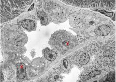
What type of cells are pictured here? (X) |
Collecting ducts |
|
|
LM characteristics of collecting ducts |
-Nuclei close together (small cells) -Sharp apical surface + wide lumen (few microvilli - usually) -Pale cytoplasm (few mitochondria) -Borders between cells visible (few basolateral infoldings) |
|
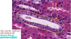
Outlined structure? Arrows? |
Outlined structure = collecting duct Arrows = peritubular capillaries |
|
|
Tubules found in the deep medulla? |
Thin limbs of loop of Henle Collecting ducts Capillaries = vasa recta |
|
|
Both the thin limbs and vasa recta have _____ eqpithelium |
simple squamous |
|
|
How to differentiate between thin limbs + vasa rectum? |
Thin limbs have slightly thicker squamous cells + vasa recta may have RBCs |
|
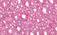
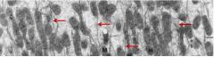
ID the following tubules in the deep medulla |
-Red: collecting ducts - small cuboidal cells, pale cytoplasm, borders between cells visible -Blue: thin limbs of loop of Henle - cells are squamous, but slightly thicker than endothelial cells (no RBCs in lumen) -Purple: vasa recta - flat endothelial cells, RBCs in lumen |
|
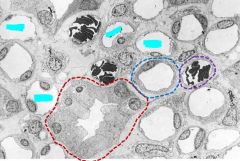
ID the following tubules in the deep medulla |
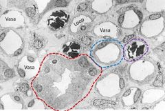
Red = collecting ducts Blue = thin limbs of loop of Henle Purple = vasa recta (note there are also some vasa recta w/out RBCs) |