Reading...
![]()
Play button
![]()
Play button
![]()
Use LEFT and RIGHT arrow keys to navigate between flashcards;
Use UP and DOWN arrow keys to flip the card;
H to show hint;
A reads text to speech;
6 Cards in this Set
- Front
- Back
|
A case presentation of tricuspid atresia
|
A case presentation of tricuspid atresia (important): a newborn shows signs of cyanosis, dyspnea at birth, examination reveals holosystolic murmur (characteristic)audible along (L) sternal border, single 2nd heart sound, (L) ventricular heave. CHEST X-RAY: pulmonary undercirculation, square-shaped heart.
|
|
|
EKG:
|
EKG: characteristic (L) axis deviation, (L) ventricular hypertrophy; almost absent (R) ventricular force.
|
|
|
Next step in management
|
Next step in management: hyperoxia test reveals Pao, less than 100 (normal more than 150 mm Hg) when the patient is receiving 100% oxygen.
|
|
|
MOST LIKELY DIAGNOSIS: tricuspid atresia what is absolute feature
|
MOST LIKELY DIAGNOSIS: tricuspid atresia Please remember, (L) axis deviationis absolute feature of tricuspid atresia. It can also be seen in endocardial cushion defect. In endocardial cushion defect. (R) ventricular force is present Tricuspid atresia in older child presents with above features and polycythemia, clubbing, easy fatigability, cyanosis, extertional dyspnea.
|
|
|
TREATMENTMEDICAL: SU
Keywords: cyanosis, holosystolic murmur, left RGICAL: |
TREATMENT:
(a) MEDICAL: immediately give PGE, (prostaglandin) to keep duct open, so blood flows from aorta to pulmonary artery, oxygen forhypoxia, Sodium bicarbonate for metabolic acidosis. (b) SURGICAL: nodefinitive surgeryisavailable. 1ST STAGE: balloon septostomy is helpful in some patients if gradient between (R) atrium and (L) atrium to increase pulmonary blood flow, or anastomosis of aorta and pulmonary artery. Modified Blalock-Taussig shunt is preferred procedure. It consists of a Gore-Tex conduit anastomosed side-to-side from subclavian artery to homolateral branch of pulmonary artery. It isprocedure ofchoice. 2ND STAGE: bidirectional Glenn shunt (anastomosis between superior vena cava and pulmonary arteries) is performed between 4 and 12 months of age when patients have shown signs of outgrowing a previous aorto-pulmonary shunt. 3RD STAGE: modified Fontan operation (cavopulmonary isolation procedure involves anastomosis between inferior vena cava to pulmonary artery via a baftle that runs along lateral wall of right atrium or via a homograft or Gore-Tex tube running outside heart) is performed between 1.5 and 3 years of age. This procedure reduces incidence of postoperative pleural effusion and right atrial dilatation because blood flows by a more direct pathway into pulmonary arteries. Failed Fontan circuits: heart transplantation is procedure of choice. Keywords: cyanosis, holosystolic murmur, left axis deviation. |
|
|
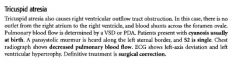
tricuspid atresia
|
|