Reading...
![]()
Play button
![]()
Play button
![]()
Use LEFT and RIGHT arrow keys to navigate between flashcards;
Use UP and DOWN arrow keys to flip the card;
H to show hint;
A reads text to speech;
82 Cards in this Set
- Front
- Back
|
What is the purpose and use of a panoramic ?
|
to evaluate impacted teeth and eruption patterns (root angulations); to detect lesions, diseases, jaw conditions and evaluate trauma
|
|
|
What is a pano NOT useful in diagnosing?
|
caries, periodontal disease, periapical lesions
|
|
|
In a pano, the tubehead and film rotate in the _____ direction of eachother
|
opposite
|
|
|
Radiographic technique that allows imaging of one layer at a time (while blurring other structures in other planes)
|
Tomography
|
|
|
The focal trough is also known as…
|
the "image layer"
|
|
|
What is similar to a camera focusing on an object and the background is blurred?
|
the focal trough
|
|
|
Usually the focal trough is _____ in the anterior region and _____ in the posterior region.
|
narrow, wide
|
|
|
What is the focal trough?
|
the 3D, curved zone where structures are clearly demonstrated on the film
|
|
|
If the teeth are set behind the notch, how will the film appear?
|
a fat anterior
|
|
|
If the chin is too far down, how will the film appear?
|
"Grinch Smile"
|
|
|
What is the function of the collimator?
|
to restrict the size and shape of the x-ray beam
|
|
|
What does the collimator in a pano xray machine look like?
|
a lead plate with an opening in the shape of a narrow vertical slit
|
|
|
Which direction is the panoramic tubehead?
|
fixed in a slightly upward position
|
|
|
The tubehead always rotates ____ the patient, and the receptor (film) always rotates _____the patient.
|
behind, in front of
|
|
|
What are the 3 head positioners in a pano machine?
|
1. Chin rest and /or forehead rest 2. Notched bite block 3. lateral head supports
|
|
|
Which of these can be altered and which can NOT be altered: exposure time, kVp, and mA
|
exposure time can NOT be altered. mA & kVp can be adjusted.
|
|
|
What is the screen film sensitive to?
|
light (fluorescence) emitted from intensifying screens
|
|
|
Some films are sensitive to _____ light, while others are sensitive to _____ light
|
green, blue
|
|
|
Where is the film placed?
|
between 2 intensifying screens
|
|
|
How does the film get exposed?
|
A screen film is placed between 2 intensifying screens in a cassette holder. When the cassette holder is exposed to xray, the screens convert the xray energy into light, which, in turn, exposes the screen film.
|
|
|
What color light do calcium tungstate screens emit?
|
blue light
|
|
|
What color light do rare earth screens emit?
|
green light
|
|
|
Which screens are faster (how much?) and requires less radiation?
|
Rare earth screens are 4 times faster and require less radiation
|
|
|
What is used to hold the film and intensifying screens together?
|
the cassette
|
|
|
The front side of the cassette is composed of _____ (allows xrays in) and the backside is usually composed of _____.
|
plastic, metal
|
|
|
What are the 3 Equipment Preparation steps?
|
1. Load pano cassette in darkroom 2. Follow infection control procedures for patient's bit block 3. set exposure factors and adjust the machine to general height for patient
|
|
|
What are the 2 Patient Preparation steps?
|
1. Place a lead apron (WITHOUT) a thyroid collar . Long side in back. 2. Make sure all metalic objects are removed from patient
|
|
|
What are the 6 Patient Positioning steps?
|
1. Instruct patient to sit or stand as straight as possible 2. Instruct patient to bit on bite-block 3. Position midsagittal plane perpendicular to the floor 4. Position Frankfort plane parallel to the floor 5. Instruct patient to place tongue on roof of mouth and close lips around bite-block 6. Instruct patient to remain still during exposure
|
|
|
What is caused by metallic or radiodense objects that show up on film?
|
Ghost images
|
|
|
What is a radiopaque cone shaped artifact?
|
Lead Apron Artifact
|
|
|
What happens if the patient's lips are closed around the bite-block, and the tongue is not up?
|
A dark/radiolucent band/band will show up that can obscure the image
|
|
|
What happens if the Frankfort plane is up?
|
1. The hard palate and the floor of the nasal cavity will look superimposed over the maxillary teeth 2. Loss of detail in max. incisor region (blurred and magnified) 3. a "reversed smile line" appears (frown)
|
|
|
What happens if the Frankfort plane is down?
|
a. Mand incisors appear blurred, loss of detail in anterior apical region 2. condyles may not be visible 3. An exaggerated smile will appear (Grinch smile :) )
|
|
|
What happens if the anterior teeth are positioned forward of the focal trough?
|
anterior teeth appear skinny and out of focus
|
|
|
What happens if the anterior teeth are positioned behind the focal trough?
|
anterior teeth appear fat and out of focus
|
|
|
What happens if the Midsagittal plane is turned?
|
results in unequal magnification
|
|
|
What happens if the patient isn't standing tall and straight?
|
the spine will appear radiopaque down the center of the film and obscure diagnostic information
|
|
|
What are some advantages of panoramic images?
|
1. Field size 2. Simplicity 3. Patient Cooperation 4. Minimal exposure 5. Great for completely edentulous patients
|
|
|
What are some disadvantages of panoramic images?
|
1. Image quality 2. Focal trough limitations 3. Distortion 4. Equipment Cost
|
|
|
What is the most common extraoral radiograph?
|
panoramic
|
|
|
What size films will not be available as we move to digital radiographs?
|
occlusal (size 4)
|
|
|
What is a cephalostat?
|
"craniostat" A special extension arm and device that holds the film and positions the head when taking and extraoral image on an intraoral x-ray machine
|
|
|
What is the device that is used to reduce the amount of scatter radiation?
|
a grid
|
|
|
What is Lateral Jaw Radiography and who is it valuable for?
|
used to examine the posterior region of the mandible, valuable for children and pt's with limited jaw movement
|
|
|
What are the 2 types of Lateral Jaw Radiographs?
|
1. Body of Mandible 2. Ramus of Mandible
|
|
|
What is viewed on the body of the mandible radiograph?
|
mand premolar and molar regions, and inferior border of the mandible
|
|
|
Where is the cassette placed, and the xray source on the body of the mandible radiograph?
|
The cassette is centered over the body of the mandible (the patient must hold the cassette in place). The xray source is located on the opposite side of the face.
|
|
|
To what degree is the head tipped in both the body of the mandible radiograph and the ramus of the mandible radiograph, as well as the beam?
|
the head is tipped 15 degrees. The beam is directed -15 to -20 degrees
|
|
|
What is viewed and what is the purpose of the ramus of the mandible radiograph?
|
the purpose is to evaluate impacted 3rd molars, large lesions and fractures that extend into the ramus of the mandible. It views the ramus from the angle of the mandible to the condyle.
|
|
|
When is skull radiography used?
|
oral surgery and orthodontics
|
|
|
What are the 5 projections of skull radiography?
|
1. Lateral Cephalometric Projection 2. Posteroanterior Projection 3. Waters Projection 4. Submentovertex Projection 5. Reverse Towne Projection
|
|
|
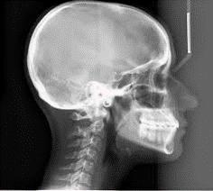
What projection is used to view the bones of the face, skull and soft tissue profile of the face?
|
Lateral Cephalometric Projection
|
|
|
How should the Frankfort plane be and where is the cassette positioned in the lateral cephalometric projection?
|
The Frankfort plane should be parallel to the floor, and the cassette should be positioned against the left side of the patient's face
|
|
|
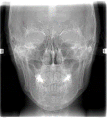
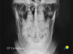
Which projection is used to view the frontal and ethmoid sinuses, the orbits and the nasal cavity?
|
Posteroanterior Prjection
|
|
|
How is the patient's head positioned as well as the cassette in the Posteroanterior Projection?
|
The long axis of the cassette is placed vertically. The patient's forhead and nose both touch the cassette (Frankfort plane is NOT parallel to floor)
|
|
|
What projection is used to view the maxillary sinuses, as well as the frontal and ethmoid sinuses, orbits and nasal cavity?
|
Waters Projection
|
|
|
How should the patient be positioned in the Waters Projection?
|
The chin is elevated and touches the cassette. The tip of the nose should be 1/2 to 1 inch away from the cassette.
|
|
|
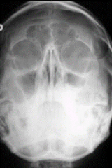
What projection is used to view the condyles and their position, the base of the skull, the zygomatic arch and the sphenoid and ethmoid sinuses?
|
Submentovertex Projection
|
|
|
How is the patient positioned for the submentovertex projection?
|
The top of the skull touches the cassette and the Frankfort plane is perpendicular to the floor.
|
|
|
If the zygomatic arch is the target of interest in the Submentovertex Projection…..
|
the exposure time is reduced to 1/3 the normal exposure time.
|
|
|
What projection is used to view the condylar neck and ramus?
|
Reverse Town Projection
|
|
|
How is the patient positions in the Reverse Town Projection?
|
Patient faces the cassette, mouth wide open, chin resting on the chest, and forehead touching the cassette
|
|
|
What is viewed on TMJ Radiography?
|
Only the bones can be viewed, not the entire joint-structure soft tissue
|
|
|
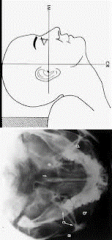
What is viewed on the Transcranial Projection (Lindblom Technique)?
|
the superior surface of the condyle and the articular eminence
|
|
|
What is the angle of the beam when using the Transcranial Projection?
|
The beam is directed 2" above and .5" behind the opening of the ear canal at a +25 degree (downward) and +20 degree (forward) angle of the OPPOSITE TMJ you wish to capture
|
|
|
Which tubehead can be used for the transcranial projection?
|
the intraoral tubehead
|
|
|
Which of the following describes a use of panoramic film?
a. diagnosis of caries b. evaluation of periodontal disease c. evaluation of impacted molars d. evaluation of periapical disease |
c. evaluation of impacted molars
|
|
|
The zone in which structures are clearly demonstrated on a panoramic radiograph is termed the:
a. focal trough b. rotation center c. ghost image d. midsagittal plane |
a. focal trough
|
|
|
Rare earth intensifying screens are recommended in panoramic radiography because:
a. Rare earth screens emit a blue light b. Rare earth screens provide a more diagnostic image c. Rare earth screens require less exposure time d. the images convert faster in automatic processors |
c. Rare earth screens require less exposure time
|
|
|
A thyroid collar is not recommended because:
a. it blocks the xray machine and obscures information b. There is a relatively low dose of radiation to the thyroid gland c. It is impossible to sterilize it d. all of the above |
a. it blocks the xray machine and obscures information
|
|
|
Which projection is best for examining the maxillary sinus?
|
Waters projection
|
|
|
Which projection is best for examining the zygomatic arch?
|
Submentovertex Projection
|
|
|
Which projection is best for examining fractures of the condylar neck?
|
Reverse Towne Projection
|
|
|
Which projection is best used for examining the soft tissue profile of the face?
|
Lateral Cephalometric Projection
|
|
|
Which projection is best for examining fractures of the mandibular body?
|
Lateral Jaw Projection
|
|
|
Which projection is best for examining the condyle and articular eminence?
|
Transcranial projection
|
|
|
Which projection is best for examining a large lesion of the ramus?
|
Lateral Jaw Projection
|
|
|
Lateral Jaw Projection
|
|
|
Posteroanterior Projection
|
|
|
Waters Projection
|
|
|
Submentovertex Projection
|
|
|
Reverse Town Projection
|