Reading...
![]()
Play button
![]()
Play button
![]()
Use LEFT and RIGHT arrow keys to navigate between flashcards;
Use UP and DOWN arrow keys to flip the card;
H to show hint;
A reads text to speech;
61 Cards in this Set
- Front
- Back
|
The coronoid process is visible on what film
|
Max. molar
|
|
|
The anterior nasal spine is visible on what film?
|
Max central-lateral
|
|
|
The nasolabial fold is visible on which film?
|
Max. canine
|
|
|
The lingual foramen is visible on which film?
|
Mand. central-lateral
|
|
|
The coronoid process is (radio-opaque, radiolucent)
|
RO
|
|
|
The internal oblique ridge is located on the (mandible/maxilla) and is also known as what?
|
mandible, mylohyoid ridge
|
|
|
The mandibular canal (or inferior alveolar canal) stretches from mandibular foramen to what?
|
mental foramen
|
|
|
The mandibular canal contains what vessels?
|
inferior alveolar A, N and V
|
|
|
What is the name for the RO structure that is superior to the mylohyoid ridge and is located on the external surface of the mandible?
|
external oblique ridge
|
|
|
The mental fossa is (above/below) the mental ridge and is a depression on the (internal/external) surface of the mandible
|
above, external
|
|
|
The mental foramen is a (RO/RL) structure between the canine and what other tooth in the (mandible/maxilla)?
|
RL, 2nd premolar, mandible
|
|
|
True or False, the inferior border of the mandible is seen when a radiograph is correctly angulated
|
False, only seen with excessive vertical angulation
|
|
|
The lingual foramen is a (RO/RL) structure located within what?
|
RL, mental spine (or genial tubercle)
|
|
|
What is pneumatization of the maxillary sinus?
|
Downward extention of floor of max sinus due to LOSS OF TEETH or AGING
|
|
|
What structure in the maxilla is often mistaken for pathology?
|
Lateral fossa (or incisive fossa)
|
|
|
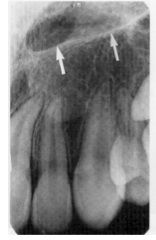
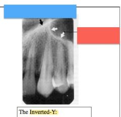
The "inverted Y" on a canine film is made by what two RO structures?
|
lateral wall of nasal fossa and anterior border of max. sinus
|
|
|
The floor of the nasal fossa is also what other structure?
|
hard palate
|
|
|
The anterior mandible bone pattern is as follows: (geometric/round) bone marrow spaces, and trabeculae forming a (coarse, smooth) pattern
|
round, coarse
|
|
|
The posterior mandible bone pattern is as follows: (small/large) bone marrow spaces, (vertically/horizontally) oriented trabeculae, area just below roots of (incisors/canines/premolars/molars) may not have trabeculae
|
large, horizontal, molar
|
|
|
The posterior maxilla is similar radiographically to the anterior maxilla except that it has (smaller/larger) bone marrow spaces
|
larger
|
|
|
The anterior maxilla has (thin/thick) and (many/few) trabeculae, forming a fine, round, and (dense/spaced) pattern with (small/large) bone marrow spaces
|
thin, many, dense, small
|
|
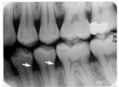
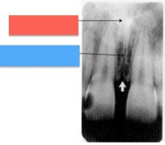
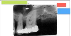
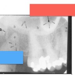
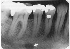
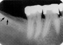
What is indicated by the white arrows?
|
cervical burnout
|
|

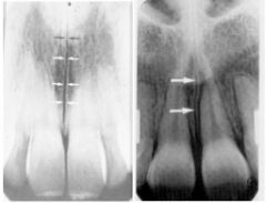
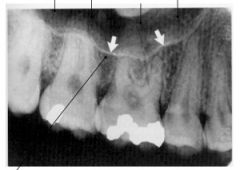
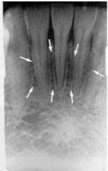
The white arrows are pointing to what structures in both pictures?
|
apical foramen
|
|
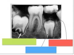
What is indicated by the red, green, and blue boxes?
|
green - developing root (#31)
blue - developing premolar (#29) red - Primary tooth (T) with resorbing roots |
|

What RO structure is indicated by the red box?, RL structure by the green box?
|
red - lamina dura
green - pdl |
|
The red boxes indicate what type of bone?
|
Alveolar crestal bone
|
|

The white arrows are pointing to what RL structure?
|
Nasopalatine foramen OR incisive foramen
|
|
The white arrows are pointing to what RO structures?
|
Lateral walls of nasopalatine canal
|
|
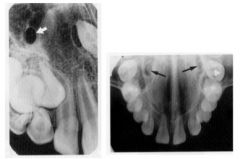
The black arrows are pointing to what RL structures?
|
Superior foramina of the nasopalatine canal
|
|
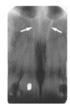
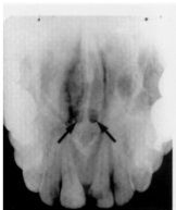
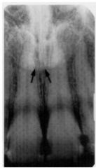
The arrows in both pictures are pointing to what structure?
|
Intermaxillary suture OR median suture OR mid-palatal suture
|
|
The red and blue box are pointing to what two structures?
|
red - anterior nasal spine
blue - intermaxillary suture (or median suture/mid-palatal suture) |
|
What 2 structures are indicated by the blue and red box?
|
blue - nasal fossa
red - anterior nasal spine |
|
The white arrows indicate what structure?
|
floor of nasal fossa (or hard palate)
|
|
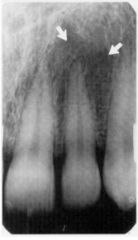
What RL structure is indicated by the white arrows?
|
Lateral or Incisive Fossa
|
|
What structures are indicated by the colored boxes?
|
blue - lateral wall of nasal fossa
red - anterior border of max. sinus |
|
What structure is indicated by the white arrows?
|
floor of max sinus (or inferior border of max sinus)
|
|
What structure is indicated by the colored boxes?
|
red - malar process
blue - septum green - floor of max sinus |
|
What structure is indicated by the green box?
|
septum
|
|
What structures are indicated by the colored boxes?
|
green - zygomatic process of max
red - septum blue - floor of max sinus |
|
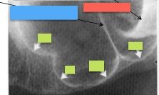
What structures are indicated by the colored boxes?
|
blue - coronoid process
red - zygomatic process of the maxilla |
|
What structures are indicated by the colored boxes?
|
blue - pterygoid plates
green - hamular notch red - hamular process |
|
What structure is indicated by the white and black arrows? (same structure)
|
zygomatic process of maxilla (malar process)
|
|
What structures are indicated by the colored boxes
|
green - zyg process of max
orange - floor of max sinus |
|
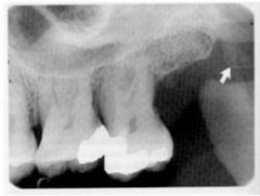
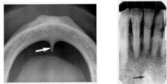
What structure is indicated by the white arrow (left) and black arrows (right)? (same structure)
|
nasolacrimal canal
|
|
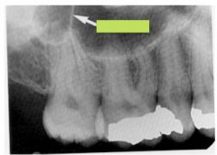
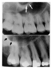
What structure is indicated by the black arrows?
|
nasolabial fold
|
|
What structure is indicated by the black arrows?
|
soft tissue of nose
|
|
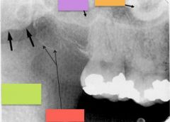
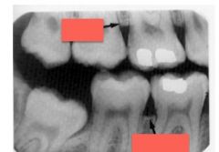
What structures are indicated by the colored boxes?
|
purple - floor of max sinus
orange - malar green - coronoid process red - max tuberosity |
|
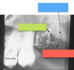
What structure is indicated by the white arrow?
|
Coronoid process
|
|
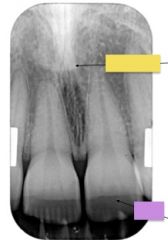
What structures are indicated by the colored boxes?
|
yellow - tip of nose
purple - lip line |
|
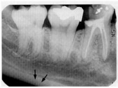
What structure is indicated by the black arrows?
|
inferior border of mandible
|
|
True or False, these two radiographs are the same structure. (Then, identify what they are)
|
True. mental spine (or genial tubercle)
|
|
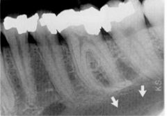
What RL structure is indicated by the white arrow?
|
lingual foramen
|
|
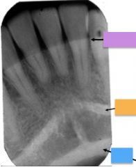
What structures are indicated by the colored boxes?
|
purple - lip line (or tongue shadow)
orange - mental ridge blue - lower border of mandible |
|
What structure is indicated by the massive amount of white arrows?
|
mental fossa
|
|
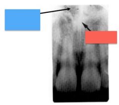
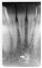
What RL structure is indicated by the white arrow?
|
mental foramen
|
|
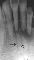
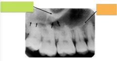
What thin RL structures are indicated by the black arrows?
|
nutrient canals
|
|
What RO structure is indicated by the black arrows?
|
external oblique ridge
|
|
What structure is indicated by the 3 white arrows?
|
mylohyoid ridge (internal oblique ridge)
|
|
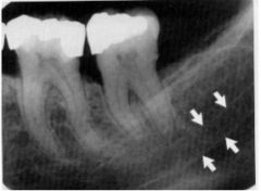
What slightly RL structure is surrounded by the white arrows?
|
Mandibular canal (inferior alveolar canal)
|
|
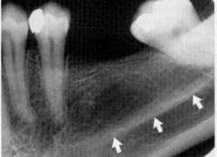
What RL area is indicated by the white arrows?
|
submandibular fossa
|
|
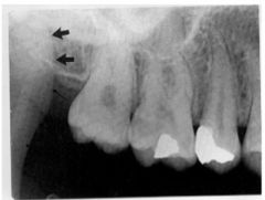
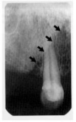
What RO structure is indicated by the black arrows?
|
Coronoid process
|