![]()
![]()
![]()
Use LEFT and RIGHT arrow keys to navigate between flashcards;
Use UP and DOWN arrow keys to flip the card;
H to show hint;
A reads text to speech;
97 Cards in this Set
- Front
- Back
|
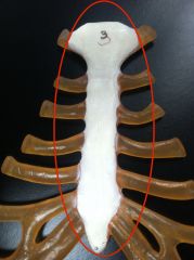
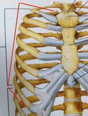
Sternum A flat bone Part of the axial skeleton |
|
|
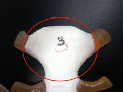
Manubrium Top portion of sternum
|
|
|
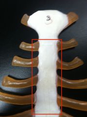
Body (of the sternum) |
|
|
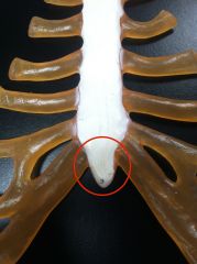
Xiphoid process Sharp tip of sternum |
|
|
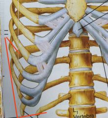
True ribs Attached to sternum w costal cartilage |
|
|
False ribs Attached to sternum indirectly by cartilage of rib of it |
|
|
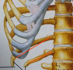
Floating ribs Don't attach to sternum (Included in number of false ribs) |
|
|
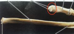
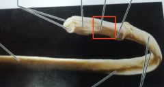
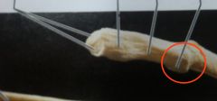
Head (of rib) Slightly thicker, attached to vertebrae |
|
|
Neck (of rib) Below head |
|
|
Tubercle Bump below neck of the rib |
|
|
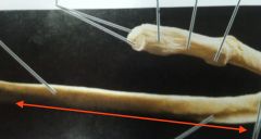
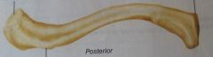
Shaft Long & uniform in thickness btwn costal cartilage and tubercle Has a dull and sharp side |
|
|
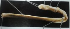
Rib (general picture) |
|
|
Clavicle Collar bone |
|
|
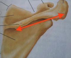
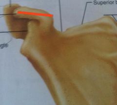
Spine of scapula (spinous process) On the posterior side |
|
|
Acromion process Extension of spinous process Scapula |
|
|
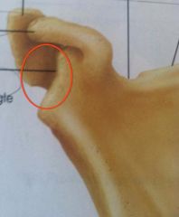
Coracoid process Scapula (shoulder blade) |
|
|
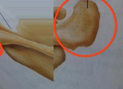
Glenoid cavity (fossa) Very shallow depression for the joint made w/ humerus |
|
|
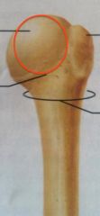
Head Medial projection of proximal epiphysis Humerus |
|
|
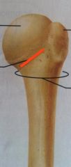
Anatomical neck Shallow groove right below head Humerus |
|
|
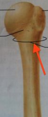
Surgical neck Start of diaphysis, where proximal fractures are likely to occur Humerus |
|
|
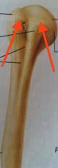
Greater & lesser tubercles Humerus |
|
|
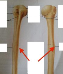
Deltoid tuberosity Humerus |
|
|
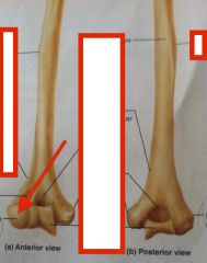
Capitulum Humerus |
|
|
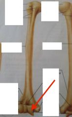
Trochlea, "pizza cutter" Humerus |
|
|
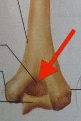
Medial & lateral epicondyles Humerus |
|
|
Coronoid fossa Humerus |
|
|
Olecranon fossa Humerus |
|
|
Humerus Upper arm bone |
|
|
Ulna Medial bone of forearm |
|
|
Radius Lateral bone of forearm |
|
|
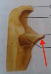
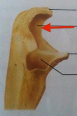
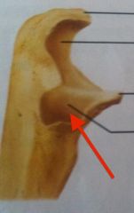
Olecranon process Ulna (elbow) |
|
|
Coronoid process "like a crown" Ulna |
|
|
Trochlear notch Ulna |
|
|
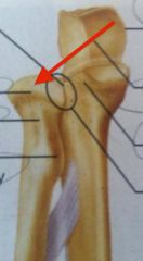
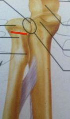
Radial notch Ulna |
|
|
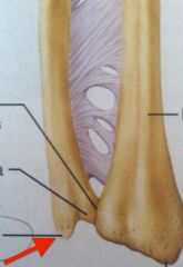
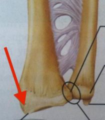
Styloid process Ulna |
|
|
Head Radius |
|
|
Neck Radius |
|
|
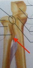
Radial tuberosity Radius |
|
|
Styloid process Radius |
|
|
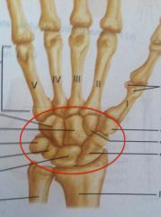
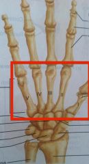
Carpals Short bones of the wrist |
|
|
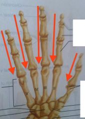
Metacarpals Palm of hand |
|
|
Phalanges Finger bones Three per finger, two in thumb |
|
|
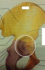
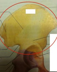
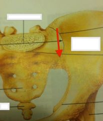
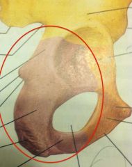
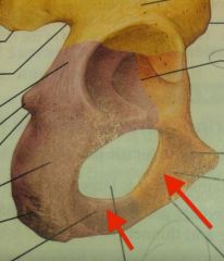
Acetabulum Deep fossa for the head of femur Divided like a pie into 3 imaginary pieces for Ilium, Ischium, Pubis Os Coxa (Coxal Bones) |
|
|
Os Coxum Coxal bone |
|
|
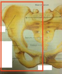
Ilium bone of os coxa |
|
|
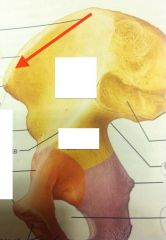
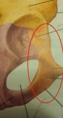
Iliac crest |
|
|
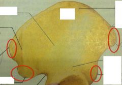
Iliac spines Two pointing anteriorly Two pointing posterior (superior and inferior ones) |
|
|
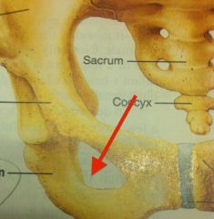
Sacroiliac joint Closes the posterior of the pelvis |
|
|
Ischium bone |
|
|
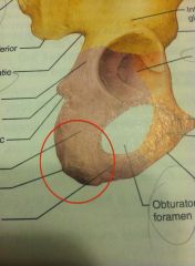
Ischial tuberosity Main body of ischium |
|
|
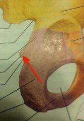
Ischial spine posteriorly pointing |
|
|
Pubis bone Two of these bones close pelvis at front w/ pubic symphysis |
|
|
Ramus of ischium Ramus of pubis |
|
|
Obturator foramen |
|
|
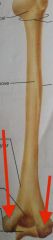
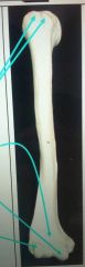
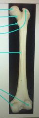
Femur Thigh bone |
|
|
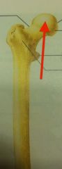
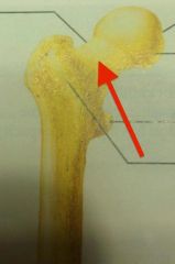
Head of femur Points medially |
|
|
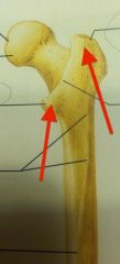
Neck of femur |
|
|
Greater and lesser trochanters Femur |
|
|
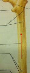
Linea aspera Ridge along diaphysis of femur |
|
|
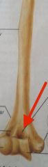
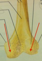
Medial and lateral condyles Femur |
|
|
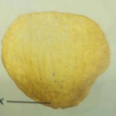
patella Assoc w knee joint Suspended by muscles |
|
|
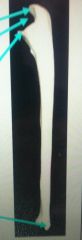
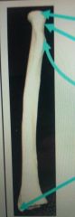
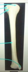
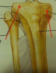
Tibia bone Medial calf bone |
|
|
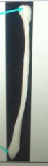
Fibula bone Lateral calf bone |
|
|
Medial and later condyles Tibia |
|
|
Tibial tuberosity Under your knee Tibia |
|
|
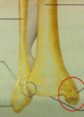
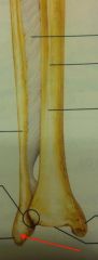
Medial malleolus on medial side Tibia |
|
|
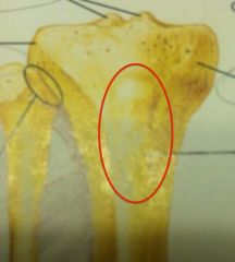
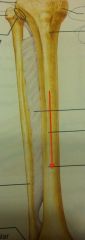
Anterior crest Why hitting your shin is so painful Tibia |
|
|
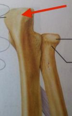
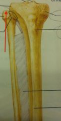
Head of fibula |
|
|
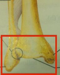
Lateral malleolus Fibula |
|
|
"ankle bones" of tibia and fibula |
|
|
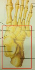
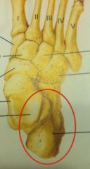
Tarsals Small ankle bones |
|
|
Talus Main bone of the ankle Connected to tibia and fibula |
|
|
Calcaneus Large heel bone |
|
|
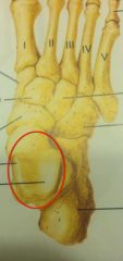
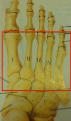
Metatarsals Most of arch and balls of feet |
|
|
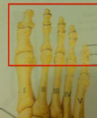
Phalanges Toe bones big toe has two bones, other toes have three |
|
|
Two types of cartilaginous joints |
Sympheses and synchondroses - Disc of fibrocartilage (intervertebral discs and pubic symphysis) - Hyaline cartilage (epiphyseal plate; also costal w/ 1st rib) |
|
|
Describe diagram of synovial joint |
Two-layered membrane of articular (joint) cartilage on top and bottom Right and left sides are synovial membrane which produces synovial fluid (reduces friction) Articular capsule usu reinforced w/ bursae & ligaments Fibrocartilage pads (articular discs) may be there Articular capsule is techn. on L and R sides |
|
|
What kind of joint has all 6 types (ball and socket, plane, hinge, pivot, condylar, saddle) |
Synovial |
|
|
Hip joint is what kind of joint |
Ball and socket |
|
|
Moement of hip joint is limited by what |
Deep socket Strong reinforcing ligaments These account for its exceptional stability |
|
|
Knee joint is what kind of joint |
Hinge |
|
|
Shoulder joint is AKA? |
Glenohumeral joint |
|
|
Where is the temporomandibular joint located |
Anterior to the ear (mandible to the temporal) |
|
|
Bursitis |
Inflammation of the patellar bursa (from hard blow to the knee) |
|
|
Sprain |
Ligaments that reinforce a joint are damaged by over stretching / torn away Heals slowly bc of its poor blood supply (lig and tend are dense CT) |
|
|
Dislocations Usu accompanied by what |
Bones are forced out of their normal position in the joint cavity Torn or stressed ligaments, inflammation "reduction" returns the bone to its orig position |
|
|
Bursae |
Flattened, fibrous sacs lined w/ synovial membranes & containing synovial fluid Common where ligaments, muscles, skin, tendons, or bones rub together |
|
|
Tendon sheath |
Elongated bursa, wraps around a tendon |
|
|
Stability of synovial joint determined by |
Articular surfaces and ligaments |
|
|
Muscle tone accomplished by |
Muscle tendons acting as stabilizing factors, and they're kept tight by muscle tone |
|
|
Nonaxial means what |
Joint has slipping / gliding movements only |
|
|
Uniaxial |
Movement in one plane; bi and multi |
|
|
2 muscle attachments across a joint movement is described how |
Origin (attachment to immovable bone) Insertion (attachment to movable bone) Along transverse, frontal, or sagittal planes |
|
|
Cartilage injury |
Snap and pop of overstressed cartilage |
|
|
Subluxation |
Partial dislocation of a joint |
|
|
Tendonitis |
Inflammation of tendon sheaths from overuse |
|
|
Gouty arthritis |
Deposition of uric acid crystals in joints and soft tissues, followed by inflammation response |