![]()
![]()
![]()
Use LEFT and RIGHT arrow keys to navigate between flashcards;
Use UP and DOWN arrow keys to flip the card;
H to show hint;
A reads text to speech;
130 Cards in this Set
- Front
- Back
|
_____ attention is where, what, and when we allocate our attention to. ("focalization" and "concentration") |
Selective attention |
|
|
What is the action of attention? |
Privileged processing |
|
|
Capacity Models
Why don't hands-free laws don't really do anything for improving safety when driving? |
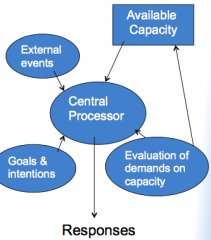
We have a limited capacity in our attentive state. The capacity limitations when talking on the phone are not where your hands are. Maintaining vigliance on the road & maintaining vigilance on a cellphone conversation both drop cognitive capacity severely. |
|
|
What does "change blindness" demonstration tell us about our attention? |
Our experience of the world is extermely limited because of capacity limitations of our attention |
|
|
During a "change blindness" test, why is it that we pay attention to certain things more than other things? |
We have certain priority signals that's in type of gradient.
e.g. we pay more attention to people>fruit>sky>etc. |
|
|
The ______ model links alertness (attention state) to performance of some kind of task |
Yerkes-Dodson Law model |
|
|
According to the Yerkes-Dodson model, predict what will happen to optimal perfromance if the arousal is either too high or too low |
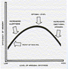
Optimal performance won't be as good. There's a sweet spot for optimum performance |
|
|
In the Yerkes-Dodson model, rising alertness _____ performance |
Improves performance |
|
|
In the Yerkes-Dodson model, too much stress _____ performance |
Impairs performance |
|
|
The relationship between arousal, attentive state, and cognitive performance depends on ______ |
The type of task you're doing |
|
|
Yerkes-Dodson Law
Difficult tasks are optimized at _____ arousal levels
e.g. Taking a midterm |
Low arousal levels |
|
|
Yerkes-Dodson Law
Vigilance tasks are optimized at _____ arousal levels
e.g. Monitioring and requires quick response |
High arousal levels |
|
|
What did scientist find in studying female rats performance in mazes? |
Female rats performed optimally at different stress levels depending on their cycle. Stress hormones, which are influenced by this cognitive state, can mediate optimal performance |
|
|
Neural markers of attentive state
In humans, how can we monitor the vigilance state? |

By looking at the omnibus EEG signal |
|
|
"Vigilance state" is reflected in the synchronization of _____ brain networks
|
Large-scale brain networks |
|

Neural markers of attentive state
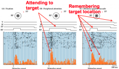
What does this image show and what is one of the things that this EEG picks up very nicely? |
These are electro recordings from EEG, and it's god at picking up "volley of activity" between networks |
|
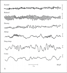
Which part of these EEG signals show "high attentive state"? |
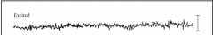
The high frequency part ("excited") |
|
|
In "high attentive states," what's dominating the EEG signal? |
Low scale oscilation networks that are going back and forth (high enery frequency). This means that the local brain networks (e.g. info within the visual, auditory, or motor systems) are what's dominating the overall brain activity. Therefore, the EEG signal is dominated by the "sensory & cognitive systems" that are prepped for action. Thus, this allows us precise attention focus |
|

Which part of these EEG signals show "low state of vigilance"? |
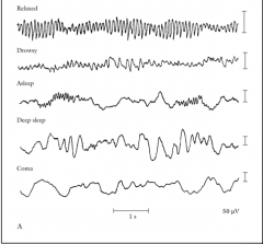
The low frequency parts. ("relaxed" all the way down to "coma") |
|
|
In "low state of vigilance," what's dominating EEG signal? |
Large scale oscilation networks that are going back and forth. Therefore, we're looking at "sleepy" or "inattentive" states where these local networks within systems are not prepped for input. Even input that's high priority may not get through |
|
|
______ is a neurotransmitter tightly linked to alert states.
e.g. "sleep-wake" cycles, physiological responses, brain metabolism, attention & arousal |
Norepinephrine |
|
|
Neuromodulators
Neurons are more likely to fire if norepinepherine is _____
|
High. Norepinipherine in the brain has excitatory effects on cortical function |
|
|
Neuromodulators
Why does norepinepherine always has a net effect of excitation? |
Because norepinepherine inhibits inhibitory net. No matter which neurons are being targeted, the net effect is alway excitation. |
|
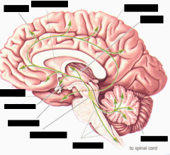
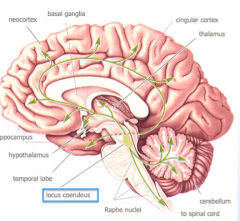
Norepinpherine is located in the "locus coeruleus", which is a small bilateral nucleus (aka "blue spot). Point at the location of the "locus coeruleus." |
|
|
|
Neuromodulators
The ______ has long axons that can excite the entire neocortex, thus making it highly interconnected with about every other part of the brain. |
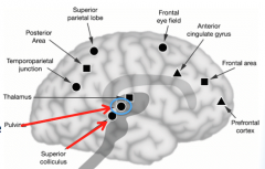
Locus coeruleus. It's also heavily connected with the frontal lobe, posterior parietal lobe, pulvinar (part of the thalamus), and superior colluculus |
|
|
_____ is the mechanism by which some inputs get privileged processing while others are ignored
e.g. "focalization" & "concentration" |
Selective attention |
|
|
The _____ process is the funneling in of input to some narrow band and only some make it through to our attention/awareness |
Attention bottleneck (a.k.a. filter) |
|

In the "dichotic listening experiment", why don't we get input from the other speakers when you just pay attention to only one of the speakers? |
This is becasue of selective attention limitations. We gain fidelity with the one we pay attention to, while the other things we don't pay attention to gets filtered out |
|
|
If you have lesser capacity limitations, does that mean that you have a better active process of selecting? |
No. They are two separate mechanisms. |
|
|
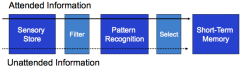
The _____ model proposes attended information goes all the way through the pathway, unattended information gets blocked by a sensory filter. Attention bottleneck happens right at the sensory store |
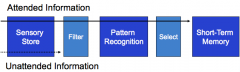
Early selection model; early experiments made scientists believe that attention was building a filter that operated on units such as location, loudness, pitch, & color
e.g. dots, concentric lines, things that are red, high pitch, low pitch ("dichotic listening experiment") |
|
|
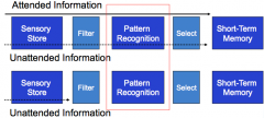
The _____ model argues that all inputs get processed, but some in a weaker form than others |
Late selection model
e.g. cocktail party effect (real world "dichotic listening experiment") |
|
|
In both models, no matter which model you assume, what we have conscious awareness of are the _____ not the features |
Patterns |
|
|
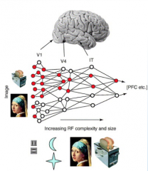
______ propose multiple possible filters, with the level of processing depending on the complexity of the stimulus and the nature of the task. Either way, what we experience is the ______
Ex. If task is simple, you can filter it early. If task is complicated (like faces), you're gonna have to organize it all and throw some out later |
Bottleneck theories; object |
|
|
Can "attention" be mapped in the brain? |
No. There is no map for attention. |
|
|
There are many areas that control the brain known as "______", which include both cortical and subcortical areas |
Brain systems/networks |
|
|
Animals that don't have the same neo cortical organization as us still have _____ systems |
Attention systems |
|
|
_____ is the signaling of important events towards which attention will/should be allocated |
Alerting; it is a map of the world that important stuff in it
e.g. Something important over there, something important over here, etc |
|
|
_____ alerting signals are "goal directed" and are high in priority because of the shape of your current attention filter. This 'top down' process allows us to focus on certain things we are trying to accomplish, thus making things more important for our attention
e.g. Where's Waldo, Cocktail effect, Searching for the imporatnt things on the board |
Endogenous alerting signals; top-down (frontal cortex down to sensory)
|
|

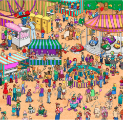
The efficiency of your search when playing "Where's Waldo" can be improved by ______ filtering, and not ______ filtering. |
Narrow filtering (e.g. looking for red and white shirts); Broad filtering (e.g. people)
|
|
|
_____ alerting signals are stimulus driven and can grab your attention very easily
e.g. green scarf in red context |
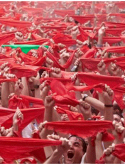
Exogenous alerting signals |
|
|
Endogenous and Exogenous alerting cues denote items of high _____ because these cues are of important to you and match your fitler, or because things out there are very different in their physical properties |
Salience; highly context dependent (depends on your goals, or what the surrounding is) and extremely flexible |
|
Explain the "biased competition model" |
It's the idea of competition between inputs in order to get through the "attentional bottleneck". Neural ensembles that are encoding the different inputs (face & toaster) are actually in competition with each other because they share nodes with one another. Thus, they compete in "mutually suppressing" each other in order to disambiguate shared neurons in those ensemble codes. Therefore, the image shows high fidelity with the toaster than the face. The competition also improves the signal to noise of a high priority event. |
|

Things that are high _____ salience, and high _____ salience almost always wins the the biased competition model. |
Stimulus salience; Goal-directed salience |
|
|
In certain scenarios, "biased competition" can also be won by _____ |
Chance |
|
|
"Mutual inhibition" competition of neural ensembles that are coding the objects happen in ______, and not in the attention control centers |
Sensory cortex
e.g. Happening in V1, but we really have evidence of this happening in V4 |
|
|
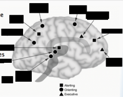
_____ are the areas in the brain with the information as to whether locations in the world are different or important. They're also known as "high alerting signals" |
Salience maps;
- Must have topography (must have a space-based coordinate system) - Must be modality independent (the alerting system should be able to signal something important out there regardless of how the input came in; via auditory, vision, touch, etc) - Must be capable of coding specific features (ex: the specific shirt of Waldo) - Must be flexible (what's important now might change in a moment; ex: green scarf is important because surrounded by red scarves, but won't be important later when surroun |
|
|
Alerting signals (salience maps) are not in _____ because that tells us about the input comng in, while salience maps evaluate the salience of the inputs in the "context" of the other stuff that's coming in. |
Sensory cortex
- Note: salience maps are very strongly connected to sensory cortex and motor system |
|

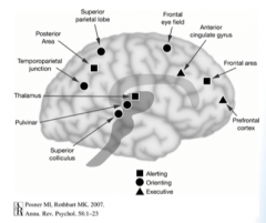
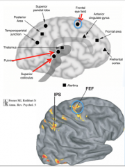
Find the "posterior parietal cortrex" (1st alerting system) |
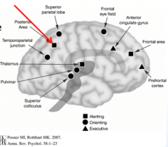
Evidence show that salience maps are found in the "posterior perietal cortex" and it is mapped |
|
|
Posterior parietal cortex has neural activity that strongly linked to ______ salience. Therefore, it's connected with ______ cues that capture attention |
Stimulus driven salience; exogenous |
|
|
The posterior parietal cortex provides input to many ______ areas |
Motor planning areas |
|
|
_____ areas provide sensory inputs for attentionally guided movements
e.g. eye movements, reaching, head turning, etc. |
Dorsal stream brain areas |
|
|
The "multiple object tracking" exeriement tells us that the kind of neural signaling that comes from stimulus driven brain system is _____ |
Capacity limited
Ex: Multiple object tracking |
|
|
In the "multiple object tracking" experiment, performance gets bad if the balls gets too close becasue this exceeds the _____ of attention. Perfromance also gets bad if the balls move too fast because this exceeds the _____ of attention |
Spatial resolution of attention; temporal limits of attention
Note: The spatial and temporal limits of attention are far more restrictive than our sensory limits; Our ability to discriminate things is much more restricted by our attentional awareness bottlenecks than by our sensory systems |
|

Find the "frontal cortex" (2nd alerting system) |
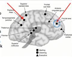
The blue circle. |
|
|
Frontal cortex has neural activity that strongly linked to ______ salience. In order for the prefrontal cortex to encode current goals, it must be strong connected with the ______ |
Goal-directed salience; working memory
- frontal cortex neurons have very complex, flexible tuning properties depending on what's relevant/important |
|

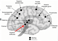
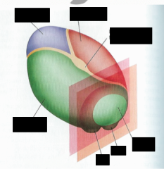
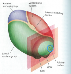
Find the Thalamus LGN/MGN (3rd alerting system) |
The blue circle. |
|
|
The _____ is the relay station from peripheral sensory systems to neocortex. It uses the context from feedback to evaluate the stuff coming in, which is effectivey, salience. |
Thalamus |
|
|
In the Thalamus, prioritization of input signals is based on _____ (on the basis of what's recently been happening.) Some signals are more alerting than others based on _____ |
Cortical activity; context |
|
|
Damage to the thalamus strongly mutes _____ |
Alerting signals |
|
|
_____ is the execution of attention shifts. This comes online early in development, gets refined over time
|
Orienting (motor system of attention system) |
|
|
Orienting is most closely linked to ______ networks |
Motor networks;
e.g. occularmotor and reaching network |
|
|
Explain the "premotor theory of attention"; and explain the alternative |
Goal-directed attention and motor planning are one of the same; there's no distinction between them. The alternative theory is that in humans, goal-directed attention and motor planning are strongly linked; however in other species, these two are decoupled |
|
|
______ attention are shifts of attention that without any motor movements
Ex: Think of eveasdropping |
Covert motion |
|
|
In covert shifts of attention, the _____ is built in the brain, therefore if the professor tells you to fixate in the middle of the screen and then move your eyes to the left and then around, your oculomotor areas are actually planning eye movements to all those places, but cancelling them |
Motor plan |
|

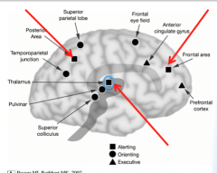
Locate the "superior colliculus" (1st oculomotor system) |
|
|
|
The "sensory layers" of "superior colliculus" primarily gets its input from the _____ system and from the _____. |
Magno system; FEF (frontal eye field) |
|
|
Superior colliculus neurons have neurons involved in planning eye & head movements that are _____. |
Stimulus-driven; therefore, the "superior colliculus" integrates the fastest visual input with motor planning
think exogenous cues (stimulus-driven) |
|
|
"Blindsight" patience are able to attentionally guide behavior without awareness. For instance, they're able to correctly guess the location of the light, although they are not able to perceive it. How are they able to do this? |
These patience are able to correctly guess the location of the light because the input is still able to get into the superior colliculus, thus allowing them attentionally guided behavior without awareness |
|

Locate the "pulvinar" (2nd oculomotor system) |
The blue cirlce |
|
Locate the pulvinar |
The pulvinar has fast motor planning from coarse analysis of inputs. But the informtion that's coming in is not very refined. So attentionally guided eye moevements on not very much input. |
|
|
Proper function of "pulvinar" is important for proper execution of _____. It gets most of its input from _____ |
Eye movements; superior colliculus |
|

Locate the "frontal eyefield" (FEF) (3rd oculomotor system) |
The blue circles. |
|
|
The FEF are coding for both planned and executed eye movements. Therefore, the eye movements are ______ atentionally guided behaviors |
Goal-directed |
|
Locate the "superior parietal lobule"/"intraparietal sulcus" (IPS) (4th oculomotor system) |
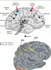
The blue circles. |
|
|
The "superior parietal lobule" is the first area in parietal cortex that's not ______ |
Sematotopic |
|
|
The "superior parital lobule" in the IPS is important for _____ attentionally guided movements. |
Planning |
|
|
Does the IPS have specific regions for eye movements, reachign, and grasping? |
Yes |
|
|
Damage to the "superior parietal lobule" impairs the ability to generate ______ |
Purposeful movements |
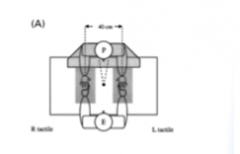
|
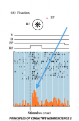
In this experiment, a monkey is asked to fixate on a point (FP). Then after a while an exogenous cue (flash of light) happens. Immediately after, the LIP neurons start spiking. What does this experiment tell us about LIP neurons? |
LIP has information about "stimulus salience" within its receptive field (RF) |
|
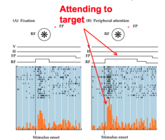
The same experiment is done as the previous one except this time, the monkey was paying attention to the location where the exogenous cue was, there was a boost in the firing of the LIP neurons. What does this tell us about LIP neurons? |
LIP neuron can tell us where the stimulus is (maps) and whether or not the monkey is paying attention to that location |
|
In the same monkey experiment, the flash comes on in the RF, but now the monkey is told that when the FP goes off, make an eye movement. And indeed, the monkey makes an eye movement. What does this tell us about LIP neurons |
LIP neurons can tell us that the monkey remembers the target's location and plan its action. And it's holding onto that plan until it gets a cue (flash of light) to do it, and then it executes it |
|
|
Can LIP neurons be stimulated by an electrode to cause them to fire and trigger eye movements? |
Yes. LIP also has maps for stimulus salience and motor sytems |
|
|
The "dorsal" component of the orienting network is specialized for control of attention across _____
|
Spatial orientation; bilateral |
|
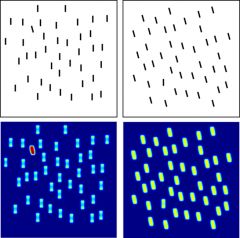
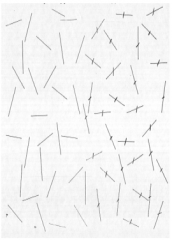
In the "visual search paradigm" of horizontal and vertical lines, the search time did not depend on the amount of distractors because it was a _____ search |
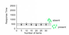
Pop out search/bottom-up/exogenous
Performance is based on the level of difference of the stimulus. Slope is 0 throughout |
|
|
In the "visual search paradigm" with the 5's and 2's, the type of search is a _____ search because you're using some type of attentional filter and you have to match if the input is fitting your attentional filter or not |
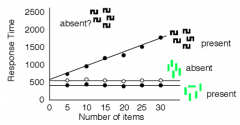
Serial search/top-down/endogenous/conjunction
Performance is based on the amount of distractors |
|
|
What has a faster response time, exogenous search, or endogenous search and why? |
Monkey have a faster response time with the exogenous search because it has lesser features to follow |
|
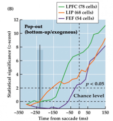
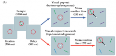
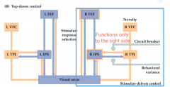
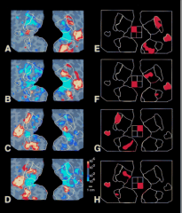
What does this image tell us about LIP and FEF in "pop-out" searches? |
LIP is predicting the onset of an eye movement because it's sensitive to salience (something is different out there) and the FEF is not. The information that's guiding the behavior primarily goes to the parietal cortex. |
|
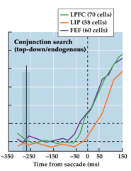
What does this image tell us about LIP and FEF in "serial" searches? |
The FEF tells you about the attentionally guided behaviors and the LIP can not. The LIP neuron doesn't even know the location of the target until after the monkey has already made an eye movement. |
|
|
Why isn't there a primary feedforward attention pathway? |
Becauase it's a circuit that can go anywhere depeding on context. For instance, when your behavior is being driven by exogenous signals (very differen things), it goes through the parietal cortex first. When your behavior is being driven by endogenous signals (stimulus requires sophisticated analysis) then it's the frontal lobe |
|
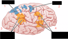
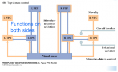
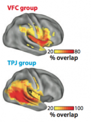
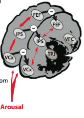
Name the brain areas: 1, 2, 3, 4 |
1. IPS (intra parietal sulcus) 2. FEF (frontal eye field) 3. TPJ (temporal parietal junction) 4. VFC (ventral frontal cortex) |
|
|
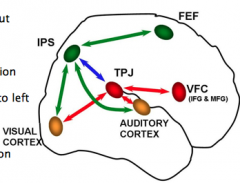
The _____ attention control network in general is associated, with top-down (endogenous control of attention), with bottom-up (exogenous control of attention) |
Frontal parietal |
|
|
The "ventral" component of the orienting network is specialized for control of attention across _____ |
Nonspatial demands; right hemisphere dominant
e.g. vigilance (monitoring), reorienting |
|
|
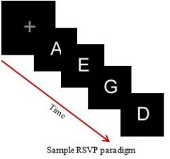
In vigilance tasks with "RSVP" experiments, subjects tend to be good at identifying T2 (assigned letter (X) after T1), only if T2 is immediately after T1. This is known as _____ |
Lag one sparing; as soon as you see that white letter, your attention gates open for about 200ms, and if T2 is immediately after T1, T2 is able to make it into the gates. Then the gates close doesn't open until about 400ms |
|
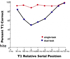
Explain attentional blink in this RSVP chart |
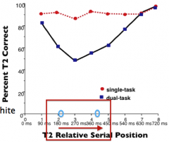
For about 200-400ms after a target is detected, your attentional resources are engaged in the anlysis of that first target, thus creating an attentional blink and missing the information in between |
|
Dual task rapid serial visual searh presentatios (RSVP) reveal the _____ of attention in the "ventral attention stream" |
Temporal limitations |
|
In reorienting tasks with "spatial cueing paradigm," explain why response times are much faster in valid cued vs. invalid cues |
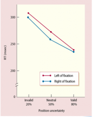
The difference between the valid and invalid cue is the time associated with reorienting your attention. On invalid trials, attention must be reoriented away from the cued position, which in this case, is about 50ms |
|
|
Explain "inhibition of return" |
It's the idea that if you've already looked there, you won't look there again |
|
|
The ____ is not involved in oculomotor system, however is involved in vigilance and RSVP tasks and is strongly connected oculomotor system |
TPJ |
|
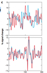
This image shows brain acitivity during a T1 task. What is one important thing to note about this image? |
The brain response before the trial starts is at zero, and then during monitoring, it's acutally going down and then going back up. In other words, quiet during tasks and active during rest known as "default mode network" and the TPJ is one of them |
|
|
______ neglect is a disorder of space-based attention, which impairs the ability to explore contralesional space. A failure in “looking, listening and exploring”, but visual, somatosensory, auditory, and motor are all intact. It's usually caused by a stroke
Example: Eye movements during visual search from a neglect patient |
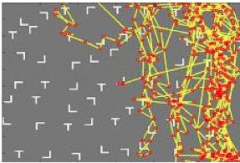
Hemispatial neglect; found in the parietal, therefore, failure to explore the left part of space because salience is downgraded. |
|
|
Explain how neglect is modality independent in neglect patience |
Neglect patience treat the left side of their world as if it doesn't exist. For instance, they have a tendency to use their left hand; they have downgraded salience of touches on the left side of their body; they don't respond to auditory cues that are localized to the left side of space; etc. However, they can be cued to do so, but they can't do it endogenously |
|
This "line cancellation" test show that neglect patience have _____ & _____ deficits |
Spatial and nonspatial deficits |
|
|
When neglect patience are given RSVP tasks and spatial cueing paradigm tasks, researchers found that patience have _____ and imparied on _____ tasks |
Long attentional blink (~1400ms); reorienting tasks (~200ms) > tends to be bilateral |
|
|
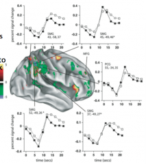
The VFC group tends to be severely impaired in the _____ field, while the acute neglect TPJ group tends to be fine in the |
Ipsilateral field; Ipsilateral field |
|
|
Explain the "mental imagery" study with neglect patience |
Neglect patience were asked to imagine landmarks that they know in Rome and to also name places that were around those land marks. Turns out that people were only able to name the places that were on the right side of those land marks, and when they were asked to imagine themselves standing on the other side of those landmarks, they were able to imagine the new placs that were originally on their left side |
|
Explain the "prism study" |
Neglect patience were intially tested with motor tasks (pointing straight ahead with eyes closed (motor), drawing a daisy (visuo-motor), & imagining and naming cities around landmarks (mental imagery task)). For a few minutes, they wore prism goggles that flipped their contralteral and ipsilateral fields of vision. They were brought in again 24 hours later to be tested with the same intial tasks, and they performed better than before; however, the only improvement that retained is the drawing of the daisy. |
|
|
Neglect is associated with damage to the _____ attention network, but primarily it's associated with damage to the _____ attention network |
Frontoparietal attention network; Ventral attention network |
|
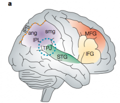
Circle all the areas that can result in neglect |
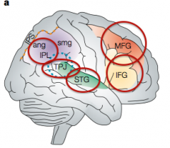
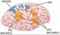
1. IPL 2. STG 3. IFG 4. MFG 5. TPJ
If you tinker with anyone of this system, it all goes out of wack in all the same way |
|
|
Hemispatial neglect patience all have a lesions in the _____ |
Midbrain |
|
|
What are the mystery conditions of neglect? |
1. Neglect is a spatial deficit, but damage is to the part of the brain that controls nonspatial parts of attention 2. Neglect damage is in the right side of the brain for the vast majority of patience
|
|
What is the 1st theory of neglect? |
The first theory is known as "right hemisphere specialization" and it's the idea that the right hemisphere is some how specialized for attention control while the left is for language. Therefore there is some form of asymetry between the right and left himesphere so the right hemisphere is getting more input than the left. Not really a theory. We know that the dorsal netowork are bilateral, only the ventral aspects of attention that are right lateral. Some evidence that these locus coeruleus are asymmetric; therefore, there is some evidence that the arousal systems is feeding more strongly into the right hemisphere than the left |
|
What is the 2nd theory of neglect? |
The second theory is known as "hemispheric asymmetry" and it is the idea that both hemispheres of the brain control attention, however, the left hemisphere only controls attention in the contralateral side, while the right hemisphere controls attention in both sides. Thus if there's damage in the right hemisphere, attention can still be allocated to that ipsilateral side to the right, but we lost attention to the left. Researchers propose that the dorsal attention network (which is mapped) gets input largely from the ventral attention network, and damage to the input system (ventral) causes direct nonspatial deficits + downstream spatial deficits to the left side of the brain
|
|
What is the 3rd theory of neglect? |
The third theory is known as "hemispheric attention" and it is the idea that both hemispheres control control attention, but it's controlled by a competitive process between the left and right hemispheres; and particularly we're talkinb about in the dorsal systems. It's based on a balance in cortical excitation and inhibition that maintains attentional control. The proposal is that the damage to the right TPJ results in inhibition to the left TPJ and that creates out of control TPJ, which is pushing all the attentional inputs to the right |
|
|
______ is how we reveal coordinated brain activity among different brain areas and it reveals highly correlated brain activity within the attention networks |
Functional connectivity |
|
When an individual has had a stroke to the TPJ (ventral attention network), the right and the left are _____ from each other |
Decoupled, the right and left sides of the brain are no longer engaged in that patterned brain activity that's cohesive and synchronized. Therefore, the more they're decoupled, the more severe the neglect |
|
|
______ is the failure to detect contralesional stimulus. When there's compeiting inputs, the contralesional one gets downgraded; they lose that bias competition |
Extinction; they only report one stimulus when both fingers are wiggling |
|
Give an example of Tactile extinction |
Subject placeds hand under the table. Experimenter touches one of the hands and asks the subject to report the hand that was touch. Subject reports correctly, however, when both are touched at the same time, only the ipsilateral side is reported and the contralteral side loses the competition |
|
|
______ patience have bilateral damage to the parietal lobes where both of the attentional systems are gone. These individuals exeprience severe extinction and very disorganized orienting. So failure to be able to use attentional signal to guide motor behaviors |
Balint's syndrome |
|
|
_____ is the inability to attend to more than one event at a time; can't report and 2 simultaneous events. _____ is the inability to reach towards a target; inability to guide eye movements to an object. _____ is the Inability to orient eye gaze towards a target |
- Simultagnosia/Extinction - Optic ataxia - Oculomotor apraxia |
|
|
_____ is a term used for driving inputs that go through the sensory system that are being altered depending on the control |
Gating or modulating |
|
|
When subjects are attending to particular regions of space, the corresponding region in early visual cortex ____ in bold response and is ____ specific. In other words, those neurons that are tuned to just to that part of space, their firing rate increases. |
Increases in bold response; location specific |
|
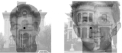
Why isn't there binocular rivalry in this superimposed image? |
There isn't enough retinal disparity |
|
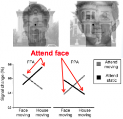
When subjects were told to fixate on the face and not the house, the brain response for the FFA was boosted. When the subjects were told to fixate on the house and not the face, the brain response for the PPA was boosted. What might be an explanation for this? |
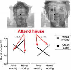
This is known as the "interaction effect": brain resonse is boosted in the area most selective for that object |
|
|
When subejcts were asked to simultaneously listen to noise and speech, we see that "interaction efect" also happens in the _____ system. This can also happen with other senses |
Auditory system; brain response is boosted in the area that is selected for attention |
|
|
What does it mean when neurons are firing negatively? |
It means that neurons are just firing less frequently when you're not attending to that input |
|
|
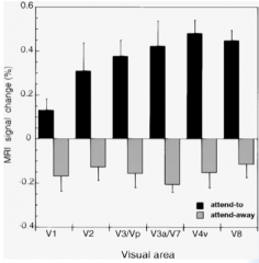
The modulatory effect of attention _____ as you get to higher cortical areas because higher sensory and association areas are largely driven by attention |
Increases
e.g. V1, V4, V8 |
|
|
At the "neuronal level" scientists are looking at the _____ of the tuning functions. For insatnce, subjects are told to attend to tilted lines |
Shaping |
|
More fMRI activation in right visual cortex when attending to the _____
More fMRI activation in left visual cortex when attending to the _____ |
Left; Right
Same ERP response |
|
In this ERP you see a wave of excitation that dissipates and then reappears. This is known as _____ of attention |
Volley of attention
The stimulus comes into the eyes, the input goes up the primary feedforward visual pathway then you get this surge of a driving response in your early visual cortex that gets quiet, but overtime that input bounces back and the response gets boosted again from feedback processing. Therefore attention is not sustained over time, but it's oscillating over time. |
|
|
How does the attentional control system improve salience? |
Attentional control systems improve salience because modulatory effects of attention help shape the input in order to boost the salience of stuff that's important. Therefore, the input that's coming in from the neural signals have stronger firing rates becasue you're paying attention to them. Thus, salience is improved through the attentional control system
|