![]()
![]()
![]()
Use LEFT and RIGHT arrow keys to navigate between flashcards;
Use UP and DOWN arrow keys to flip the card;
H to show hint;
A reads text to speech;
27 Cards in this Set
- Front
- Back
|
what size aortic aneurysm should be repaired (in men and in women)? |
men - >5.5cm women - >5cm rapid grown 1cm/yr symptomatic - back pain, abdominal pain, distal embolisation (AFP June 2013) |
|
|
true or flase - risk of aortic aneurysm rupture increases with age, and screening men over the age of 65yr decreases mortality |
true. unfortunately there is no recommended screening program for men age >65yr for aneurysm in australia |
|
|
what pharmacological therapies have been shown to reduce peripheral vascular disease symptoms and overall cardiovascualr risk? |
1. ramipril 10mg/day, statins, clopidogrel |
|
|
what risk factors need to be addressed is someone with peripheral vascular disease? |
same as for all CVD - cholesterol, blood pressure, diabetes, smoking. slowly increase physical activity |
|
|
how do you diagnose peripheral vascular disease? symptoms, signs, investogations |
symptoms - intermitant claudication, pain in legs when elevated relieved when put down signs - cool limb with dry skin and lack of hair, diminished or absent peripheral pulses, ulceration, gangrene starting at the toes investigations - ankle/brachial index of <0.9. ABI >1.3 indicated non compressible calcified vessels and the test in invalid. doppler USS, angiograhy |
|
|
what is buerger's disease? also known as thromboangitis obliterans |
this disease usually occurs in youger men who smoke. it looks very similar to atherosclerotic disease but its cause by inflammation of the arteries and veins in the legs. it presents with severe cludication and rest apin leading to gangrene. treatment is the same as for PVD, they must stop smoking! |
|
|
what is raynaud's disease/phenomenon |
pallor to the fingers or toes from vasoconstriction, then redness due to hyperaemia. can last for hours. Can get burning pain or numbness particularly in the rewarming stage. usually precipitated by cold and relieved with heat. can be associated with systemic sclerosis so look for underlying condition. |
|
|
the following symptoms can be caused by what? a cosmetically displeasing appearance – venous dilatation - pain - ankle swelling - skin pigmentation - venous eczema - Limb heaviness or ache that occurs after prolonged standing and eases on walking or elevation. |
varicose veins |
|
|
what investigation do you chose for varicose veins? |
duplex USS |
|
|
which patients should get surgical treatment for varicose veins? |
All patients with venous eczema or ulceration, evidence of chronic venous insufficiency, thrombophlebitis, bleeding, or severe discomfort, should be referred to a vascular team for assessment. |
|
|
which patients are suitable for conservative treatment for varicose veins? |
As long as a patient has easily palpable foot pulses or an ankle-brachial index over 0.6, it is safe to fit Class 2 below-the-knee compression stockings. These will provide great relief for the symptoms of chronic venous insufficiency, and will control most varicose veins. |
|
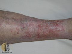
Itchy red, blistered and crusted plaques; or dry fissured and scaly plaques on one or both lower legs. Orange-brown macular pigmentation due to haemosiderin deposition. Atrophie blanche (white irregular scars surrounded by red spots) |
venous eczema |
|
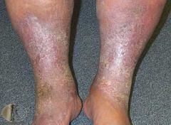
“Champagne bottle” shape of lower leg (narrowing at the ankles) and induration |
lipodermatosclerosis and venous eczema |
|
|
whats the treatment for venous eczema? (general principles) |
treat the leg swelling, treat the eczema, treat the varicose veins. |
|
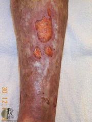
what's this? |
venous leg ulcer |
|
|
what are the 3 general principles for chronic wound management? |
Define the aetiology. Control factors affecting healing. Select appropriate local environmental management (dressings). |
|
|
what are the 3 most common risk factors for venous ulcers ? |
The three most common risk factors for VLUs are a history of obesity, past deep vein thrombosis (DVT) and poor mobility resulting in venous stasis |
|
|
what's the treatment for venous ulcers on legs? |
exclude arterial disease. then graduated pressure stockings, optimise nutrition, treat infection if present |
|
|
what are some patient factors that affect wound healing? |
Health status - Good arterial and venous circulation: anaemia impairs oxygen transport Immune function - Normal immune function helps to cleanse the woundReduced function increases the risk of infection Comorbidities - DiabetesRheumatoid arthritisOther diseases Age-related changes to skin - Loss of hair follicles, sebaceous glands, receptors, Reduced blood supply, Increased fragility, Dryness, Thinning Nutrition |
|
|
what are some external factors that affect wound healing? |
Mechanical stress - PressureFrictionShearing forces Debris - Slough, Necrotic tissue , Eschar, Scab , Dressing residue, Sutures Dessication - Drying of the wound surface results in death of surface cells Maceration - Excess moisture retards healing and damages the peri-skin Infection Smoking |
|
|
new heart murmur + fever + splinter haemmorhages in nails or osler's nodes = ? |
infective endocarditis |
|
|
fever, myalgia, night sweats, weight loss, fatigue, arthralgia can also be insidious symptoms of...? |
infective endocarditis |
|
|
risk factors for infective endocarditis |
abnormal cardiac endothelium allowing for bacteria to adhere and grow (eg. abnormal valves form rheumatic heart disease, congnital valve disease, mitral valve prolapse, calcification of the aortic arch, VSD, PDA, prosthetic valce); presence of organisms in the blood stream (eg. dental procedures, IVDU, any other infection, IVC, PPM) |
|
|
how to diagnose infective endocarditis? |
3 sets of blood cultures, echocardiography, CXR may show signs of heart failure. other bloods such as LFT, FBE, CRP helpful but not for diagnosis |
|
|
there are 3 common presentations of pericardial disease. these are...? |
acute pericarditis, pericardial effusion, constrictive pericarditis |
|
|
acute pericarditis lasts <6 weeks, causes chest pain whic is substernal and shapr, and is relieved with sitting forward and worse with lying down, also worse with respiration. what is the diagnostic investigation? |
ECG - will show ST elevation in anterior, lateral and inferior leads. Later in the illness the ST section will normalise and T wave inversion may occur. Cardiac enzymes may be elevated if these is also myocarditis |
|
|
what causes acute pericarditis? |
coxsackie and echoviruses infections and myocardial infarction are the most common causes. other causes - bacterial esp. with pneumonia and sepsis; uraemic; TB; fungal; malignant; autoimmune. |