Reading...
![]()
Play button
![]()
Play button
![]()
Use LEFT and RIGHT arrow keys to navigate between flashcards;
Use UP and DOWN arrow keys to flip the card;
H to show hint;
A reads text to speech;
127 Cards in this Set
- Front
- Back
|
What is obstructive lung disease? Differential?
|
Obstructive lung disease is any disease that causes increased resistance in the airways to air flow. The mechanism that increases resistance is the narrowing of an airway.
FACES: foreign body obstruction, asthma, chronic bronchitis/bronchiectasis, emphysema, small airway disease (bronchiolitis). |
|
|
What principal factor determines the radius of an airway? What 2 factors can manipulate this?
|
Smooth muscle in the airway determines the radius. PaC02 and the automonic nervous system can change the airway.
PS: bronchoconstriction S: bronchodilation Low C02: bronchoconstriction (body thinks you don't need to ventilate as much) |
|
|
How does expiratory flow change from a normal lung, normal forced expiration lung, to an obstructed lung?
|
Normal Expiration: alveolar pressure > atm pressure, this is driving force to move gas out of lungs. As gas moves, the pressure drops due to resistance but airway stays open because transmural is + (and Ptm is + because Pip is negative).
Normal forced expiration: different because Pip becomes +, from the ab muscles. The pressure drop occurs faster, but transmural pressure = 0 (equal pressure point) in upper airways that can't be compressed. Obstructed lung: the pressure drop occurs EVEN faster because of the high resistance, so EPP happens in lower airways get can be compressed. |
|
|
Why is expiratory flow limited in COPD (emphysema)?
|
In COPD, alveolar elastic recoil is decreased and there is a loss of radial traction (alveolar tethering). This results in lower alveolar pressure, so the pressure drop occurs even faster (and in a collapsible airway).
|
|
|
What is restrictive lung disease? DIfferential?
|
Restrictive lung disease means there is a decreased distendability of the lungs, pleura, or thoracic wall. Think LOW compliance (stiff). There is not impairment to airflow, it just takes more pressure to distend a similar volume.
PAINT pleural effusion, alveolar filling (pneumonia, ARDS, pulmonary edema, alveolar hemorrhage), interstitial (sarcoidosis or pulm fibrosis), neuromuscular, thoracic cage (obesity, kyphosis, scoliosis). |
|
|
What is spirometery used for? What factors determine standard values?
|
Spirometry is used to find FEV1, FVC, peak flow, MVV. Take a full breath, then forcefully expire. You can generate a volume vs time and flow vs volume loops. Normal values are influenced by race, sex, height, and age.
|
|
|
What are the hallmarks of obstructive lung disease?
|
Obstructive
-FEV1 low, FVC low, ratio is low -TLC may be high due to hyperinflation (usually only in COPD-E & sometimes asthma) -RV/TLC may be high due to air trapping -DLCO only low in COPD-E -bronchodilators only help FEV1 with asthma |
|
|
What are the hallmarks of restrictive lung disease?
|
Restrictive
-FEV1 and FVC are low, but ratio is normal -TLC is decreased for all -DLCO is low with alveolar filling and interstitial -MIP is low with neuromuscular |
|
|
How can you measure lung volumes? Any disadvantages?
|
Use body plethysmography or helium dilution.
helium dilution: patient breaths in helium, equilibrium is achieved, measure helium concentration to calculate lung volume. Problem is that in really diseased patients, obstruction may limit helium from reaching certain areas and volume can be underestimated. body plethysmography: patient breathes in mouthpiece in a box, volume at FRC is determined by analyzing pressures and change in volumes. This is most accurate way. |
|
|
How do you measure diffusion capacity? How do you interpret this?
|
Use carbon monoxide, take a max inspiration and hold for 10 seconds. Measure expired air and calculate how much CO was taken up. If DLCO is low, there is a problem with gas exchange. If it's a gas exchange issue, think alveoli/interstitium/capillaries. If DLCO is low and other PFTs are normal, think pulmonary vascular disease.
|
|
|
Is diffusion is reduced, what types of obstructive and restrictive lung diseases should be in your differential?
|
obstructive: emphysema
restrictive: alveolar filling or interstitium |
|
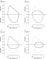
Identify each volume flow loop and give a DD.
|
From top left going clockwise
1: normal 2: normal inspiration, low expiration: tracheomalacia 3: reduced inspiration & expiration: tracheal tumor or foreign body 4: reduced inspiration, high expiration: vocal cord paralysis |
|
|
What is type 2 respiratory failure? What are the 4 signs?
|
Type 2 respiratory failure is also known as ventilatory failure, and it is due to hypercapnia (too much C02).
-tachypnea -decreased mental status -hyperinflation- -acidemia |
|
|
What are the four causes of hypercapnia?
|
Remember, PaC02 = VC02/(RRx(VT-VD))
1. increase C02 production: fever, seizure, sepsis 2. decrease RR: drugs, brainstem lesion, obesity 3. increase VD: anything that distends alveoli/cuts off perfusion 4. decrease VT: anything that decreases compliance or increases resistance |
|
|
How do you manage hypercapnia?
|
-Treat underlying cause
-Non invasive positive pressure ventilation -Intubation or mechanical ventilation |
|
|
What is type 1 respiratory failure? What are the 4 signs?
|
Type 1 respiratory failure is characterized by hypoxemia.
1. tachypnea 2. alkalemia 3. cyanosis 4. agitation/changed mental status |
|
|
What is the normal A-a gradient? How does it change with age? What does an elevated A-a gradient suggest?
|
Normal is <12, but it increases with age. (Age+4)/4 should give appropriate gradient.
Increased gradient suggests -V/Q mismatch -shunt -diffusion issue |
|
|
What are the 5 causes of hypoxemia?
|
-#1 is alway V/Q mismatch!
-shunt -alveolar hypoventilation -decreased PI01 (maybe from high altitude or fire) -diffusion abnormality |
|
|
Mechanism and key features of hypoxemia due to
V/Q mismatch |
Mechanism:
Normal V/Q units cannot compensate for reduced oxygen content (CcO2) from mismatched units Increased A-a gradient Improves with 100% FiO2 Clinical scenarios (many): Pneumonia Congestive heart failure |
|
|
Mechanism and key features of hypoxemia due to
shunt |
Mechanism
Blood from the right heart does not come in contact with ventilated alveoli Poor improvement with 100% oxygen Clinical scenarios Intrapulmonary Arteriovenous malformation Severe alveolar filling process (ie, pneumonia) Foreign body obstruction Intracardiac (right → left) |
|
|
Mechanism and key features of hypoxemia due to
hypoventilation |
Mechanism
Increased PACO2 reduced PAO2 Normal A-a gradient Clinical scenarios: Can’t breathe (neuromuscular) Won’t breathe (CNS depression) |
|
|
Mechanism and key features of hypoxemia due to
reduced PI02 |
Mechanism:
Low atmospheric pressure low PAO2 Normal A-a gradient Clinical scenarios: High Altitude Air travel |
|
|
Mechanism and key features of hypoxemia due to
diffusion abnormality |
Increased A-a gradient
Can improve with increased FiO2 Oxygenation often gets worse with exercise With exercise, RBC transit time decreases Abnormal alveolar capillary membrane impedes oxygen diffusion Clinical scenario: pulmonary fibrosis |
|
|
What conditions are associated with a decreased response to hypoxia?
|
Narcotic addicts
Carotid endarterectomy: scrap plaque off and damage carotid bodies Cyanotic congenital heart disease Altitude native Arnold-Chiari syndrome: brain is displaced, pressure on breathing centers |
|
|
Define the following terms
apnea, hypopnea and respiratory disturbance index |
apnea: cessation of airflow for greater than 10 seconds
hypopnea: reduction of airflow by > 30%, desat by >4% respiratory disturbance index: number of apneas + hypopneas per hour while sleeping |
|
|
Obstructive sleep apnea – definition, pathophysiologic causes, symptoms, risk factors, associated features, and complications
|
OSA: repeated episodes of airway obstruction, which decreases 02 sat, can wake you up from sleep, causes loud snoring, and catecholamine release.
RDI must be > 5, along with associated symptoms More prevalent in men, risk factors include obesity, endocrine issues, snoring, craniofacial abnormalities, and family history Associated features: depression, daytime sleepiness, GERD, loss of libido, anxiety, cognitive deficits Treatment: lose weight, avoid EtOH, get surgery, CPAP *PFTs during daytime will be normal, blood gases should be normal unless they also have OHS |
|
|
What is CSA?
|
CSA is central sleep apnea, meaning there is no signal from the brainstem. It is NOT an obstruction issue. No flow and no effort. Can be due to a variety of neurological causes and CHF.
Treatment: treat underlying causes, resp stimulants, or ventilation *SIDS and is Congenital Central Hypoventilation Syndrome are forms of CSA |
|
|
What is OHS?
|
Obesity associated hypoventilation. Must be morbidly obese, you get awake arterial hypercapnia (>45 mmHg) with no other causes. Patients present with fatigue, morning headaches, mood changes, and dyspnea.
There is also possible leptin resistance, hormone that suppresses appetite and stimulates ventilation. Treat with respiratory stimulants (bad side effects), or ventilation. |
|
|
How is obstructive sleep apnea different from central sleep apnea?
|
OSA: no flow with effort, decreased resp drive during sleep and while awake
CSA: no flow and no effort, decreased resp drive during sleep only |
|
|
What is is most common cause of death for pts with neuromuscular disease? How does respiratory muscle involvement vary across diseases?
|
Most common cause of death = respiratory failure.
Neuropathies have less severe/frequent involvement with respiratory muscles, myopathies more severe/frequent. Overall, degree of involvement varies significantly (progressive, reversible, relapsing). |
|
|
How are respiratory muscles affected by obstructive lung disease?
|
Obstruction leads to hyperinflation. Hyperinflation shortens the length of the muscle fibers, so the MIP decreases and less force is generated. Also, even though muscles aren't weakened, the structure of the thoracic cage has changed. This means the muscles can be dysfunctional due to the changed relationship w/abdomen and chest wall. WOB goes way up.
|
|
|
What causes muscle fatigue/weakness?
What is muscle fatigue? What is muscle weakness? |
The balance between capacity (ability to generate a force) and load determines muscles weakness and fatigue. Too high of a load with normal capacity can lead to fatigue, just as low capacity with normal load can.
muscle fatigue: your muscles can't generate a force (capacity) to deal with the load but improve at rest weakness: capacity of muscle is impaired (regardless of load) no improvement at rest |
|
|
What motions are generated from contraction of the diaphragm?
|
Diaphragm shortens and moves down during contraction ,pushes ribs out and up. As it moves down, pleural pressure decreases and abdominal pressure increases. Apposition occurs at the lower 1/3 of the rib cage, which pushes out because of the increased abdominal pressure (not pleural!). Lung volume also increases because of insertional forces (lifts).
|
|
|
What is Hoover's sign?
|
Hoover's sign = inward motion of the lower ribcage during resting inspiration. Occurs due to hyperinflation/diaphragm shortening (seen in obstructive diseases). Zone of apposition goes away, abdominal pressure can't influence lower ribs anymore so pleural pressure does.
|
|
|
What is thoracoabdominal paradox?
|
thoracoabdominal paradox: thorax moves outward as the abdomen moves inward, due to the diaphragm moving into the thorax during inspiration. This creates negative abdomen pressure, which is why it moves in.
If bilateral, see bad orthopnea due to gravitational forces. Overall effect = decreased lung volume and decreased compliance |
|
|
How does contraction of the scalenes assist in respiration? Why is it important with cervical cord injury? Parasternals?
|
These are primary muscles of respiration, they lift the upper ribs in a pump handle motion (upward and outward expansion).
cervical cord injury: diaphragm alone would cause expiration, but scalenes prevent this. Spinal cord injury below the upper cervical spine means scalenes are preserved, no upper ribcage paradox. *same is true for parasternals which move rib cage upwards |
|
|
What is function of intercostals in respiration?
|
EIC: inspiration, move ribs upwards
IIC: expiration, moves ribs down Nerve supply is T1-T12, paralysis causes inward paradox at first, but stiffness over time due to ankylosis will decreases the paradox. |
|
|
What is the function of abdominal muscles during inspiration?
|
Primary muscles of forced expiration, contraction pulls ribs down, displaces diaphragm into thorax to reduce lung volume. Can help w/inspiration during hyperventilation.
Supplied by T7-L1, so pts with tetraplegia or paraplegia lose expiratory action while upright because flaccid abs shorten the diaphragm > increases resting lung volume, but decreased VC. Classic sign is dysnpea while upright. |
|
|
What other muscles can aid in respiration? How can this help patients with COPD?
|
Muscles that normally maintain posture can help out, COPD pts should lean forward and brace arms.
|
|
|
What three factors (muscle groups) ultimately lead to respiratory failure?
|
can't ventilate (respiratory muscles)
can't cough (abs, upper airway muscles, and resp muscles) increased risk of aspiration (weak upper airway muscles) |
|
|
What are the classic signs of NM disease? What will PFTs look like?
|
Dyspnea, tachypnea (rapid shallow breathing), Hoover's sign, thoracoabdominal paradox, clear lung fields.
PFTs look like restriction..low FEV1 and FVC but normal ratio, decreased TLC, normal DLCO. FEV1 decreases more in supine. MIP and MEP are lower. |
|
|
Define emphysema and chronic bronchitis.
|
Emphysema and chronic bronchitis are part of COPD.
emphysema: respiratory bronchiole become enlarged and alveoli get destroyed. chronic bronchitis: smoker's cough and excess sputum production. Over time lumen narrows, smooth much proliferates. |
|
|
What are the risks factors for COPD? What type of genetic predispositions exist? What is alpha-1-antitrypsin deficiency?
|
#1 risk factor is tobacco. Other risk factors include any type of hazardous exposure (pollution, occupational, etc). Host factors that predispose include hyper responsive airways, HIV, BPD, Tb, low socioeconomic status.
genetics: can have increased elastase/MMP (breaks down collagen), or increased risk of oxidative injury A1A deficiency: can't fight elastase, get emphysema early on, PiZZ is worst (but treatable with anti-elastase) |
|
|
What are the 3 mechanisms behind COPD?
|
chronic inflammation (CD8 lymphocytes, alv macrophages, neutrophils): Irritant activate these cells, CD8 breaks down alveoli, macrophages activates neutrophils dump enzymes to destroy alveoli and cause mucus.
proteinase/antiproteinase imbalance (and factors that contribute): more elastase than antielastase oxidative stress: effects of toxic oxygen radicals can increase mucus, recruit more neutrophils, lower antiproteinases |
|
|
What is the mechanism behind limited expiratory flow in COPD?
|
increased resistance related to changes of chronic bronchitis: mucus fills lumen and inflammatory cells thicken airway
decreased radial traction: alveoli get destroyed, tethering is lost, airways collapse in expiration decreased elastic recoil: elastin in walls gets destroyed, less alveolar driving force |
|
|
What are the physical effects of hyperinflation in COPD?
|
Decreased diaphragm function, neuromuscular capacity goes down. Takes much more work for hyperinflated lung to exhale the same load (remember shortened fiber length as well).
|
|
|
How does the pathophysiology of COPD explain its signs and symptoms?
|
dyspnea: results from increased WOB, decreased diaphragm function, hypoxemia
wheeze: airflow obstruction cough/sputum: increased mucus production weight loss/weakness: inflammation and lower p02 tachypnea: increased WOP, hypoxemia, increased dead space hoovers, TA paradox, accessory muscle use: hyperinflation and diaphragm dysfunction pulmonary hypertension/cor pulmonale: increased vascular resistance |
|
|
What is the classic PFT pattern for COPD?
|
decreased FEV1, FVC and FEV1/FVC; increased TLC and decreased DLCO (if emphysema present); decreased MIP if significant hyperinflation
|
|
|
What factors contribute to hypoxemia and hypercapnia in COPD?
|
Hypoxemia secondary to V/Q mismatch; hypercapnia secondary to increased dead space (other factors that can contribute to hypercapnia are decreased diaphragmatic capacity and abnormal control of breathing).
|
|
|
What is the treatment for COPD?
|
Treatment: smoking cessation improves survival; oxygen therapy improves survival (if hypoxemia, PaO2 < 55 or <60 if there is concomitant pulmonary hypertension); short acting bronchodilators on an as needed basis for increased dyspnea; long acting bronchodilators and inhaled corticosteroids improve symptoms and quality of life; Pulmonary rehab (principally an exercise and reconditioning program) improves QOL, exercise capacity and symptoms, and decreases exacerbations.
|
|
|
What cell types are involved in asthma? What cell mediators are involved? Describe the events that lead to inflammation.
|
(TH2 lymphocytes, mast cells, eosinophils)
(IL-4, IL-5, IL-13, histamine, leukotrienes) Airway inflammation: TH2 lymphocytes get overproduced, sends inflammatory cells into the airways. IgE goes up and mast cells get activated. Eosinophils release toxins that damage epithelium & nerves. |
|
|
What host & environmental factors are involved in asthma?
|
atopy is MOST important (predisposition to allergies, high levels of IgE), obesity, AHR=airway hyperresponsiveness, indoor allergens, pollution, smoking.
Genetics are at play (influence asthma development and severity), but not Mendelian. |
|
|
What are the hallmarks of asthma?
|
There are 3 hallmarks of asthma..
inflammation, hyperresponsiveness, and obstruction. inflammation: all severities, small and large airways AHR: exaggerated response to stimuli, can improve with Rx obstruction: result of thickening, mucus, etc |
|
|
What are the characteristics of early and late phase and implications for treatment?
|
Early Phase:mast cells, think of bronchospasm only.
Late phase:recruited inflammatory cells & mediators, think bronchospasm/edema/inflammation. Need steroids to prevent late phase. TH2/eoisinophils/mast cells too. Sensory nerves exposed, more bronchospasm. (e.g. anti-inflammatory therapy important for minimizing the late phase) |
|
|
What is the pathophysiology of nocturnal asthma and exercise induced bronchoconstriction?
|
Nocturnal: Circadian rhythms impacts lung function, causes decrease of bronchodilating and increase of bronchoconstricting substances. Supine position also contributes because lung volume goes down. Lower catecholamines, more histamine/inflammatory.
Exercise induced: 80% of pts get increased bronchoconstriction during exercise, cold dry air is bad. Evaporating fluid triggers constriction. No late phase rxn. Treat w/albuterol ahead of time. |
|
|
How is asthma treated? What are the goals of long term management?
|
Treat by getting rid of triggers, use short and long acting bronchodilators, steroids, LT antagonists, treat related problems. Anti-IgE Malizumab can help with atopic patients.
Goal is to maintain control of symptoms, be able to exercise/live normally, prevent adverse events. |
|
|
What is bronchiectasis? Why does it occur? What is the cause of majority of cases? What population is it more common in?
|
Bronchiestasis: acquired, females > males. dilation and destruction of bronchial walls leads to mucus retention, infection, inflammation. Majority of cases are idiopathic, associated w/infxn early in life.
|
|
|
What is the CFTR mutation? How does it cause CF?
|
CFTR: cystic fibrosis transmembrane regulator, control's CL movement in and out. CFTR mutation associated with inflammatory response (increased IL-8, TNF-alpha – increase recruitment of neutrophils) even before there is infection. delta F508 most common.
skin: Cl- can't get into skin, high chloride lung/intestine/pancreas: Cl- can't get out of cell, low chloride in the lumen, mucus gets dehydrated/viscous Degenerating neutrophils and bacteria release double-stranded DNA- adds to the viscosity of sputum. |
|
|
What is the criteria for diagnosing a pt with CF?
|
CF (sweat chloride positive in 98%, genetic testing for CFTR), meconium doesn't pass. Sweat chloride is gold standard
|
|
|
What is the pathogenesis of CF in the lungs?
|
Defective CFTR leads to inflammation and infection..lumen is dehydrated, mucus builds up, good growth medium for bacteria.
IL8 and TNF-a are increased, independent of infection. (typical organisms, Staph aureus, H flu, B cepacia and mucoid Pseudomonas aeruginosa – the latter associated with increased mortality) |
|
|
What are the clinical findings of CF?
|
PFT pattern (may be mixed obstructive/restrictive). Bronchiectasis (non-CF) is primarily a respiratory disease while CF is a multisystem disease (sinusitis, bronchiectasis, exocrine panc insufficiency, meconium ileus/bowel obstruction, hepatobiliary disease, infertility).
|
|
|
How do you treat CF?
|
Airway toilet (help clear secretions) via chest physiotherapy, antibiotics (included inhaled aminoglycosides, tobramycin), DNase (chops ds-DNA into ss-DNA making it less viscous), beta-2-agonists (not anticholinergics as they may precipitate colonic obstruction), hypertonic saline (to rehydrate apical fluid layer and improve mucociliary clearance), NSAIDs (anti-inflammatory) and Azithromycin as anti-inflammatory (if patient colonized with Pseudomonas), Lung Transplant when FEV1<30% predicted. Newer treatments focusing on potentiating effect of CFTR.
|
|
|
How is bronchiectasis associated with CF different from non-CF associated?
|
With CF, multiple organ systems are also affected. Bronchiectasis is simply the pulmonary manifestaton.
|
|
|
What do PFT's look like for the following diseases?
OSA PHTN Mixed |
OSA: normal PFTs, only affected during sleep
PHTN: normal PFTS, except a lower DLCO Mixed: low FEV1, FVC, ratio. Low TLC. |
|
|
What are the common features of interstitial lung disease?
|
Progressive dyspnea, nonproductive cough, bilateral infiltrates on radiography, decreased compliance/stiff, abnormal gas exchange, inflammation, fibrosis.
Rapid and shallow breathing, shallow to minimize work but compensate by increasing RR. |
|
|
What is the pathogenesis of ILD? In idiopathic pulmonary fibrosis?
|
Triggered by antigen or injury (radiation, meds, aspiration, smoking). Epithelial cells activated, T cells activated. T cells > cytokines, B cells get activated, auto antibodies can cause injury to capillaries or epithelium. Process unchecked = fibrosis.
IPF: direct injury means cells release TGF beta, fibroblasts and myofibroblasts lay down collagen, clearance mechanisms (caveolin) gets blocked. |
|
|
What is the most common cause of abnormal gas exchange in ILD? What happens in exercise?
|
V/Q mismatch, even though diffusion is a problem. Lost alveoli (less V), can't distend the lung well (less V). Dead space also goes up, large cystic spaces with ventilation but not perfusion, also see pulmonary vascular disease.
Exercise limits time for diffusion, 02 saturation goes down (C02 not affected until it's really bad). Once it is severe enough, low diffusion even at rest. |
|
|
What are the complication associated with ILD?
|
Respiratory failure due to high WOB, many have exacerbations that lead to more reduction in fxn, pulmonary vascular disease can lead to pul hyper tension and RHF. Pneumothorax and PE (some disease make pt hypercoaguable, like lupus). Lung cancer (not sure why). Infxn risk is high because lung structure is so abnormal.
|
|
|
What are the clinical findings of ILD?
|
Dyspnea, slowly progressive (WOB increased, hypoxemic). Non-productive cough, airways are distorted and cough receptors activated.
Cyanosis (due to hypoxemia), alopecia/rash/sclerodactyly/muscle weakness (clue for systemic diseases), tachypnea, tachycardia, crackles/rales on end inspiration, squeaks at end of inspiration (bronchioles involved), edema in lower extremities. |
|
|
What are some key radiographic findings for ILD?
|
fibrosis: see blood vessels going to pleura and honeycombing or cystic spaces, reticular opacities
nodular opacities: granulomas or malignancies ground glass infiltrates: active inflammation, pneumoitis or alveolitis |
|
|
What is usual interstitial pneumonia?
|
Can lead to pulmonary fibrosis. No cause = IDF. Association with smoking, pollution, aspiration. Injury to epithelium causes collagen formation. See reticular infiltrates, honey combing starting at base. Lung looks like it is at different stages (temporal & spatial heterogeneity). Relentlessly progressive, with no treatment!
|
|
|
What is desquamative interstitial pneumonia?
|
Seen in smokers. See ground glass infiltrates (inflammation), lungs filled with alveolar macrophages (hemosiderin = orange) and damage is diffuse. Stop smoking and give anti-inflam, you'll get better. Presentation is same as pulmonary fibrosis.
RB-ILD is similar!! |
|
|
What is cryptogenic organizing pneumonia?
|
BOOP! Presents like pneumonia, shortness of breath, fever, non-productive cough. Patchy migratory infiltrates. No fibrosis, get chunks of granulation tissue in bronchioles (NOT granulomas) which extends into alveoli. Fibrotic plugs. Can get better w/treatment or on it's own, can be idiopathic, no relation to smoking!
|
|
|
What is sarcoidosis?
|
Don't know cause. Non-caseating granulomas (chronic inflammatory cells + multinucleated giant cells) in multiple systems. Can turn into fibrosis. Hilar adenopathies. Increased ACE. Can diagnose with bronchoscopy. Must exclude mycobacteria & fungal infxns. Very treatable with steroids.
|
|
|
What is chronic eosinophilic pneumonia?
|
Non-smokers. Tons of eosinophils in alveoli, can have fever and wheezing (> 50% have asthma). Wash out lung and analyze the cells to see if there are eisoinophils. Steroids make it much better.
|
|
|
What is pulmonary Langerhans cell histocytosis aka histiocytosis X aka eosinophilic granulomas?
|
Birbeck granules (lollipops) and Langerhan's cells (coffee beans). Similar presentation to ILD, but a disease of smokers in 40s-50s. Best treatment is to stop smoking. Bone and brain can get involved.
|
|
|
What is Wegener Granulomatosis?
|
Multiple nodules on gross lung. Vasculitis, diffuse necrotizing granulomas. Acute onset. Considered pul-renal, abnormal C-ANCA.
|
|
|
What is acute interstitial pneumonia?
|
Clinically similar to ARDS but no precipitating event. DIffuse damage to alveoli. Diffuse fibroblastic proliferation in interstium & alveolar space, thick hyaline membrane.
|
|
|
What are some of the treatments for ILD?
|
Hypoxemic: give oxygen, need vaccinations, rest depends on specific disease.
Smoking cessation, remove from occupation, glucocorticoids for inflammatory (nodules & ground glass), do NOT give if they have infxn. Fibrosis = lung transplant. |
|
|
How does the blood supply of parietal and visceral pleura compare? How does lymphatic drainage compare? How do nerves compare?
|
Parietal pleura is supplied by systemic circulation. Visceral pleura is supplied by bronchial arteries (systemic) and pulmonary veins (remember low pressure). Pressure in parietal capillaries > visceral capillary.
Parietal: stoma openings drain to IVC, plugging means no drainage. Visceral drains into hilar lymph nodes, does not drain pleural space. Visceral has no nerve fibers, parietal is richly innervated. Feel pleuritic pain from intercostal nerves. Cental pleura = phrenic nerve, get referred pain. |
|
|
What are the 2 functions of the pleura?
|
1. reduce friction, the fluid acts as a lubricant
2. absorb gas to prevent build up. Low total gas pressure in capillaries, gas moves from pleura to capillary. |
|
|
What is a transudate vs an exudate pleural effusion? What are the most common examples of each?
|
Transudate: disruption of Starling forces
-CHF: increased hydrostatic/capillaries causes edema -hypoproteinemia: decreased oncotic -atelectasis: reduced intrapleural pressure -ascites: transdiaphragmatic flow Exudate: disruption of the pleural surface (capillary leakage, inflammation, etc) -malignancy: drainage blocked -pneumonia -empyema -Tb etc! |
|
|
Explain the clinical manifestations of pleural effusions?
|
Dyspnea: fluid makes it hard to distend lung (restrictive), or fluid pushes diaphragm down. Depends on size of effusion and function of the lung.
Chest pain: not caused by pleural effusion, but inflammation or tumor does. Less movement of chest wall on side of effusion, dullness to percussion, absent breath sounds on lung. |
|
|
Why are protein and LDH important in the differential of pleural effusions?
|
If fluid has 1+ of the following, must be exudate (high protein and high LDH).
pleural fluid protein/serum protein > 0.5, pleural fluid LDH/serum LDH >0.6 pleural fluid LDH > 2/3's normal serum LDH |
|
|
How is CHF related to pleural effusion?
|
Most common cause. See cardiomegaly, pulmonary venous pressure has gone way up. Usually bilateral. Don't need to sample fluid, know that it is transudate. Only thoracocentisis if pain or fever, sample to see what is up.
|
|
|
What is hepatic hydrothorax?
|
Cirrhosis > ascites > increase in portal venous pressure. Get fluid in peritoneum, defects in diaphragm allow fluid to move into pleural space. Most common on the right side 90%. Fever or pain should cause you to sample to see what else could be going on. Just treat underlying disease. Transudate.
|
|
|
How is pneumonia related to pleural effusion?
|
Parapneumonic effusion: intense inflammatory response due to pneumonia, makes it leaky. Exudate is sterile.
Empyema: organisms or pus from pneumonia are leaking into pleural space. High WBC, see exudate, low glucose because of metabolic activity, LDH is high, pH is low due to metabolic activity. Treat small effusion with antibiotics, treat large effusion with catheter. |
|
|
1. How is TB related to pleural effusion?
2. How is PE related to pleural effusion? |
1. Rupture into pleural space. Hypersensitivty rxn occurs, get lymphocytic rich exudate (not eiosinophils). Culture is not always postive, biopsy would be helpful to find necrotizing granulomas.
2. PE: hemorrhagic exudate or transudate |
|
|
How is malignancy related to pleural effusion?
|
Most common in lung cancer, breast cancer. Many mechanisms. Drugs, blockages that lead to pneumonia. Exudate, see lymphocytosis and RBCs. Low glucose due to lots of cells, no treatment for this.
Treatment: drainage and rapid reaccumulation..put in a catheter. |
|
|
What is a chylothorax?
|
Thoracic duct is on vertebral column, disruption in thorax cause chyle to leak out (lymph). Exudate but milky, lots of triglycerides. Due to tumor or trauma.
|
|
|
How do we treat pleural effusions?
|
Transudate: treat disease
Exudate: treat disease and drain Hemothorax: get blood out (too good of a culture medium for bacteria) |
|
|
What is a pneumothorax?
-simple -tension -secondary -iatrogenic |
Too much gas, can't be reabsorbed. Positive pressure raises, get restrictive lung disease. In highly compliant lungs, small pressure may cause collapse. In stiff lungs, much harder to cause collapse.
simple: youngish smokers/men with small blebs. Complain of chest pain and dyspnea. Drain air with catheter, trachea displaced same side. tension/traumatic: crazy high pressure in space, heart/trachea pushed to other, inverted diaphragm, cause shock and severe resp. distress secondary: occurs with COPD/CF/asthma/HIV/PF, due to alveoli rupturing. Drain air but need more therapy (pleuradesis) iatrogenic: trying to put a cather in, lung biopsy, etc |
|
|
How do we diagnose pneumothorax?
|
Acute onset of chest pain and dyspnea. Big PT means low breath sounds, percussion sounds like a drum. Tension PT can see trachea shifted. Small PT can treat with 100% 02, large PT needs chest tube to evacuate air.
|
|
|
Who is at risk for asbestos exposure?
|
Ship workers, steel industry, paper products, brake linging repair, chemical/plastics industry, installing/destroying products with asbestos. Relationship btw exposure and asbestosis is temporal and dose dependent.
|
|
|
What are the clinical/patho features of asbestosis?
|
Interstitial lung disease, (+exposure to make diagnosis), asbestosis is different than asbestos exposure. See honeycombing on CT and reticular infiltrates (fibrosis). See asbestos bodies (ferruginous) on pathology + identical to PF. Macrophages rupture from needles. Calcified pleural plaques (most common marker, NO clinical significance) and pleural effusion. Mesothelioma almost always due to asbestos but lung cancer >. Occurs 10-20 yrs after exposure.
|
|
|
What is silicosis?
|
Not common anymore but a type of pneumocosis, from mining or cutting rocks, sand blasting. Dust particles don't get phagocytosised by alveolar macrophages. Asymptomatic, adenopathy calcified (egg shell). Used to be risk factor for Tb, macrophages can't fight!
*Coal Workers Pneumoconiosis is clinically similar. |
|
|
What is hypersensitivity pneumonitis?
|
Lymphocytes are involved (Type 3/4 hypersensitivity), caused by organic dusts. More CD8, NO eiosinophils! Poorly formed non-necrotizing granulomas. Can be acute, subacute, chronic.
acute: 4-8 post exposure, resolves w/steroids & exposure removal chronic: similar to pulmonary fibrosis, need history Actinomyces, avian proteins, humidifiers. |
|
|
What causes direct tissue injury (RAD) for pulmonary diseases?
|
Different from hypersensitivity, but treat like it is asthma.
Silo Fillers: inhalation of N02, get acute pulmonary edema, quick rxn Metal fume fever: vaporized metal during welding can cause flu like illness. reactive airway dysfunction syndrome: caustic vapor (cleaning agent), large airway injury (because you don't inhale deeply), inflammation results and can lead to long time disease. |
|
|
What jobs can lead to occupational asthma? What is occupational asthma?
|
Auto body shops, bakers, lumberjacks. Hard to tell if it is occupational or just asthma. Makes up 1-5% of all asthma cases. Can't have other triggers or regular asthma. More severe when go home/throughout the day, symptoms improve on vacation. Takes a few weeks to start up! Best way to diagnose is peak flow monitoring!
|
|
|
What is respiratory failure?
|
1/3 of ICU pts have resp. failure. It can be acute, rapid onset, life threatening or it can be chronic/progressive. Can be hypoxemic = type 1 (Pa02 < 60 mmHg) or hypercapnic & hypoxemic = type 2 (PaC02 > 45 mmHg).
|
|
|
What is hypoxemic respiratory failure?
|
V/Q mismatch and shunt are most common causes, severe when Pa02 < 60. Pa02/Fi02 ratio below 300. Decreased 02 leads to catecholamine release, tachycardia, and hyperventilation.
|
|
|
What does oxygen delivery depend on? What happens when it decreases?
|
D02 = Ca02 x the cardiac output.
*Ca02 = 1.34 x Hgb x Sa02 + .003 Pa02 As oxygen delivery decreased, tissue maintains normal metabolism at first, but then it can't extract more oxygen. Further decreases in delivery = cell death/organ failure. Goal is to increase delivery. |
|
|
What are common causes of type 1 respiratory failure? What is the general aim of treatment?
|
Anything that causes hypoxemia, most common is V/Q mismatch. Otherwise consider shunt, diffusion abnormality, decreased Fi02, decreased MV02.
In general, goal is to improve ventilation to affected area or reduce perfusion to a poorly ventilated area. |
|
|
What are the mechanisms of hypercapnic respiratory failure?
|
Increase in dead space, increase in c02 production (but not on it's own), decrease in minute ventilation (won't breath or can't breath, drug overdose/stroke/problem with muscles). Could also have imbalance btw. load and capacity. Causes: drug OD, spinal cord injury, acute NM disease (Myasthenia, Guillan-Barre, botulism), COPD, bad asthma (last two increase dead space).
|
|
|
What is chronic respiratory failure?
|
Mechanism is similar to acute. Pa02 goes down, erythopeotin goes up, more Hb is produced and more oxygen delivery happens. If PaC02 goes up, renal bicarb increases, bicarb goes up, prevents a fall in pH. Main idea is our body can adapt physiologically to prevent end organ damage.
|
|
|
How do we treat respiratory failure with oxygen (different types of delivery systems)? What is the goal of all systems?
|
1. nasal cannula (most common), 100% deliver but diluted by the room air so the Fi02 is variable. Must humidify!
2. face mask: less comfortable, higher flow rate and 02 concentration than nasal cannula, Fi02 is variable. C02 can build up. Want to maximize 02 delivery, keep above 90% sat and Pa02 above 60 (flat part of curve), but not too high because it can be toxic. Acute requires more oxygen/higher flow than chronic. |
|
|
What are complications of oxygen therapy?
|
Generation of free radicals that injure cells..don't give more than 60% for 24 hrs (unless they really need it). In peds, premature babies are really affected by retrolental fibroplasia (vascular proliferation in the retina which causes blindness) & bronchopulmonary dysplasia (lung development disrupted, alveoli don't form normally). Adults & peds get injury to epithelium (tracheitis, cilia injury, acute lung injury), absorptive atelectasis, hypercapnia with COPD (blood flow disrupted, areas with low V/Q or high V/Q that act like dead space). Always use lowest concentration to achieve goals.
|
|
|
What are other treatments besides maximizing oxygen delivery for respiratory failure?
|
Avoid hypercapnia by avoiding sedatives, preventing fever, treating underlying disease.
|
|
|
When should we use mechanical ventilation?
|
Endotracheal intubation when other oxygenation isn't working, acute type 2 failure (to guarantee the minute ventilation), WOB is too much, lung is too far collapsed, or patient has limited respiratory drive. You can give 100% oxygen, and positive pressure helps with lung expansion, machine takes over the WOB so muscles can rest.
|
|
|
What is ARDS? What do you see on x-ray or CT? What do you need to make the diagnosis?
|
Clinical syndrome with acute type 1 respiratory failure, constellation of findings. Due to inflammation injury to alveoli, permeability goes up and causes pulmonary edema. Chest x-ray shows diffuse process of white (alveolar filling), denser at bases than apex. CT shows heterogenity (pleural effusion, consolidation, and normal lung). Must have acute onset (one week of known insult), abnormal radiology (not explained by other processes), respiratory failure (not explained by CHF or fluid overload), & hypoxemia (moderate to severe based on Pa02 to Fi02 ratio).
The lower the ratio, the worse it is. |
|
|
What are clinical manifestations of ARDS? Differential? Common causes?
|
Depends on what the cause is. Pnemonia = cough, sepsis = fever, pancreatitis = abdominal pain. Dyspnea & respiratory distress.
Could also be cardiogenic pulmonary edema, bilateral pneumonia, alveolar hemorrhage, eosinophilic pneumonia. Sepsis, pneumonia, aspiration, but over 60 possible causes. Direct = pulmonary cause, indirect = extrapulmonary. |
|
|
What is pathophysiology of ARDS?
|
Initial injury, early exudative phase, proliferative, late fibrotic phase.
1. Injury leads to inflammation and permeability goes up. Macrophage release cytokines, neutrophils recruited. 2. exudative: further injury by PMNs causes exudate to leak , overwhelm lymphatics and then transudate leaks. Alveolar filling wrecks type 2 pneumocytes & surfactant > increased collapse of alveoli > shunt (perfusion w/out ventilation). Hyaline membrane thickens. 3. fibroproliferative: either rapid resolution w/minimal damage or fibrosis. |
|
|
What is treatment for ARDS? What are the benefits?
|
First, try to treat the cause, then MV.
Mechanical ventilation: provide resp support during recovery, let the lungs heal. Maintain oxygenation, don't let ventilator do harm. Keep TV LOW!! (6 mL/kg vs 12 mL/kg) and use PEEP. Benefits = high concentrations of 02, and PEEP to correct atelectasis/move fluid into interstitium. Other treatment includes... Supportive care: put in prone position, fluid management, nutrition, prohylaxis for DVT or GI bleeds. Drugs: sedatives and neuromuscular blockades to improve ventilation, corticosteroids don't work except maybe in severe... |
|
|
What is PEEP? What is relationship btw tidal volume and PEEP?
|
PEEP = positive end expiratory pressure, does a lot to improve gas exchange and open up diseased parts of lung. Want just to right amt, not too much and not too litte.
As you increase the tidal volume, some of lungs open up that cause small changes in pressure for large changes in tidal volume. Over distension occurs, and then a small change in tidal volume causes major pressure change. Main idea = treat with smaller tidal volume (6 mL/kg of body weight). |
|
|
What are physiological complications of mechanical ventilation?
|
Positive pressure ventilation can
-reduce venous return and decrease cardiac output (too much intrathoracic pressure) -can cause acute respiratory alkaosis if there is too much ventilation -increase in pulmonary vascular resistance due to the pressure |
|
|
What are the causes of hypoxemia in ARDS? What would cause C02 to rise in ARDS? Why do we see restrictive physiology?
|
Shunt (extreme V/Q mismatch). Shunt because perfusion is normal at first, but no ventilation due to alveolar filling and collapse.
Increased dead space due to damage of capillaries endothelium, some parts of lung maintain ventilation but perfusion is down. If dead space goes up, PaC02 goes up. Restriction due to fluid compressing lungs, hard to expand so decreased compliance. Takes more pressure to open collapsed alveoli & fill fluid filled. Later on, fibrosis decreases compliance. |
|
|
What are the negative effects of mechanical ventilation (ie. barotrauma, volutrauma, biotrauma, alectetrauma)?
|
Barotrauma means pressure from ventilator can cause leakage into pleural space
Volutrauma = overdistension causes stretch injury biotrauma = release of inflammatory mediators atelectrauma = opening/closing too many times will destroy alveoli |
|
|
What is neonatal respiratory distress syndrome?
|
50% of neonates born at 26-28 weeks.. Premature infants produce insufficient quantities of surfactant. When born they develop hypoxemic respiratory failure. The disease can be prevent by administering corticosteroids prior to birth – promotes maturation of Type II cells and ability to produce and secrete surfactant. After birth, exogenous surfactant can be administered into the lung.
|
|
|
How is pulmonary hypertension defined? Pulmonary venous hypertension?
|
Pulmonary hypertension = mean PA of > 25 at rest or > 30 with exercise. The post capillary wedge pressure must be < 15. If the PCWP is > 15 mmHg, then it is pulmonary venous hypertension.
|
|
|
How can you calculate the pressure in pulmonary arteries? How can you calculate the vascular resistance? What factors can increase resistance?
|
PA = PVR x CO + PCWP
*PVR = (PA - PCWP)/ CO *This should make sense, because resistance depends on the pressure drop/flow. Resistance increase: increase viscosity (polycythemia), decrease radius |
|
|
What factors lead to vasocontriction of blood vessels?
|
Either increase constriction or decrease ability to dilate.
vasoconstrictors: increase thromboxane, endothelin vasodilators: decrease NO, prostacyclins hypoxic vasoconstriction: can lead to smooth muscle proliferation and medial thickening genetic: BMPR2 gene leads to smooth muscle proliferation/thickening (TGF B increase) |
|
|
What is cor pulmonale?
|
Changes in the structure or function of the right ventricle, must be due to dysfunction of the repsiratory system. Can be acute (fast increase in PVR causes failure) or chronic (RV hypertrophy to accomodate increased PVR).
|
|
|
How is pulmonary hypertension classified?
|
Group 1: Phtn with normal PCWP and increased PVR. Think idiopathic, connective tissue disease, ASD or VSD (l to r), cirrhosis, HIV/drugs.
Group 2: PVhtn, high PCWP (> 15) but normal PVR. Think left side heart problems, metabolic issues (obesity, OSA, systemic HTN > LV hypertrophy) Group 3: chronic hypoxia or parenchymal lung disease, can be obstructive or restrictive Group 4: acute issues, either PE or maybe schistosomiasis Group 5: randos |
|
|
What are the clinical features of pulmonary hypertension? How is it diagnosed? How is it treated?
|
Features: Dyspnea, syncope, edema, JVD, tricuspid regurg, loud P2, symptoms worse on exertion.
Diagnose: EKG, CXR, ECG, right heart catheter, PFTs, blood gas, V/Q scan, CT angiogram. Treat: causative factors 1st, anticoagulants, Ca2+ blockers to reduce smooth muscle contraction, vasodilators (endothelin anatagonist, prostacyclins, PDE5 inhibitors). |
|
|
What is a pulmonary embolism? What are the pathophysiological consequences?
|
PE: some vascular obstruction that causes acute increase in dead space, usually from deep leg vein. Leads to hypoxemia due to V/Q mismatch, wide A a gradient, atelectasis (surfactant goes down). PVR goes way up, get Phtn. If > 50-70% of circulation is blocked, think severe Phtn/acute cor pulm/systemic shock.
|
|
|
What are clinical features of PE? How do we diagnose PE? How do we treat PE?
|
Clinically: Dyspnea, chest pain, hemoptysis, syncope, tachypnea, crackles/rales, tachycardia, fever.
Must search for risk factors using Virchow's triad: stasis, inflammation, hypercoagubility. CXR, V/Q scan, CT angio, lower extremity ultrasound. Treat with heparin/coumadin to bust clots. |
|
|
What is pulmonary edema? What are the pathophysiological consequences?
|
Pulmonary edema is when fluid flows from capillaries into alveolar spaces and interstitium, due to Starling force disruption.
Think left side heart problems (flow backs up and pressure goes up), volume overload, hypoalbuminemia (low protein in vessel means fluid flow to high protein outside), lymphatic blockage, damage to capillaries (ARDS or acute interstitial pneumonia). |