![]()
![]()
![]()
Use LEFT and RIGHT arrow keys to navigate between flashcards;
Use UP and DOWN arrow keys to flip the card;
H to show hint;
A reads text to speech;
75 Cards in this Set
- Front
- Back
|
What are "fortification illusions"? |
Visual displays experienced by (20% of) migraine sufferers |
|
|
Can some animals see with NO light? |
No, light is needed to see - some can see with dim illuminations |
|
|
What wavelength range does visible light fall under? |
380-760nm (Red to Violet light, ROYGBIV) |
|
|
What is the iris? |
Donut-band of contractile tissue, controls the amount of light that reaches the retinas |
|
|
What is the pupil? |
Hole in the iris where light enters |
|
|
Definition of sensitivity vs. acuity? |
Sensitivity: Ability to detect presence of objects Acuity: Ability to detect details of objects NOTE: There is a trade-off between them |
|
|
a) What is the lens? b) What is it adjusted by? |
a) Focuses light on the retina b) Adjustments made by the ciliary muscles |
|
|
What is "Accommodation"? |
The process of adjusting the lens configuration to bring an image into focus |
|
|
What is binocular disparity? What kinds of species have it? |
Definition: The difference in position of the same image on the two retinas Humans have it (For 3D depth perception) vs. Species with 2 eyes on sides of their head, allowing them to see every location around them |
|
|
What is the order of structures that light passes through? |
Pupil, then lens, then reaches the retina |
|
|
What is the retina? |
The back of the eye, converts light to neural signals and conducts them towards the CNS (Participates in signal processing) |
|
|
What are the 5 layers of retina neurons? |
1) Receptors 2) Horizontal cells 3) Bipolar cells 4) Amacrine cells 5) Retinal ganglion cells RHBAR |
|
|
Which of the retina cells are specialized for lateral communication (I.e. Collateral branching)? |
Horizontal cells Amacrine cells |
|
|
Problem: Incoming light is distorted by retinal tissue it has to pass before reaching the receptors How is this solved? |
The fovea: Specialized for high-acuity vision, there is a thinner retinal ganglion layer here |
|
|
Problem: Must be a gap in the receptor layer, for ganglion cell axons to leave the eyeball (I.e. the blind spot) How is this solved? |
"Completion": Information used from surrounding receptors around the blind spot to fill in the "gaps" in the images |
|
|
What is the process of "filling in" the areas of the blind spot called? |
"Surface interpolation"? |
|
|
What are the two receptor types in the eyes? Animals active in what time of day only, use which receptor? |
Daytime: Cone-only retinas Nighttime: Rod-only retinas |
|
|
What is duplexity theory? |
Idea that cones and rods mediate different kinds of vision |
|
|
What is photopic vision? |
Cone-mediated vision - provides high-acuity colour perception |
|
|
How many cone receptors converge on a single ganglion? How to stimulate? |
Just a few -- you need GOOD lighting, dim lighting is not sufficient |
|
|
What is scotopic vision? |
Rod-mediated -- lacks colour/detail, but greater level of sensitivity |
|
|
How many rods converge on a ganglion cell? How to stimulate? |
Hundreds on a single cell - dim light is fine |
|
|
What is the distribution of rods/cones throughout the retina? |
In the fovea -- ALL cones Rod density is greatest, 20deg from the foveal center Cone density declines significantly at the boundaries of of the foveal indent |
|
|
What is the photopic spectral sensitivity curve? |
Measured in humans by relative brightness of lights shone directly onto the fovea |
|
|
What is the scotopic spectral sensitivity curve? |
Measured in humans by relative brightness of light shone onto retinal peripheries - at an intensity too low to activate the cones there |
|
|
What is a spectral sensitivity curve? |
A graph displaying the relative brightness of lights of the same intensity, but with different wavelengths |
|
|
What is the Purkinje effect? |
Peak sensitivity of the human eye tends to shift towards the blue end of the spectrum, when low levels of light are present (More rods activated than cones) |
|
|
What is temporal integration? |
Visual perception is "summation" of all recent visual information - eyes are constantly scanning for new information |
|
|
a) What are fixational eye movements? b) What are the 3 kinds? |
a) Involuntary eye movements to allow for continuous scanning of an area b) Tremors, drifts, saccades (small, jerky movements) |
|
|
What is visual transduction? |
Conversion of light to neural signals (via visual receptors) |
|
|
a) What is rhodopsin? b) What happens with continuous exposure to light? C) If returned to dark? |
a) The photopigment of rods b) Gets bleached, can't absorb light c) Returns to normal |
|
|
Physiologically, how does rhodopsin respond to light? |
Darkness: Sodium channels partially open - allows for steady flow of glutamate, and slight depolarization of rods Light: Cascade closes sodium channels, rods are hyperpolarized, and glutamate release is reduced |
|
|
What type of receptor is rhodopsin? |
G-protein receptor |
|
|
What is the retina-geniculate-striate pathways? (RGSP) |
The path that carries visual information to the brain |
|
|
What nucleus of the thalamus are retinal signals carried to, before reaching the primary visual cortex? |
The lateral geniculate nuclei |
|
|
What is distinct about the left/right sensory pathways of the visual system? |
Signals from left visual field reach the right primary visual cortex - Right eye: Temporal hemiretina - Left eye: Nasal hemiretina Signals from right visual field reach the left primary visual cortex - Right eye: Nasal hemiretina - Left eye: Temporal hemiretina |
|
|
Describe the lateral geniculate nuclei |
Each nucleus has 6 layers - 3 each get input from 1 of the 2 eyes |
|
|
What is the striate cortex? |
The striped pattern of neurons as they reach the primary visual cortex (from the geniculate nuclei) |
|
|
Where do the neurons of the visual system terminate in the brain? |
Deep in cortical layer 4 |
|
|
What is retinotopic organization? What is the exception? |
Each level of the visual system is organized like a map of the visual system BUT, in the primary visual cortex, 25% is dedicated to foveal input analysis - disproportionate representation |
|
|
What are the 2 channels of communication that flow through the lateral geniculate nucleus? Where do both project to? |
Parvocellular layers (P): Runs through top 4 layers, neurons have small cell bodies - Responsive to colour/pattern detail - Input from cones Magnocellular layers (M): Runs through the bottom 2 layers, neurons have large cell bodies - Responsive to movement - Input from rods Projection: Visual cortex, cortical layer 4 |
|
|
What is a visual edge? |
The place where 2 areas of a visual image meet |
|
|
What is contrast enhancement? Mach bands? |
When looking at an edge, the darker side appears darker, and the brighter side, brighter An illusion that illustrates this |
|
|
What are the receptors in the eyes of horseshoe crabs that are connected with a lateral neural network? |
Ommatidia receptors |
|
|
Why does contrast enhancement occur, explained with ommatidium? |
If an ommatidium is illuminated, it inhibits the neighbouring cells (I.e. Lateral inhibition) Intensity (I.e. of a darker colour) increases firing, and so there is more firing/lateral inhibition with darker colours, but less with a lighter colour |
|
|
When Hubel/Wiesel studied visual receptive fields, what 3 levels did they study of the RGS system? |
1) Retinal ganglion cells 2) Lateral geniculate neurons 3) Striate neurons of cortical layer 4 |
|
|
When Hubel/Wiesel studied visual receptive fields, what 4 commonalities did they find? |
1) At each level, foveal area receptive fields were smaller than those at the periphery 2) All neurons had circular receptive fields 3) Neurons were monocular (Had a receptive field in ONE eye...obviously) 4) There was a boundary of an ON! and OFF! area (Some cells were on-center, others off-center) |
|
|
Describe the receptive fields of simple cells (I.e. NOT RGS system, just the rest of the neurons in the primary visual cortex) |
Receptive fields with ON! and OFF! sections - therefore unresponsive to diffuse light BUT, the visual fields are composed of straight lines |
|
|
How are "complex cortical cells" different from simple cells? |
a) More numerous b) Larger receptive fields c) Binocular (Respond to stim. in either eye) d) The cell cannot be divided into ON! and OFF! regions |
|
|
How are "complex cortical cells" similar to simple cells? |
a) Both have rectangular receptive fields b) Both respond to straight-line stimuli in a CERTAIN ORIENTATION c) Both are unresponsive to diffuse light |
|
|
What is retinal disparity? |
Some cells fire best when the stimulus is presented at the same time to both eyes in 2 different positions on the 2 retinas |
|
|
Is primary visual cortex organized columnarly or horizontally? |
Columnar organization: If electrode is advanced through the visual cortex layers, vertically, each cell will have: a) Receptive area in same location of visual field b) Respond best to straight lines of light in the same orientation c) Ocular dominance, dominated by the same eye |
|
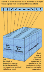
What does this show? |
Functional columns for eye each Each columns is further clustered into neurons with preference for straight line stimuli of a particular direction |
|
|
Explain neuroplasticity of the visual system? |
The visual cortex neuron's response is dependent on the stimuli in its receptive field, and also the larger scene the stimuli is coming from Basically, the receptive field can change based on the stimulus and the scene |
|
|
What is a hue? |
The correct term for a colour - the combination of wavelengths it reflects into the eye |
|
|
What is component theory (trichromatic theory)? |
Proposes that there are 3 kinds of colour receptors (cones) with different spectral sensitivities, and colour of a stimulus is encoded by mixing 3 different wavelengths in certain proportions |
|
|
What is opponent-process theory? |
Two different classes of cells in the visual system for encoding colour....and one for encoding brightness Red/Green, Blue/Yellow, Black/White Cells code for opposing colours by depolarizing/hyperpolarizing in a certain direction |
|
|
What are complementary colours? |
A pair of colours that produce white/gray when combined equally CANNOT exist together Afterimage of staring at one colour is the complementary colour |
|
|
There are trichromats (3 colour vision photopigments) and dichromats, but what can you see with 4 photopigments? |
UV light - some birds/fish have this ability |
|
|
What is colour constancy? |
The ability of an object to stay the same colour, despite major changes in the wavelengths of light it reflects (I.e. Yellow shirt is always yellow) Occurs as long as object is part of a scene |
|
|
What is reflectance? |
The proportion of light that a surface reflects |
|
|
What is retinex theory? |
The colour of an object is determined by its reflectance Visual system perceives colour of an object by comparing to adjacent surfaces (Basically we see colours and understand them by comparing to adjacent colours) |
|
|
What are dual-opponent colour cells? |
Certain cells respond with ON! if the center is stimulated with one colour, and the surrounding, the opposite colour - Rich in cytochrome oxidase - Concentrated in "peg-like" columns, or "blobs" in the primary visual cortex (but NOT deep cortical layer 4, but also below deep cortical 4) - Blobs found in ocular dominance columns - The cell basically responds to contrasting wavelengths in the visual field |
|
|
Where is the primary visual cortex located? |
Posterior region of the occipital lobes |
|
|
What are the two areas of the secondary visual cortex, and where are they located? |
Prestriate cortex: Band of tissue in the occipital lobe, surrounds the primary visual cortex Inferotemporal cortex: Cortex of the inferior temporal lobe |
|
|
Where is the visual association cortex? (IMPORTANT) |
POSTERIOR PARIETAL CORTEX |
|
|
What is a scotoma? |
Area of blindness in the visual field |
|
|
How do you test for a scotoma? |
A perimetry test - TINY light shone over parts of the eye, and a button is held when the person sees the light or not - maps out the visual field |
|
|
What does it mean to be hemianoscopic? |
Have a scotoma that covers half of the visual field |
|
|
What is blindsight? How does it happen? |
Ability of people with scotomas to respond to stimuli in the scotoma - Striate cortex not COMPLETELY destroyed - remaining islands of cells can mediate visual abilities without conscious awareness - Some visual pathways ascend directly to secondary visual cortex |
|
|
How do we map the areas of the visual cortex? How many functional areas of the macaque monkey? How many in humans? |
PET or fMRI 30 Areas monkey 12 areas human |
|
|
What are the 2 major pathways through which information is sent from the primary visual cortex to the secondary visual cortex, then association cortex? |
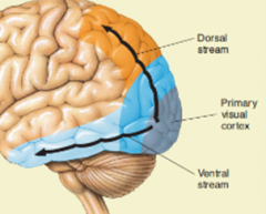
Dorsal stream: 1ary visual cortex --> dorsal prestriate cortex --> posterior parietal cortex Ventral stream: 1ary visual cortex --> ventral prestriate cortex --> inferotemporal cortex |
|
|
Difference between the dorsal and ventral visual streams? |
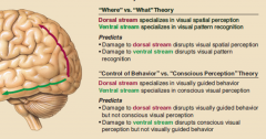
Dorsal: "Where" objects are - the spatial stimuli responder - Directs behavioural interactions with objects Ventral: "What" objects are - the classes of objects stimuli responder - Mediates conscious perception of objects |
|
|
What is prosopagnosia? Result of damage to what area? |
Visual Agnosia (failure of recognition, unattributable to a sensory deficit) - for faces Can SEE faces, but looks like a jumble of parts - Damage is to the fusiform face area (border between occipital and temporal lobes) - ventral stream |
|
|
a) What is akinetopsia? b) What can trigger it? c) Associated with damage to what area of the cortex? |
a) Inability to see movement progress, in a smooth fashion b) Large doses of anti-depressants c) Middle temporal (MT, or V5 area) area - Function of MT is motion perception |