Reading...
![]()
Play button
![]()
Play button
![]()
Use LEFT and RIGHT arrow keys to navigate between flashcards;
Use UP and DOWN arrow keys to flip the card;
H to show hint;
A reads text to speech;
50 Cards in this Set
- Front
- Back
|
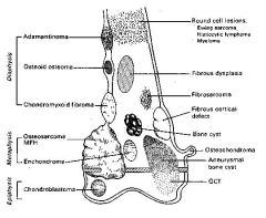
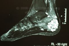
Locations of Bone Tumors
|
|
|
|
Radiographic evaluation of a bone tumor helps determine rate and aggressiveness: list some qualities of an aggressive tumor
|
-moth eaten, permeative patterns of bone destruction
-cortical involvement or breakthrough -periosteal reaction (codmans) -large tumor > 6cm -NO sclerotic margins -wide zone of transition (no sharp border) -no trabeculation (no new bone formation) - |
|
|
radiographic signs of slow or benign tumor growth
|
-small tumor size, <6 cm
-rarely a periosteal reaction or cortical involvement -usually geographic in appearance -sclerotic margins that show a narrow zone of transition -trabeculations present (new bone formation) |
|
|
In a 1 yo pt, what bone tumor is MC
|
metastatic neuroblastoma
|
|
|
In a 1-10 yo pt, what bone tumor is MC
|
Ewings sarcoma - has spiculated cortical reaction located in diaphysis
|
|
|
In a 10-20 yo pt, what bone tumor is MC
|
aneurysmal bone cyst - fast growing so it expands into the cortex
|
|
|
In a 10-30 yo pt, what bone tumor is MC
|
osteosarcoma - lamellated onion skin periosteal reaction
|
|
|
In a skeletally mature - 50yo pt, what bone tumor is MC
|
Giant cell tumor - at the end of the bone (epiphysis)
|
|
|
In a 30-60 yo pt, what bone tumor is MC
|
chondrosarcoma, lymphoma, fibrosarcoma
|
|
|
In a 50-80 yo pt, what bone tumor is MC
|
multiple myeloma, metastasis
|
|
|
what lab studies can be used for bone tumors
|
-alkaline phosphatase (>85 in bone disease)
-CBC (Ca is >11 in bone neoplasm) |
|
|
what diagnostic tests can be used for bone tumors
|
-bone scans (increased uptake of the nucleotide reflects osteoblastic activity)
-CT -MRI -blood tests (Alk Phos, CBC) |
|
|
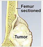
which type of biopsy removes the entire lesion and what type of tumors is it performed on
|
-excisional biopsy used for benign lesions
|
|
|
which type of biopsy removes a sample only of the lesion
|
-incisional biopsy used for malignant lesions
|
|
|
-benign osteoblastic lesion
-well demarcated -less then 1 cm -zone of reactive bone formation -night pain -point tenderness over lesion -common in the outside of the bone (cortex) |
-osteoid osteoma
-aspirin may relieve the symptoms |
|
|
list 11 benign bone tumors common in the distal leg and foot
|
1. osteoid osteoma
2. osteoblastoma 3. enchondroma 4. osteochondroma 5. non ossifyng fibroma 6. chondromyxoid fibroma 7. aneurysmal bone cyst 8. simple bone cyst (unicameral) 9. giant cell tumor 10. fibrous dysplasia 11. chondroblastoma |
|
|
list 5 malignant bone tumors cmmon in the distal leg and foot
|
1. osteosarcoma
2. chondrosarcoma 3. fibrosarcoma 4. ewings sarcoma 5. multiple myeloma |
|
|
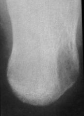
-dull aching, localized pain NOT releived by aspirin
-MC on anterior talus -tumor is gritty and friable |
-osteoblastoma
|
|
|
-benign hyaline cartilage with speckled calcification
-growth in the medullary cavity -seen in mets and phalanges -metaphysis location |
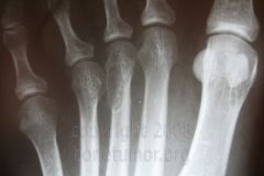
Enchondroma
-multiple enchondromas is Ollier's |
|
|
-benign, slow growing mushroom shaped hyaline cartilage
-sharply pedunculated or sessile tumor |
osteochondroma
|
|
|
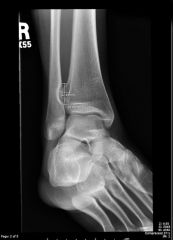
-primary tumor that initiates an AV fistula
-pt under 30 -blow out stage |
aneurysmal bone cyst
|
|
|
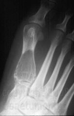
-long fluid filled cyst
-fallen fragment sign (piece of cortical bone falling from the cyst) |
simple bone cyst (unicameral)
|
|
|
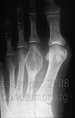
-contains multinucleated osteoclast type giant cells
-soap bubble lesions |
giant cell tumor
|
|
|
-ground glass appearance
-well defined sclerotic rim -haphazardly arranged trabecula of woven bone |
fibrous dysplasia
|
|
|
-geographic lucency with spotty calcifications
|
chondroblastoma
|
|
|
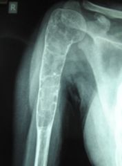
-agressive mesynchymal tumor in which neoplastic cells produce bone matrix
-common with lung metastases -blastic mass with permeative pattern and codmans triangle |
osteosarcoma
|
|
|
-agressive mesenchymal tumor in which neoplastic cells produce bone matrix
-large mass with permeative pattern and codmans reaction |
osteosarcoma
|
|
|
-produces neoplastic cartilage
-bone destruction puncuated by mottled densities |
chondrosarcoma
|
|
|
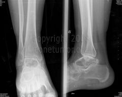
ewings sarcoma; moth eaten appearance
-common in the calcaneus and femur -10-15 yo |
|
|
-multiple myeloma
-40-70 yo -oval lesions |
|
|
what are the MC sources of metastatic cancer
|
-breast, lung, prostate, kidney, thyroid
|
|
|
compare a benign soft tissue mass and a malignant soft tissue mass
|
-benign soft tissue mass looks like derivative tissue, small, cystic filled and moveable
|
|
|
list 5 common benign soft tissue tumors
|
1. ganglion cyst
2. plantar fibroma 3. lipoma 4. neurofibroma 5. neurolemoma |
|
|
most common benign soft tissue tumor
|
lipoma; subcut, soft, moveable mass
|
|
|
subcutaneous thickening of the plantar fascia
|
plantar fibroma
|
|
|
what colored fluid comes out of a ganglion cyst
|
straw colored fluid
|
|
|
popcorn bone tumor
|
enchondroma
|
|
|
onion skinning tumor
|
ewings sarcoma
|
|
|
list the 4 radiographic clues about the growth rate of a bone lesion
|
1. destructive pattern
2. size and shape 3. cortical involvement 4. periosteal reaction |
|
|
list the diff types of destructive patterns and how they relate to growth rate
|
-geographic (small circles with sclerotic margins, slow growth)
-moth eaten (small lesions across an area, fast) -permeative (small streaks in the bone, fast) |
|
|
acute osteopenia can have a permeative or moth-eaten appearance; so how does it differ from a fast growing malignancy
|
-it involves multiple bones
|
|
|
which type of lesion is longer (rather then wider) and fills the medullary canal bc it takes the path of least resistance
|
-slow growing lesion
|
|
|
which lesions penetrate or break through the cortex and invade the soft tissue
|
-agressive lesion
|
|
|
which is more agressive growth; constant periosteal reaction or interrupted periosteal reaction
|
-interrupted periosteal growth
|
|
|
trabeculation is only found in certain tumors; list them
|
-giant cell tumor (fine trabeculation)
-aneurysmal bone cyst (horizontal fine trabecula) -chondromyxoid fibroma(thick trabecula) -hemangioma(radiating trabecula) -non ossifying fibroma(soap bubble trabecula) |
|
|
name a specific continuous periosteal reaction
|
-onion skin or spiculated (hair on end)
-this is still an agressive lesion, but least agressive |
|
|
name a specific interrupted periosteal reaction
|
-Codmans triangle
-sunburst |
|
|
stippled or speckled matrix production
|
-chondroblastic tumors (enchondroma, osteochondroma, chondroblastoma, chondrosarcoma)
|
|
|
solid matrix production
|
-osteblastic tumors (osteoid osteoma, osteoblastoma, osteosarcoma)
|
|
|
oval, metaphyseal, blow out lesion with soap bubble trabeculation
|
-ABC (aneurysmal bone cyst)
|