Reading...
![]()
Play button
![]()
Play button
![]()
Use LEFT and RIGHT arrow keys to navigate between flashcards;
Use UP and DOWN arrow keys to flip the card;
H to show hint;
A reads text to speech;
76 Cards in this Set
- Front
- Back
|
What electromyogram (EMG) can be used for (in connection to muscles)?
|
Can be used to monitor the level of activation of muscle cells in skeletal muscle.
|
|
|
What is a motor unit?
|
It is a motor nerve plus all the fibres that the nerve innervates.
|
|
|
What is excitation-contraction coupling?
|
The events occurring between the action potential in the muscle fiber and the contraction of the muscle fiber.
|
|
|
Myosin is a ... filament. And Actin, tropomyosin and troponin are ... filaments.
|
Myosin is a thick filament. Actin, tropomyosin and troponin are thin filaments.
|
|
|
What is the name of a large molecular weight protein that thick filaments comprise?
|
Myosin.
|
|
|
Describe the structure of myosin.
|
Myosin is a large molecular weight protein which has six polypeptide chains, including one pair of heavy chains and two pairs of light chains.
|
|
|
What forms the "tail" of myosin molecule?
|
The coiled pair of heavy chains.
|
|
|
What forms two globular "heads" of the myosin molecule?
|
The four light chains and the N terminus of each heavy chain.
|
|
|
What is the myosin molecule globular head necessary for?
|
They have actin-binding site, which is necessary for cross-bridge formation, and a site that binds and hydrolyzes ATP (myosin ATPase).
|
|
|
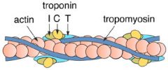
What are the thin filaments composed of?
|
Actin, troponin, tropomyosin.
|
|
|
What happens if the myosin binding sites on a thin filament are covered with tropomyosin?
|
The muscle is at rest and actin cannot interact with myosin.
|
|
|
What is tropomyosin?
|
The part of thin filaments. this is a filamentous protein that runs along the groove of each twisted actin filament. At rest it covers the myosin-binding sites of actin so the actin and myosin cannot interact, and if contraction must occur, tropomyosin moves away from the binding sites.
|
|
|
Name two types of actin.
|
G-actin - globular actin; F-actin - filamentous actin.
|
|
|
Explain the role of troponin (T,I,C) in the thin filaments.
|
Troponin is a complex of three globular proteins (T, I, C), located in the regular intervals along the tropomyosin filaments. Troponin T attaches the troponin complex to tropomyosin. Tr.I inhibits the interaction of actin and myosin by covering the myosin-binding site on actin. Tr.C is a Ca++ binding protein that plays major role in the initiation of contraction. Ca binds to TrC and tropomyosin moves away letting actin interract with myosin.
|
|
|
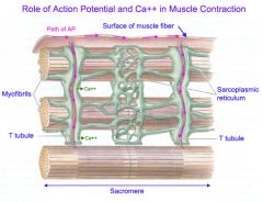
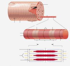
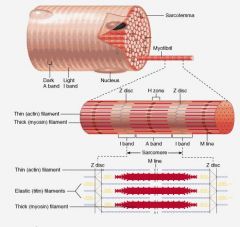
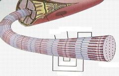
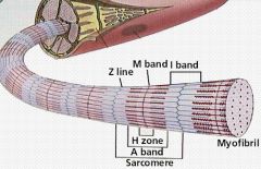
Explain the sarcomere (incl.bands).
|
The sarcomere is the basic contractile unit, and it is delineated by the Z disks. Each sarcomere contains a full A band in the center and one half of two I bands on either side of the A band.
|
|
|
Describe A band.
|
A bands are located in the center of the sarcomere and contain the thick filaments. Thick and thin filaments may overlap in the A band; these overlaps are potential sites for a cross-bridge formation.
|
|
|
Describe I band.
|
The I bands are located on either side of the A band of the sarcomere. They contain thin filaments, intermediate filamentous proteins and Z disks. They have no thick filaments.
|
|
|
Describe the M line.
|
The M line is in the middle of the sarcomere, bisects the bare zone and links the central portions of the thick filaments together.
|
|
|
Describe the bare zone.
|
The bare zone is in the middle of the sarcomere. There are no thin filaments in the bare zone, so there can be no overlap of thick and thin filaments or cross-bridge in this region.
|
|
|
What delineates the ends of each sarcomere?
|
The Z disks, that run down the middle of each I band.
|
|
|
What do the cytoskeletal proteins do?
|
They establish the architecture of the myofibrils, ensuring that the thick and thin filaments are aligned correctly and at proper distances from each other.
|
|
|
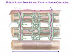
Describe transverse (T) tubules and their function.
|
T tubules are part of the sarcolemmal membrane of the muscle cell. They are responsible for carrying the depolarisation from action potentials at the muscle cell surface to the interior of the fiber.
|
|
|
What does transverse tubules and terminal cisternae make together?
|
Triad.
|
|
|
What is the name of the muscle cell membrane?
|
Sarcolemma.
|
|
|
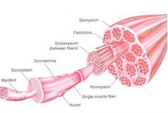
What is single muscle fiber consists of?
|
Myofibrils.
|
|
Put in labels.
|
|
|
Put in labels.
|
|
|
Fill in labels.
|
|
|
|
Which of the sarcomere bands appear to be light and which - dark?
|
I band is light and A band is dark, because I band has no thick filaments, A band does.
|
|
|
How many units of actin have binding site which myosin can interact with?
|
All of them.
|
|
|
Which proteins maintain the architecture of the myofibrils?
|
Cytoskeletal proteins, like titin (associated with thick filaments, goes from M line to Z line through the myosin, makes sure that the thick filament is positioned in the right place to give the max.active tension) and nebulin (associated with thin filaments, it extends from one end of the actin to the other, controls the length of thin filaments).
|
|
|
Which bands on the sarcomere shorten during contraction of the muscle cell?
|
H band (bare zone) and I band.
|
|
|
Which band does not change its length during contraction of the muscle?
|
A band.
|
|
|
During muscle contraction why does the sarcomere shorten?
|
Because the actin filaments slide along between the myosin filaments.
|
|
|
What is the function of the sarcoplasmic reticulum?
|
The sarcoplasmic reticulum is an internal tubular structure, which stores and releases Ca ions for excitation-contraction coupling.
|
|
|
Finish the statement:
Total force generated by a single sarcomere is proportional to .... |
... the total number of cross-bridges formed between the thick and thin filaments.
AND ... the overlap between the thick and thin filaments. |
|
|
Each cross-bridge generates force .... of other cross-bridges.
|
Each cross-bridge generates force independent of other cross-bridges.
|
|
|
What are the roles of ATP in the cross-bridge cycle?
|
- Energy source: ATP hydrolysis provides the energy for cross-bridge cycle;
- Allosteric regulator: Binding of ATP to myosin breaks the link between actin and myosin. This allows the cycle to be repeated. |
|
|
What is the function of the Calsequestrin protein in the sarcoplasmic reticulum?
|
It binds Ca ions, so Ca can be stored in sarcoplasmic reticulum.
|
|
|
Name functions of sarcoplesmic reticulum.
|
- Storage of high concentrations of Ca ions;
- rapid release of Ca ions into the myoplasm in response to an action potential; - rapid removal of Ca ions from the myoplasm. |
|
|
Ca ions is accumulated in the sarcoplasmic reticulum by the action of what?
|
Of Ca++ ATPase.
|
|
|
When the muscle fibre is at rest what is the situation of Ca ions in the ICF?
|
Intracellular concentration of Ca ions is very low, because the Ca++ ATPase pumps the Ca ions from the ICF into the the interior of the sarcolasmic reticulum.
|
|
|
How is Ca stored in the sarcoplasmic reticulum?
|
It is stored bound to the protein called calsequestrin. so the concentration of free Ca ions in the intrasarcoplasmic reticulum is extremely low.
|
|
|
What are the functions of the T tubules?
|
- The conduction of action potential from the sarcolema deep into the muscle fibre;
- The conduction of action potential to the sarcoplasmic reticulum causing the release of calcium. |
|
|
Which triad receptors are involved in sensing the action potentials and releasing Ca ions?
|
- The dihydropyridine (DHP) receptor in the T tubules (only) functions as a voltage sensor;
- The ryanodine receptor in sarcoplasmic reticulum functions as a Ca release channel. |
|
|
What makes muscle relax?
|
The removal of Ca from troponin -> inhibition of tropomyosin.
|
|
|
Define tension.
|
Tension is a force exerted on an object by a contracting muscle.
|
|
|
Define load.
|
Load is a force exerted on a muscle by the weight of an object.
|
|
|
What is isometric contraction?
|
When muscle develops tension without changing length.
|
|
|
What is isotonic contraction?
|
When muscle shortens, while load remains constant.
|
|
|
What is eccentric (lengthening) contraction?
|
When load exceeds muscle tension, load pulls muscle to a longer length.
|
|
|
There are two types of smooth muscle organisation. Name and explain them.
|
Unitary: Muscle fibres act together as a functional unit.
Multiunit: Muscle fibres act independently. |
|
|
What are the gap junctions for in the smooth and cardiac muscles?
|
They permit coordinated contraction; ions move through them ensuring signalling and communication between cells.
|
|
|
What is the Ca++ sensor in muscles?
|
In cardiac and skeletal muscle it is troponin, but in smooth muscle it is calmodulin.
|
|
|
What is the name of tension developed by simply stretching a muscle to different lengths.
|
Passive tension.
|
|
|
What is the name of tension developed when a muscle is stimulated to contract at different preloads?
|
Total tension. It is the sum of the active tension developed by the cross-bridge cycling in the sarcomeres and the passive tension caused by stretching the muscle.
|
|
|
What is the name of tension which is determined by subtracting the passive tension from the total tension? It presents the active force developed during the cross-bridge cycle.
|
Active tension.
|
|
|
The active tension developed is proportional to the number of cross-bridges that cycle. What does that mean in terms of sarcomere length?
|
The active tension is maximal when there is maximal overlap of thin and thick filaments and maximal cross-bridges. When muscle is stretched to longer lengths (the sarcomere is too) the active tension is reduced. Likewise, when the sarcomere length is reduced, the thin filaments collide with each other in the centre of sarcomere and reducing the number of possible cross-bridges, active tension.
|
|
|
Fill in blanks:
The amount of tension is determined for a muscle undergoing an ... contraction, in which the muscle is allowed to develop ... at a present ... (called ...) but is not allowed to ... . |
Fill in blanks:
The amount of tension is determined for a muscle undergoing an isometric contraction, in which the muscle is allowed to develop tension at a present length (called preload) but is not allowed to shorten . |
|
|
In isotonic contraction the ... is fixed, and the muscle is allowed to ... .
|
In isotonic contraction the force is fixed, and the muscle is allowed to shorten.
|
|
|
Why there is no striations in the smooth muscles?
|
Because the muscle the thin and thick filaments are not organized in sarcomeres.
|
|
|
Which type of smooth muscle has gap junctions between cells, which allow the electrical activity to spread quickly throughout the organ, followed by coordinated contraction?
|
Unitary smooth muscle.
|
|
|
Which type of smooth muscle has no coupling between cells, no gap junctions?
|
Multiunit smooth muscle.
|
|
|
Which type of muscle is found in the vascular smooth muscle?
|
The combination of unitary and multiunit smooth muscle.
|
|
|
In excitation-contraction coupling of the smooth muscle Ca++ binds to which protein to allow cross-bridge cycling?
|
Calmodulin.
|
|
|
To start the cross-bridge cycle in smooth muscle what does Ca++ binds to and activates it?
|
Myosin-light-chain kinase.
|
|
|
In smooth muscle process of contraction what happens after Ca++ binds and activates Myosin-light-chain kinase?
|
Myosin-light-chain kinase uses ATP to phosphorylate myosin cross-bridges, which allows them to bind to actin. Contraction happens.
|
|
|
What are other sources of Ca++ in the smooth muscle except the voltage-gated Ca channels?
|
Ligand-gated Ca++ channels in the sarcolemmal membrane and IP3-gated Ca++ channels in the sarcoplasmic reticulum membrane.
|
|
|
Why does the summation of twitches not occur in cardiac muscle?
|
Because cardiac muscle must contract just once with each heart beat and must relax fully between contractions.
|
|
|
Does the smooth muscle contract fast or slowly? What advantage does it have?
|
Slowly. It can be shortened in much greater degree than striated muscle, as a result it can produce large and sustained movements of organs such as intestine.
|
|
|
Which three pathways cause an increase in the myoplasmic Ca++ concentration in smooth muscle?
|
- Ca entry via voltage-gated Ca channels in response to cell depolarisation;
- Ca release from the SR; - Ca entry via voltage-independent Ca channels. |
|
|
What are myofibrils? Organisation and components.
|
Myofibrils are cylindrical organelles. They are bundles of actomyosin filaments (sarcomeres) that run from one end of the cell to the other and are attached to the cell surface membrane at each end. The filaments of myofibrils, myofilaments, consist of two types, thick and thin.
Thin filaments consist primarily of the protein actin, coiled with nebulin filaments. Thick filaments consist primarily of the protein myosin, held in place by titin filaments. |
|
|
What are muscle fibres? Organisation and components.
|
Muscle fibres - muscle cells. Within the cells are myofibrils. Individual muscle fibres are surrounded by endomysium. Muscle fibers are bound together by perimysium into bundles called fascicles.
|
|
|
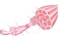
Describe organisation and components of a muscle.
|
Muscle is mainly composed of muscle cells. Within the cells are myofibrils; myofibrils contain sarcomeres, which are composed of actin and myosin. Individual muscle fibres are surrounded by endomysium. Muscle fibers are bound together by perimysium into bundles called fascicles; the bundles are then grouped together to form muscle, which is enclosed in a sheath of epimysium. Muscle spindles are distributed throughout the muscles and provide sensory feedback information to the central nervous system.
|
|
Label.
|
|
|
Label.
|
|