Reading...
![]()
Play button
![]()
Play button
![]()
Use LEFT and RIGHT arrow keys to navigate between flashcards;
Use UP and DOWN arrow keys to flip the card;
H to show hint;
A reads text to speech;
143 Cards in this Set
- Front
- Back
|
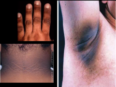
Acanthosis nigricans - The clinical hallmark is development of gray-brown, velvety plaques that may start as a dirty appearance. The hyperpigmentation is later accompanied by hypertrophy, increased skin markings, and papillomatosis.
|
|

|
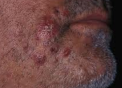
Acne vulgaris - Acne vulgaris is the most common cutaneous disorder in the United States, affecting more than 85% of adolescents. Acne is a disorder of the pilosebaceous follicle that involves proliferation of the keratinocytes at the opening of the follicle; increased production of sebum, stimulated by androgens, which combines with keratinocytes to plug the follicular opening; growth of Propionibacterium acnes, an anaerobic diphtheroid normally found on the skin; and inflammation from bacterial activity and release of free fatty acids and enzymes from activated neutrophils. Cosmetics, humidity, heavy sweating, and stress are contributing factors.
|
|
|
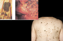
Actinic keratosis - Superficial, flattened papules covered by a dry scale. Often multiple; can be round or irregular; pink, tan, or grayish. Appear on sun-exposed skin of older, fair-skinned people. Though benign, 1 of every 1000 per year develops into squamous cell carcinoma (suggested by rapid growth, induration, redness at the base, and ulceration). Keratoses on face and hand, typical locations, are shown.
|
|
|
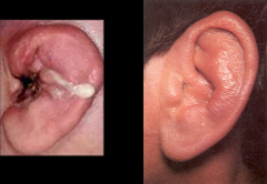
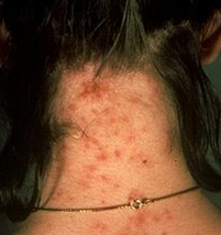
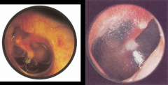
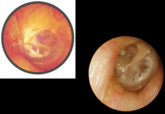
Acute otitis media - Acute otitis media with purulent effusion is caused by bacterial infection. Symptoms include earache, fever, and hearing loss. The eardrum reddens, loses its landmarks, and bulges laterally, toward the examiner’s eye. Here the eardrum is bulging, and most landmarks are obscured. Redness is most obvious near the umbo, but dilated vessels can be seen in all segments of the drum. A diffuse redness of the entire drum often develops. Spontaneous rupture (perforation) of the drum may follow, with discharge of purulent material into the ear canal. Hearing loss is of the conductive type. Acute purulent otitis media is much more common in children than in adults.
|
|
|
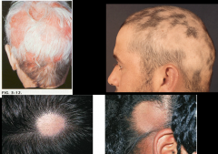
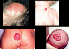
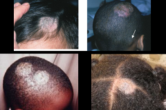
Alopecia areata - Clearly demarcated round or oval patches of hair loss, usually affecting young adults and children. There is no visible scaling or inflammation.
|
|
|
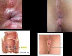
Anal fissure - An anal fissure is a very painful oval ulceration of the anal canal, found most commonly in the midline posteriorly, less commonly in the midline anteriorly. Its long axis lies longitudinally. There may be a swollen “sentinel” skin tag just below it. Gentle separation of the anal margins may reveal the lower edge of the fissure. The sphincter is spastic; the examination is painful. Local anesthesia maybe required.
|
|
|
Angioedema - A diffuse, nonpitting, tense swelling of the dermis and subcutaneous tissue. It develops rapidly, and typically disappears over subsequent hours or days. Although usually allergic in nature and sometimes associated with hives, angioedema does not itch. Recurrent large circumscribed areas of subcutaneous or mucosal edema of sudden onset, usually disappearing within 24 hours; frequently, an allergic reaction to foods or drugs
|
|
|
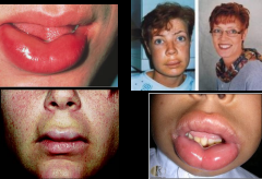
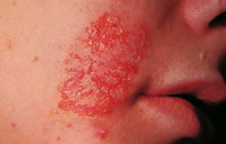
Angular cheilitis - Angular cheilitis starts with softening of the skin at the angles of the mouth, followed by fissuring. It may be due to nutritional deficiency or, more commonly, to overclosure of the mouth, as in people with no teeth or with ill-fitting dentures. Saliva wets and macerates the in-folded skin, often leading to secondary infection with Candida, as seen here.
|
|
|
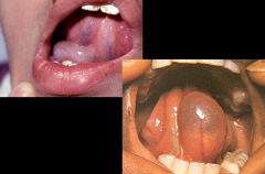
Ankyloglossia-aka tongue tied: Partial or complete fusion of the tongue to the floor of the mouth; abnormal shortness of the frenulum linguae.
|
|
|
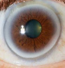
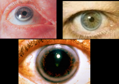
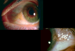
Cornel Arcus senilis - a white, gray, or blue opaque ring in the corneal margin (peripheral corneal opacity), or white ring in front of the periphery of the iris. It is present at birth, but then fades; however, it is quite commonly present in the elderly. It can also appear earlier in life as a result of hypercholesterolemia.
|
|
|
AV nicking - the phenomenon where, on examination of the eye, a small artery (arteriole) is seen crossing a small vein (venule), which results in the compression of the vein with bulging on either side of the crossing. This is most commonly seen in eye disease caused by high blood pressure
|
|
|
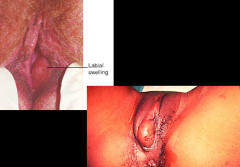
Bartholin cyst - Causes of a Bartholin’s gland infection include trauma, gonococci anaerobes like bacteroides and peptostreptococci, and Chlamydia trachomatis. Acutely, it appears as a tense, hot, very tender abscess. Look for pus coming out of the duct or erythema around the duct opening. Chronically, a nontender cyst is felt. It may be large or small.
|
|
|
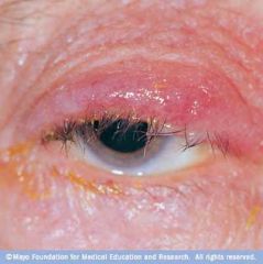
Blepharitis - an eye condition characterized by chronic inflammation of the eyelid
|
|
|
Bulla - Greater than 1 cm. A blister is a small pocket of fluid within the upper layers of the skin. Blisters can be filled with blood (known as blood blisters) or with pus (if they become infected). However, most blisters are filled with a clear fluid called serum. A blister usually forms because the outer layer of the skin has become damaged. Fluid collects under the damaged layer of skin, cushioning the tissue underneath, protecting it from further damage and allowing it to heal.
|
|
|
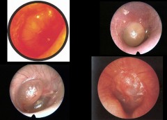
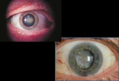
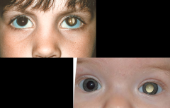
Cataract - Opacities of the lenses visible through the pupil; most common in old age.
|
|
|
Cavernous hemangioma - is a vascular disorder of the central nervous system that may appear either sporadically or exhibit autosomal dominant inheritance. deep cutaneous hemangioma with dilated vessels on gross and microscopic examination
|
|
|
Cherry angiomas - red papules caused by weakening of dermal capillary walls, which do not blanch on pressure, seen mostly in persons over 30 years of age.
|
|
|
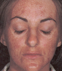
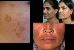
Chloasma - The mask of pregnancy is common, though not present in all pregnancies. It consists of irregular brownish patches around the forehead and cheeks, across the nose, or along the jaw.
|
|
|
Chondritis – Inflammation of cartilage.
|
|

|
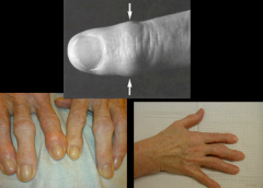
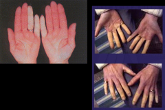
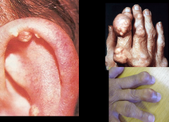
Clubbing - Clinically a bulbous swelling of the soft tissue at the nail base, with loss of the normal angle between the nail and the proximal nail fold. The angle increases to 180°or more, and the nail bed feels spongy or floating. The mechanism is still unknown but involves vasodilatation with increased blood flow to the distal portion of the digits and changes in connective tissue, possibly from hypoxia, changes in innervation, genetics, or a platelet-derived growth factor from fragments of platelet clumps. Seen in congenital heart disease, interstitial lung disease and lung cancer, inflammatory bowel diseases, and malignancies.
|
|
|
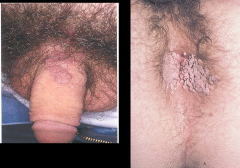
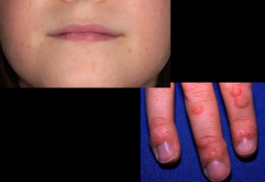
Condyloma acuminata - (genital warts) a highly contagious sexually transmitted infection caused by some sub-types of human papillomavirus
|
|
|
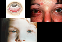
Conjunctivitis - Conjunctival injection: diffuse dilatation of conjunctival vessels with redness that tends to be maximal peripherally. Mild discomfort rather than pain. Not affected except for temporary mild blurring due to discharge, watery, mucoid, or mucopurulent
|
|
|
Corneal arcus - A thin grayish white arc or circle not quite at the edge of the cornea. Accompanies normal aging but also seen in younger people, especially African-Americans. In young people, suggests possible hyperlipoproteinemia. Usually benign.
|
|
|
Crust - The dried residue of skin exudates such as serum, pus, or blood
|
|
|
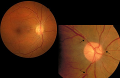
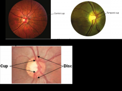
Cupping - The physiologic cup is a small whitish depression in the optic disc, from which the retinal vessels appear to emerge. Although sometimes absent, the cup is usually visible either centrally or toward the temporal side of the disc. Grayish spots are often seen at its base. When the size of the cup increases (normal cup:disc = 1:2)
|
|

|
Darier’s disease - characterized by dark crusty patches on the skin, sometimes containing pus. The crusty patches are also known as keratotic papules, keratosis follicularis or dyskeratosis follicularis. Stubborn rash which usually runs in families, on chest, neck, back, ears, forehead, and groin
|
|
|
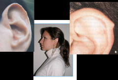
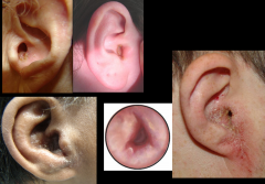
Darwin tubercle - often presents as a thickening or projection on the helix at the junction of the upper and middle thirds
|
|
|
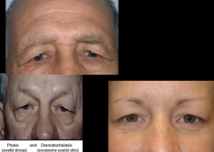
Dermatochalasis - an excess of skin in the upper or lower eyelid
|
|
|
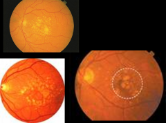
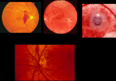
Drusen - yellowish round spots that vary from tiny to small. The edges may be soft, as here, or hard. They are haphazardly distributed but may concentrate at the posterior pole. Drusen appears with normal aging but may also accompany various conditions, including age-related macular degeneration.
|
|
|
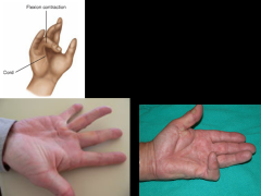
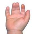
Dupuytren’s contracture - The first sign of a Dupuytren’s contracture is a thickened plaque overlying the flexor tendon of the ring finger and possibly the little finger at the level of the distal palmar crease. Subsequently, the skin in this area puckers, and a thickened fibrotic cord develops between palm and finger. Flexion contracture of the fingers may gradually ensue.
|
|
|
Ectropion - In ectropion the margin of the lower lid is turned outward, exposing the palpebral conjunctiva. When the punctum of the lower lid turns outward, the eye no longer drains satisfactorily, and tearing occurs. Ectropion is more common in the elderly.
|
|

|
Eczema - a group of medical conditions that cause the skin to become inflamed or irritated
|
|
|
Enophthalmos - the posterior displacement of the eyeball within the orbit due to changes in the volume of the orbit
|
|
|
Entropion - more common in the elderly, is an inward turning of the lid margin. The lower lashes, which are often invisible when turned inward, irritate the conjunctiva and lower cornea. Asking the patient to squeeze the lids together and then open them may reveal an entropion that is not obvious.
|
|

|
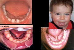
Epstein pearls - Epstein’s pearls, tiny white or yellow, rounded mucous retention cysts, are located along the posterior midline of the hard palate. They disappear within months.
|
|
|
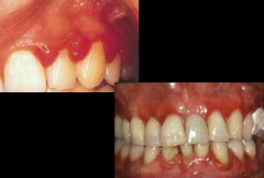
Epulis - Pregnancy Tumor (Epulis, Pyogenic Granuloma) Gingival enlargement may be localized, forming a tumor like mass that usually originates in an interdental papilla. It is red and soft and usually bleeds easily. The estimated incidence of this lesion in pregnancy is about1%. Note the accompanying gingivitis in this example.
|
|
|
Erythema multiforme - (A) Bulla, (B) target (or iris) lesion (in erythema multiforme)
|
|
|
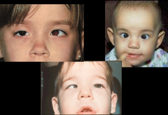
Esotropia - an inward deviation.
|
|
|
Excoriation - Linear or punctate erosions caused by scratching
|
|
|
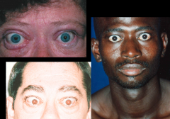
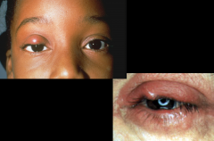
Exophthalmos - In exophthalmos the eyeball protrudes forward. When bilateral, it suggests the infiltrative ophthalmopathy of Graves’ hyperthyroidism. Edema of the eyelids and conjunctival injection may be associated. Unilateral exophthalmos is seen in Graves’ disease or a tumor or inflammation in the orbit.
|
|
|
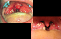
Exudative tonsillitis - This red throat has a white exudate on the tonsils. This, together with fever and enlarged cervical nodes, increases the probability of group A streptococcal infection or infectious mononucleosis. Anterior cervical lymph nodes are usually enlarged in the former, posterior nodes in the latter. If bacterial Group A strep; Viral EBV, Adenovirus
|
|
|
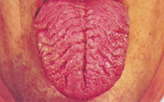
Fissured tongue - Fissures appear with increasing age, sometimes termed scrotal tongue. Food debris may accumulate in the crevices and become irritating, but a fissured tongue is benign.
|
|
|
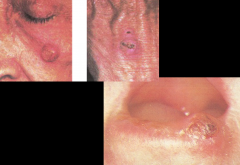
Flat Wart - (Verruca plana), a small, smooth flattened wart, flesh-coloured, which can occur in large numbers; most common on the face, neck, hands, wrists and knees;
|
|
|
Folliculitis - the inflammation of one or more hair follicles. The condition may occur anywhere on the skin. Often caused by S. aureus
|
|
|
Fordyce spots - Fordyce spots are normal sebaceous glands that appear as small yellowish spots in the buccal mucosa or on the lips. A worried person who has suddenly noticed them may be reassured. Here they are seen best anterior to the tongue and lower jaw. These spots are usually not so numerous.
|
|
|
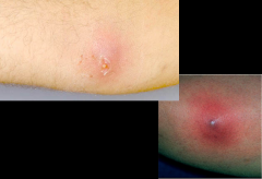
Furuncle - (boil) is a skin disease caused by the inflammation of hair follicles, thus resulting in the localized accumulation of pus and dead tissue. Individual boils can cluster together and form an interconnected network of boils called carbuncles.
|
|
|
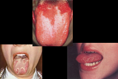
Geographic tongue - In this benign condition, the dorsum shows scattered smooth red areas denuded of papillae. Together with the normal rough and coated areas, they give a maplike pattern that changes over time.
|
|
|
Gingivitis - Common among teenagers and young adults. The gingival margins are reddened and swollen, and the interdental papillae are blunted, swollen, and red. Brushing the teeth often makes the gums bleed. Plaque—the soft white film of salivary salts, protein, and bacteria that covers the teeth and leads to gingivitis—is not readily visible.
|
|
|
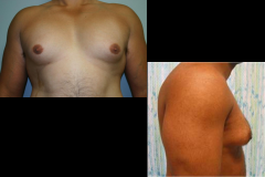
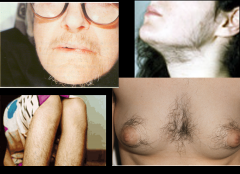
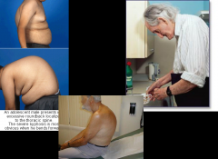
Gynecomastia - If the breast appears enlarged, distinguish between the soft fatty enlargement of obesity and the firm disc of glandular enlarge-ment, called gynecomastia.
|
|
|
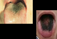
Hairy tongue - Note the “hairy” yellowish to brown or black elongated papillae on the tongue’s dorsum. This benign condition may follow antibiotic therapy; it also may occur spontaneously.
|
|
|
Halo nevi - A halo nevus is a mole that is surrounded by round, symmetric depigmented area, or a halo. The halo itself has sharply demarcated borders. There are no melanocytes, or cells that make melanin, in the halo area.
|
|
|
Heberden node - are hard or bony swellings which can develop in the distal interphalangeal joints (DIP) (the furthest joints before the tips of the fingers or toes.) They are a sign of osteoarthritis, and are caused by formation of calcific spurs of the articular (joint) cartilage.
|
|
|
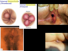
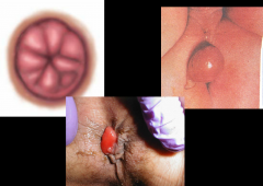
Hemorrhoid - External hemorrhoids are dilated hemorrhoidal veins that originate below the pectinate line and are covered with skin. They seldom produce symptoms unless thrombosis occurs. This causes acute local pain that increases with defecation and sitting. A tender, swollen, bluish, ovoid mass is visible at the anal margin. Internal hemorrhoids are enlargements of the normal vascular cushions located above the pectinate line. Here, they are not usually palpable. Sometimes, especially during defecation, internal hemorrhoids may cause bright-red bleeding. They may also prolapse through the anal canal and appear as reddish, moist, protruding masses, typically located in one or more of the positions illustrated.
|
|
|
Herpes labialis - infection of the lips, mouth, or gums due to the herpes simplex virus
|
|

|
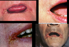
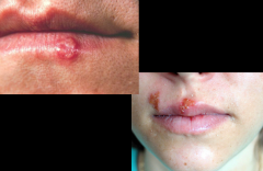
Herpes simplex – The herpes simplex virus (HSV) produces recurrent and painful vesicular eruptions of the lips and surrounding skin. A small cluster of vesicles first develops. As these break, yellow-brown crusts form, and healing ensues within 10 to 14 days. Both of these stages are visible here.
|
|

|
Herpes Zoster (Shingles)
|
|
|
Hirsuitism - male pattern hair growth in women
|
|
|
Hordeolum - infection of sebaceous glands at the base of the eyelashes. Commonly caused by S. aureus
|
|

|
Hydradenitis suppurativa - boil like lesions/abscesses on groin, neck, nipples, penis, armpit etc….scarring and chronic seepage
|
|
|
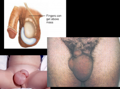
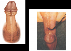
Hydrocele - A nontender, fluid-filled mass within the tunica vaginalis. It transilluminates, and the examining fingers can get above the mass within the scrotum.
|
|
|
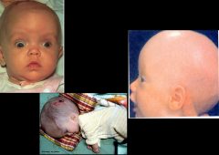
Hydrocephalus - In hydrocephaly, the anterior fontanelle is bulging, and the eyes may be deviated downward, revealing the upper sclerae and creating the setting sun sign, as shown above. The setting sun sign is also seen briefly in some normal newborns. Abnormal accumulation of cerebrospinal fluid (CSF) in the ventricles, or cavities, of the brain. This may cause increased intracranial pressure inside the skull and progressive enlargement of the head, convulsion, and mental disability.
|
|
|
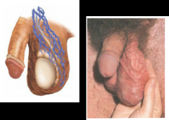
Hypospadias - A congenital displacement of the urethral meatus to the inferior surface of the penis. A groove extends from the actual urethral meatus to its normal location on the tip of the glans.
|
|

|
Impetigo - This infection is due to bacteria and can appear bullous or crusty and yellowed with some pus.
|
|
|
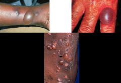
Kaposi’s sarcoma - The deep purple color of these lesions, although not necessarily present, strongly suggests Kaposi’s sarcoma. The lesions may be raised or flat. Among people with AIDS, the palate, as illustrated here, is a common site for this tumor. This malignant tumor may appear in many forms: macules, papules, plaques, or nodules almost anywhere on the body. Lesions are often multiple and may involve internal structures. On left: ovoid, pinkish red plaques that typically lengthen along the skin line may become pigmented. On right: a purplish red nodule on the foot.
|
|
|
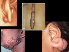
Keloid - A firm, nodular, hypertrophic mass of scar tissue extending beyond the area of injury. It may develop in any scarred area but is most common on the shoulders and upper-chest. A keloid on a pierced earlobe may have troublesome cosmetic effects. Keloids are more common in darker-skinned people. Recurrence may follow treatment.
|
|
|
Kyphosis
|
|
|
Leukoplakia - With this persisting painless white patch in the oral mucosa, the undersurface of the tongue appears painted white. Patches of any size raise the possibility of malignancy and require a biopsy.
|
|
|
Lichenification - is the thickening of skin (or epidermis) with the accentuation of the normal lines of the skin, giving rise to an appearance resembling a tree bark. It is commonly seen in chronic eczema (or atopic dermatitis), where there is constant scratching and rubbing of the skin and in lichen simplex chronicus. Thus, lichenification is often associated with pruritic (itching) disorders.
|
|
|
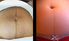
Linea nigra - a brownish-black pigmented line along the midline
|
|
|
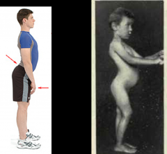
Lordosis - Too much lordotic curving is called swayback
|
|

|
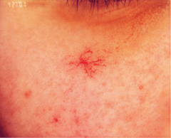
Lyme disease - caused by bacteria Borrelia
|
|
|
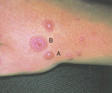
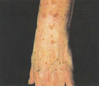
Macule - Macules on the dorsum of the hand, wrist and forearm
|
|
|
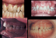
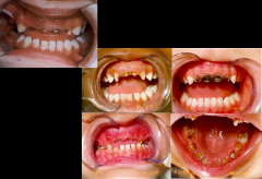
Malocclusion - a misalignment of teeth or incorrect relation between the teeth of the two dental arches
|
|
|
Mastitis - inflammation of mammary gland
|
|
|
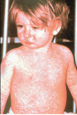
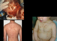
Measles - rubeola-viral infection with characteristic rash
|
|
|
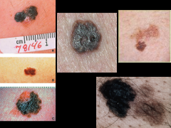
Melanoma - is a malignant tumor of melanocytes which are found predominantly in skin but also in the bowel and the eye (see uveal melanoma). It is one of the rarer types of skin cancer but causes the majority of skin cancer related deaths.
|
|
|
Melasma (cholasma) - mask of pregnancy
|
|
|
Milk bottle teeth - aka Bottle Rot, baby bottle caries
|
|
|
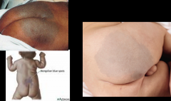
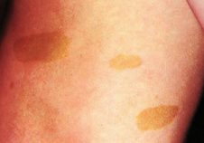
Mongolian Spot - These are more common among dark-skinned babies. It is important to note them so that they are not mistaken for bruises. A benign flat congenital birthmark with wavy borders and irregular shape. The blue color is caused by melanocytes, melanin-containing cells, that are deep under the skin. Usually, as multiple spots or one large patch, it covers one or more of the lumbosacral area (lower back), the buttocks, flanks, and shoulders.
|
|
|
Mucosal retention cyst - (Right side shows bilateral maxillary cysts: should be BLACK)
|
|
|
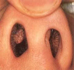
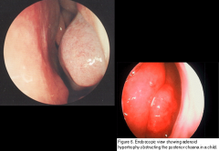
Nasal polyp - overgrowth of nasal mucosa
|
|
|
Neovascularization - Refers to the formation of new blood vessels. They are more numerous, more tortuous, and narrower than other blood vessels in the area and form disorderly looking red arcades. A common cause is the late, proliferative stage of diabetic retinopathy. The vessels may grow into the vitreous, where retinal detachment or hemorrhage may cause loss of vision.
|
|
|
Neurofibromatosis - nodules and tumors, difficult to remove b/c of NERVOUS tissue origin!
|
|
|
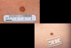
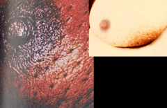
Nevus – The benign nevus, or common mole, usually appears in the first few decades. Several nevi may arise at the same time, but their appearance usually remains unchanged.
|
|
|
Nodule - Marble-like lesion larger than 0.5 cm, often deeper and firmer than a papule
|
|
|
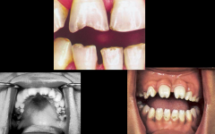
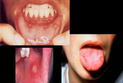
Notched teeth - Congenital syphillus, Aka Huntchinson’s teeth
|
|
|
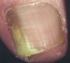
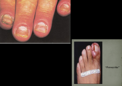
Onychomycosis - fungal infection of the nail
|
|
|
Otitis externa
|
|

|
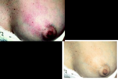
Paget’s disease - This uncommon form of breast cancer usually starts as a scaly, eczema like lesion that may weep, crust, or erode. A breast mass may be present. Suspect Paget’s disease in any persisting dermatitis of the nipple and areola. Can present with invasive breast cancer or ductal carcinoma in situ.
|
|
|
Papilloma
|
|
|
Papule - Up to 1.0 cm
|
|
|
Paronychia - A superficial infection of the proximal and lateral nail folds adjacent to the nail plate. The nail folds are often red, swollen, and tender. Represents the most common infection of the hand, usually from Staphylococcus aureus or Streptococcus species, and may spread until it completely surrounds the nail-plate. Creates a felon if it extends into the pulp space of the finger. Arises from local trauma due to nail biting, manicuring, or frequent hand immersion in water.
|
|
|
Patch - Flat spot, 1.0 cm or larger
|
|
|
Peau d’orange - associated w/inflammatory breast cancer
|
|
|
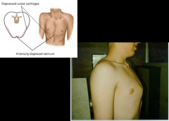
Pectus carinatum - The sternum is displaced anteriorly, increasing the anteroposterior diameter. The costal cartilages adjacent to the protruding sternum are depressed.
|
|
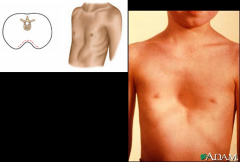
|
Pectus excavatum - Note depression in the lower portion of the sternum. Compression of the heart and great vessels may cause murmurs.
|
|
|
Pediculosis capitis - (also known as head lice infestation, "nits" and cooties
|
|
|
Pemphigus foliaceus - an autoimmune blistering disease of the skin and mucous membranes with characteristic lesions that are scaly, crusted erosions, often on an erythematous base
|
|
|
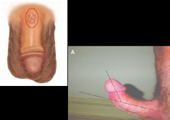
Peyronie disease - Palpable, nontender, hard plaques are found just beneath the skin, usually along the dorsum of the penis. The patient complains of crooked, painful erections.
|
|
|
Pinguecula - A harmless yellowish triangular nodule in the bulbar conjunctiva on either side of the iris. Appears frequently with aging, first on the nasal and then on the temporal side.
|
|

|
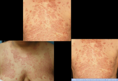
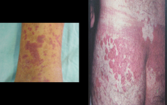
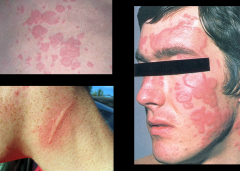
Pityriasis rosea - Reddish oval ringworm-like lesions
|
|
|
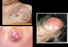
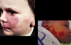
Port wine stains - is a vascular birthmark consisting of superficial and deep dilated capillaries in the skin which produce a reddish to purplish discoloration of the skin
|
|
|
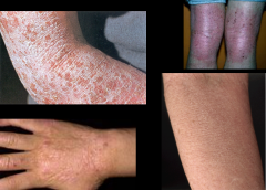
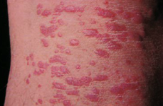
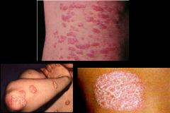
Psoriasis - Up to 1.0 cm, a disease which affects the skin and joints. It commonly causes red scaly patches to appear on the skin. The scaly patches caused by psoriasis, called psoriatic plaques, are areas of inflammation and excessive skin production.
|
|
|
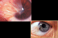
Pterygium - A triangular thickening of the bulbar conjunctiva that grows slowly across the outer surface of the cornea, usually from the nasal side. Reddening may occur. May interfere with vision as it encroaches on the pupil.
|
|
|
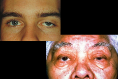
Ptosis - Ptosis is a drooping of the upper lid. Causes include myasthenia gravis, damage to the oculomotor nerve, and damage to the sympathetic nerve supply (Horner’s syndrome). A weakened muscle, relaxed tissues, and the weight of herniated fat may cause senile ptosis. Ptosis may also be congenital.
|
|
|
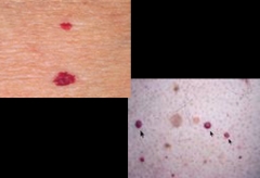
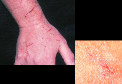
Purpura - is the appearance of red or purple discolorations on the skin, caused by bleeding underneath the skin. Small spots are called petechiae less than 1cm, while large spots are called ecchymoses greater than 1cm.
|
|
|
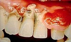
Pyorrhea - Purulent inflammation of the gums and tooth sockets
|
|
|
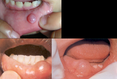
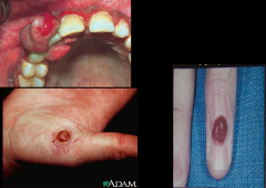
Pyogenic granuloma - Pregnancy Tumor (Epulis, Pyogenic Granuloma) Gingival enlargement may be localized, forming a tumor like mass that usually originates in an interdental papilla. It is red and soft and usually bleeds easily. The estimated incidence of this lesion in pregnancy is about1%. Note the accompanying gingivitis in this example.
|
|
|
Ranula - floor of the mouth. Ranulas present as a swelling of connective tissue consisting of collected mucin from a ruptured salivary gland duct, which is usually caused by local trauma.
|
|
|
Raynaud's disease - Vasospastic disorder causing discoloration of the fingers, toes, and occasionally other areas. Causes the nails to become brittle with longitudinal ridges. Emotional stress and cold are classic triggers of the phenomenon.
|
|
|
Rectal prolapse - On straining for a bowel movement, the rectal mucosa, with or without its muscular wall, may prolapse through the anus, appearing as a doughnut or rosette of red tissue. A prolapse involving only mucosa is relatively small and shows radiating folds, as illustrated. When the entire bowel wall is involved, the prolapse is larger and covered by concentrically circular folds.
|
|
|
Retinoblastoma - cancer of the retina
|
|
|
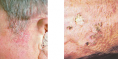
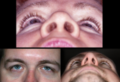
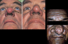
Rhinophyma - large, bulbous, ruddy appearance of the nose caused by granulomatous infiltration. This is commonly due to untreated rosacea.
|
|
|
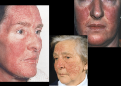
Rosacea - effects white-skinned people of NW European descent. Begins as erythema on central face and nose, cheeks, forehead. As it progresses, get telangiectasia, red domed papules and pustules, and rhinophyma.
|
|
|
Salmon patch (stork bite) - Also called the “stork bite,” this splotchy pink mark fades with age.
|
|
|
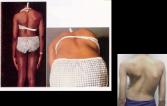
Scoliosis
|
|
|
Seborrheic keratoses - Common, benign, yellowish to brown raised lesions that feel slightly greasy and velvety or warty and have a “stuck on” appearance. Typically multiple and symmetrically distributed on the trunk of older people, but may also appear on the face and elsewhere. In black people, often younger women, may appear as small, deeply pigmented papules on the cheeks and temples
|
|
|
Serous otitis media
|
|
|
Squamous cell carcinoma
|
|
|
Stomatitis - “canker sores”: type of oral ulcer which presents as a painful open sore inside the mouth or upper throat, caused by a break in the mucous membrane.
|
|
|
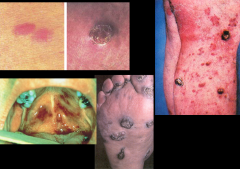
Strawberry hemangioma - tufts of extra blood vessels that occur on the surface of the skin common in childern. Those that occur deeper in skin=cavernous hemangioma; can be mixed too.
|
|
|
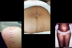
Striae - “stretch marks”
|
|
|
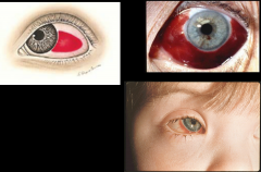
Subconjunctival hemorrhage - Leakage of blood outside of the vessels, producing a homogeneous, sharply demarcated, red area that fades over days to yellow and then disappears
|
|
|
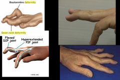
Swan neck deformity - The fingers may show “swan neck” deformities (hyperextension of the proximal interphalangeal joints with fixed flexion of the distal interphalangeal joints).
|
|
|
Syndactyly - Any degree of webbing or fusion of fingers or toes, involving soft parts only or including bone structure; usually autosomal dominant inheritance.
|
|
|
Telangiectasia - Spider-like small dilated blood vessels near the surface of the skin. More common in females, esp during pregnancy.
|
|
|
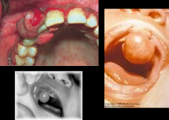
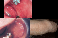
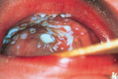
Thrush candida - Thrush is a yeast infection due to Candida. Shown here on the palate, it may appear elsewhere in the mouth. Thick, white plaques are somewhat adherent to the underlying mucosa. Predisposing factors include (1) prolonged treatment with antibiotics or corticosteroids and (2) AIDS.
|
|
|
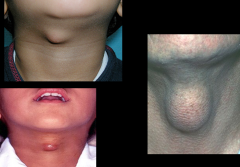
Thyroglossal duct cyst
|
|
|
Tinea Barbae
|
|
|
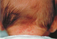
Tinea capitis - Round scaling patches of alopecia. Hairs are broken off close to the surface of the scalp. Usually caused by fungal infection from tinea tonsurans. Mimics seborrheic dermatitis.
|
|
|
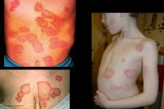
Tinea corpora (ring worm) - A fungal skin infection sometimes referred to as ringworm. Typically a scaly, red-shaped ring on the skin. Commonly seen in children
|
|
|
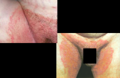
Tinea cruris - fungal infection of groin, jock itch
|
|
|
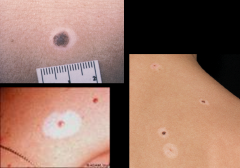
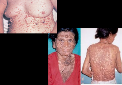
Tophi - A deposit of uric acid crystals characteristic of chronic tophaceous gout. It appears as hard nodules in the helix or antihelix and may dis-charge chalky white crystals through the skin. It also may appear near the joints, hands, feet, and other areas. It usually develops after chronic sustained high blood levels of uric acid.
|
|
|
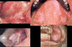
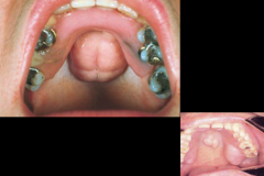
Torus palatinus - A torus palatinus is a midline bony growth in the hard palate that is fairly common in adults. Its size and lobulation vary. Although alarming at first glance, it is harmless. In this example, an upper denture has been fitted around the torus.
|
|
|
Transverse Crease
|
|
|
Turbinate hypertrophy - can cause obstructive sleep apnea and breathing problems
|
|
|
Tympanosclerosis - In the inferior portion of this left eardrum, there is a large, chalky white patch with irregular margins. It is typical of tympanosclerosis: a deposition of hyaline material within the layers of the tympanic membrane that sometimes follows a severe episode of otitis media. It does not usually impair hearing and is seldom clinically significant.
|
|
|
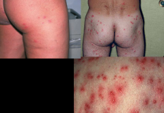
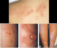
Varicella - chikcen pox; lesions more prevalent on trunk, not soles/palms. Lesion evolution: vesiclepustulescabhealing; found in multiple stages, no synchrony
|
|
|
Varicocele - Varicocele refers to varicose veins of the spermatic cord, usually found on the left. It feels like a soft “bag of worms” separate from the testis, and slowly collapses when the scrotum is elevated in the supine patient. Infertility may be associated.
|
|
|
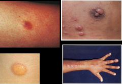
Vesicle - Up to 1.0 cm; filled with serous fluid
|
|
|
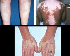
Vitiligo - In vitiligo, depigmented macules appear on the face, hands, feet, extensor surfaces, and other regions and may coalesce into extensive areas that lack melanin. The brown pigment is normal skin color; the pale areas are vitiligo. The condition maybe hereditary. These changes may be distressing to the patient.
|
|
|
Wheals/urticaria - A somewhat irregular, relatively transient, superficial area of localized skin edema
|
|
|
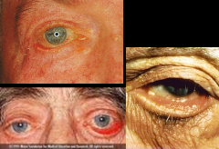
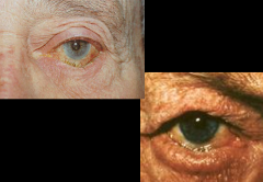
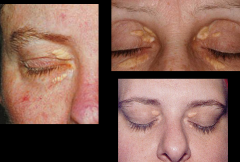
Xanthelasma - Slightly raised, yellowish, well-circumscribed plaques that appear along the nasal portions of one or both eyelids. May accompany lipid disorders.
|