Reading...
![]()
Play button
![]()
Play button
![]()
Use LEFT and RIGHT arrow keys to navigate between flashcards;
Use UP and DOWN arrow keys to flip the card;
H to show hint;
A reads text to speech;
106 Cards in this Set
- Front
- Back
|
when does gout have a peak incidence in men
|
50's
|
|
|
what is the MC cause of inflammatory arthritis in men over age 30
|
gout; hyperurecemia and deposition of urate crystals cause attacks of inflammatory arthritis (tophi can also tubular and interstitial dz in the kidneys)
|
|
|
what is the upper limit of uric acid levels (point at which gout CAN develop)
|
-above 7 mg/dL for men (above 6 for women)
-gout can dvlp at lower levels though |
|
|
why do women have lower normal values of uric acid than men (before menopause)
|
-bc the estrogen levels promote renal excretion of uric acid
|
|
|
how is uric acid formed in the body
|
-Hypoxanthine is converted to Xanthine using Xanthine Oxidase
-xanthine is coverted to uric acid using xanthine oxidase |
|
|
what enzyme do humans lack that predispose us to gout
|
-they lack uricase that oxidizes uric acid to allantoin
|
|
|
what organ excretes uric acid from the body
|
-kidney
-the rest is oxidize by bacteria in the gut |
|
|
why do urate crystals deposit in the ears and toes
|
-the lower temperatures of the peripheral structures cause the urate crystals to have decreased solubility
|
|
|
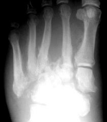
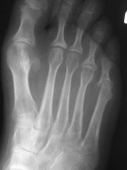
how does gout look on radiograph
|
-periarticular erosions (may have an overhanging edge or punched out lesion)
|
|
|
what are the 4 classifications of gout
|
-primary
-secondary -overproducer -unders secretor |
|
|
what is primary gout
|
-uric acid levels are high due to disorders of uric acid metabolism
|
|
|
what is secondary gout
|
gout is a minor clinical feature secondary to a number of genetic or acquired diseases
|
|
|
what class of gout to most pts with hyperurecemia have (primary, secondary, overproducer, under secretor)
|
-90% are under secretors
|
|
|
list some diseases that cause secondary gout
|
-those in which there are excessive rates of cell turnover (myeloproliferative and lymphpproliferative disorders, hemolytic anemias, psoriasis)
|
|
|
name a cause of prmary gout
|
-HGPRT deficency (part of Lesch-Nyham syndrome of spasticity, mental retardatiion, self mutilation)
|
|
|
name other commonly affected areas of the foot of gout
|
-ankle, tarsal area and knee
|
|
|
affected gout joints are red, hot, swollen; what can this be confused with
|
-cellulitis or thrombophlebitis
|
|
|
with the exception of arthritis, what is the MC complication of gout
|
-renal disease
-pts can dvlp uric acid kidney stones |
|
|
describe the bony erosions of gout on xray
|
-boney erosions tend to be round with sclerotic margin and have been described as rat bite, cyst like or punched out
-martels sign |
|
|
how can you distinguish gout from other arthrides from radiographs
|
-gout has destructive lesions in the bone that are remote from the articular surface
|
|
|
what is Martels sign
|
-gout lesions with overhanging margins
|
|
|
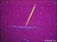
what is mandatory for a diagnosis of gout
|
-synovial fluid monosodium urate crystals
-needle shaped with negative befringence (bright yellow) |
|
|
what lab values can be elevated during a gouty attack
|
-synovial fluid leukocytes
-uric acid level (maybe) -ESR -WBC |
|
|
treatment of acute gout
|
Colchicine, NSAIDS,
|
|
|
Mechanism and dose of colchicine for acute gout
|
-inhibits inflammation, inhibit microtubule formation
-0.5 mg taken hourly until pain reduction, max 8 mg per day |
|
|
what is colchicine given in small incremental doses
|
-to limit GI issues
|
|
|
MC NSAID for acute gout
|
-indomethacin
|
|
|
Indomethacin dose for acute gout attack
|
50 mg PO tid
|
|
|
Tx choices for chronic gout
|
-colchicine, allopurinol, uricosurics (probenicid, sulfinpyrazone)
|
|
|
allopurinol dose for chronic gout
|
-300 mg po once or twice a day
|
|
|
what drugs can cause gout
|
-salicylates
-diuretics -TB meds -warfarin -nicotinic acid (niacin for high cholesterol) |
|
|
what deposits in the joints with pseudogout
|
-Calcium pyrphosphate dihydrate crystal
|
|
|
how can CPPD (calcium pyrophosphate dihydrate crystal disease) be diagnosed
|
-calcium pyrophosphate crystals in the synovial fluid
-needle or rhomboid shape -positive befringence |
|
|
disease of the nerves that leads to underlying bone and joint abnormalities
|
charcot - neuropathic osteoarthropathy
|
|
|
describe the French Theory of charcot
|
-neurovascular theory; damage to nerve centers with an alteration of sympathetic control of blood flow to bones and joints leads to persistent hyperemia and active bone resorption
|
|
|
describe the German theory of charcot
|
Neurotramatic theory; extreme progression of degenerative joint disease following loss of proprioception and protective sensation
|
|
|
MC causes of Charcot
|
-DM (#1)
-syringomyelia -tabes dorsalis |
|
|
what is syringomyelia (common cause of charcot)
|
-damage to the spinal cord due to a fluid filled sac
|
|
|
clinical presentation of charcot
|
-red, hot swollen joint
-deformed, unstable/hypermobile joint --decreased sensation -atrophy of intrinsics -diminished reflexes -spontaneous fractures/dislocations -palpable pulses -rocker bottom foot -possible ulcers over dislocated/displaced bony prominences |
|
|
list the 3 stages of charcot development
|
1. acute or developmental
2. coalescence 3. reconstruction |
|
|
describe first stage of charcot
|
-Acute or developmental: joint laxity, subluxation, osteochondral fragments
|
|
|
2nd stage of charcot
|
coalescence; absorption of debris and fusion of bony fragments
|
|
|
3rd stage of charcot
|
reconstruction: remodelling nad revascularization of bony fragments
|
|
|
DDx for charcot
|
-acute septic arthritis
-OM -CPPD crystal depostion dz -Osteonecrosis (AVN) -PVN synovitis -gout -psoriatic arthritis -RA |
|
|
conservative tx for charcot
|
-NWB, compression (jones) cast to control edema, cast immobilize for fractures, long term accomodatve footwear
|
|
|
surgical tx for charcot; when and what
|
-not performed in acute phase
-ulcer excision -exostectomy -resections -lisfranc, triple, pantalar arhtrodeses -amputation |
|
|
a spirochete is a bacteria, which tick carries this spirochete bacteria (Borrellia burgdorferi) that can cause Lyme disease
|
-Ixodid ticks
|
|
|
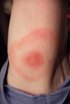
Lyme disease has three stages; early, intermediate and late; what happens in early
|
-early(3-32 d incubation): erythema chronicum migrans occurs at the site of the bite (warm, painless and bullseye)
-low grade fever -lymphadenopathy |
|
|
Lyme disease has three stages; early, intermediate and late; what happens in intermediate
|
-1-6 months after initial exposure
-arthritis, nerve or cardiac involvement (AV block) |
|
|
Lyme disease has three stages; early, intermediate and late; what happens in late
|
-CNS involvement (encepahalopathy, axonal polyneuropathy)
--arthritis |
|
|
Tx for Lyme Dz
|
Doxycycline 100 mg po bid
**during the inital 24 hours of abx therapy, some pts experience Jarisch-Herxheimer reaction characterized by high fever and worsening of symptoms) |
|
|
Lyme disease is a triphasic illness with stage 1 (intial weeks), stage 2 (weeks to months), stage 3 (months to years)..what are the main symptoms of each stage
|
stage 1: erythema chronicum migrans
stage 2: recurrent skin lesions, neurologic and cardiac abnormalities stage 3 : arthritis |
|
|
Inflammatory arthritis is inflammation of the joint lining from an immune system problem or autoimmune disease (versus degenerative arthritis): list some inflammatory arthritis
|
-RA
-lupus -ankylosing spondylitis -reiters -psoriatic arthritis |
|
|
joint pain first thing in the morning or after sitting for awhile
|
-inflammatory arthritis (like RA and Reiters)
|
|
|
joint pain at the end of the day
|
destructive arthritis (osteoarthritis)
|
|
|
immune system attacks the synovial lining of joints causing inflammation, affecting the small joints in the hands and feet first
|
-RA (synovitis is the inital symptom or sign)
|
|
|
what are some articular manifestations of RA
|
-pannus formation
-swan neck -boutonniere -hallux valgus -hammerotes -fibular/ulnar deviations -even joint space narrowing |
|
|
how does pannus in RA form and what does it cause
|
-it is the formation of proliferative and invasive granualtion tissue
-the pannus then leads to perisrticular bone and cartilage erosions and destructions |
|
|
what are some extra-articualr manifestations of RA
|
-rheumatoid nodules
-caplans nodes in lung -vasculitis -pericarditis -anemia -dry eyes and scleritis |
|
|
what is the IPJ flexion deformity in RA called
|
-boutonniere deformity
|
|
|
what is IPJ extension deformity in RA called
|
-swan neck
|
|
|
if performing sx on an RA pt, what additional preop tests are needed
|
-cervical xray due to C1-C2 dislocations
|
|
|
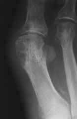
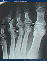
lateral deviation of the digits and erosion of met heads
|
RA
|
|
|
what is Felty's syndrome and when is it seen
|
-triad of neutropenia, splenomegaly and RA
|
|
|
morning stiffness that lasts at least 1 hour
|
RA
|
|
|
to dx a pt with RA, they need to meet 4 of 7 criteria. list the 7
|
1. morning stiffness
2. arthritis of 3 or more joints 3. arthritis of hand joints 4. symmetric arthritis (same joint on opp sides of body) 5. rheumatoid nodules 6. serum rheumatoid factor 7. radiographic changes of hand and wrist (erosions or decalcification near joint) |
|
|
name 3 diseases assoc with RA
|
-sjogrens syndrome
-amylodosis -feltys syndrome |
|
|
drug therapy for RA
|
-DMARDS (gold salts, plaquenil, methotrexate, TNF inhibitors)
-humira is a TNF inhibitor |
|
|
list 7 special considerations that need to be taken into account if operating on an RA patient
|
1. give them supplemental corticosteroids
2. discontinue aspirin or NSAIDS they are on 3. lateral cervical spine xrays 4. protect the skin during surgery 5. inc risk of infection 6. delayed wound healing 7. DVT prophylaxis |
|
|
why do RA surgical pts need periop corticosteroid supplementation
|
-pts on exogenous steroids may dvlp sudden hypotension during sx stress bc ACTH secretion is supressed
-give hydrocortisone sodium succinate or Solumedrol |
|
|
why do RA surgical pts need to discontinue NSAIDS and aspirin 7 days prior to sx
|
-to prevent bleeding complications
-lab testing should include bleeding time, PT, PTT, serum creatinine adn LFT for pts on longterm aspirin or NSAIDS |
|
|
why should RA surgical pts get lateral cervical xrays
|
-atlantoaxial subluxation (c1-C2) is present in many RA pts
-flexion of nexk during anesthesia can cause nerve damage |
|
|
where are heberdens nodes located and in what disease
|
-osteoarthritis (degenerative arthritis)
-DIPJ |
|
|
where are bouchards nodes located and in what diseae
|
-osteoarthritis
-PIPJ - Buchareds, base |
|
|
list the radiographic findings of OA
|
-uneven joint space narrowing
-joint mice (loose osseous bodies) -marginal osteophytes -subchondral sclerosis -subchondral bone cysts |
|
|
excessive and abnormal remodeling of bone that may be caused by a viral infection of osteoclasts
|
Pagets Disease of bone (osteitis deformans)
|
|
|
which bones does pagets often effect
|
-axial skeleton (pelvis, sacrum, spine, skull)
-long bones such as femur and tibia |
|
|
there are three phases of pagets
|
1. osteolytic phase
2. mixed phase 3. sclerotic phase- blastic phase |
|
|
how would a long bone with pagets look on xray
|
-increased cortical width
-increased bone length -coursening of trabeculae -bone enlargement |
|
|
what are the lab features of pagets
|
-elevated serum alkaline phosphatase (N=44-147)
-increased urinary hydroxyproline (N=50mg/24 hours) |
|
|
pagets tx
|
-biphosphonates
-calcium -vit D -calcintonin -NSAIDS |
|
|
what are the seronegative spondyloarthropathies and why are they called that
|
-group of inflammatory disorders that affect the axial skeleton
-they are sero negative meaning neg for rheumatoid factor -anklylosing spondylitis -reiters syndrom -psoriatic arthritis -enteropahtic spondylitis (crohns) -reactive arthritis |
|
|
what is bamboo spine and when is it seen
|
-spinal fusion seen in ankylosing spondylitis
|
|
|
what gene is common in seronegative spondyloarthropathies
|
HLA-B27
|
|
|
what is the classic triad of reiters
|
-conjunctivitis
-urethritis -arthritis |
|
|
when does reiters dvlp in a genetically susceptible host (one with HLA-B27)
|
-after a GU or GI infection
|
|
|
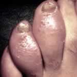
there are two characteristic lesions associated with reiters
|
-balanitis circinata (ulcers of the glans penis)
-kertoderma blennorrhagicum on soles of feet (clear vesicles on a red base that progress to macules, then small keratotic nodules) |
|
|
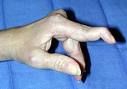
soft tissue swelling in IPJ of toes and fingers causing sausage deformities
|
reiters
|
|
|
what is the most helpful radiographic characteristic of seronegative spondyloarthropathies that distinguishes it from RA
|
-bone proliferation
(linear or fluffy periosteal proliferation) (periostitis at tendon and ligament attachment to bone-enthesitis) (intra-articular bone formation at sites of osseous erosion) |
|
|
describe the nails in psoriatic arthritis
|
-pitting
-transverse ridges -subungual hyperkeratosis -discoloration of nail -oil spot below the nail |
|
|
psoriatic arthritis is similar to RA in that it is progressive inflammatory arthritis; how does it differ
|
-psoriatic arthritis has underlying psoriasis, dactylitis and abscence of anti-cyclic citrulinated peptide antibodies (anti-CCP is present in RA)
|
|
|
resorption of the tufts of the distal phalanges of hands and feet
|
psoriatic arthritis
|
|
|
Auspitz sign
|
-bleeding occurs at sites of scale removal in psoriatic arthritis
|
|
|
what color is the synovial fluid from joints of psoriatic pts
|
lemon yellow
|
|
|
what is the name of the clinical test used for anklosing spondylitis
|
Wright-Schober; to detect the limitation of forward flexion of the lumbar spine
|
|
|
what is the radiograph hallmark of ankylosing spondylitis
|
-sacroliliac joint is sacroilitis
|
|
|
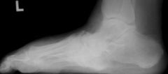
Charcot - midfoot collapse
|
|
|
Charcot
|
|
|
Blue and Yellow Gout crystals
|
|
|
Gout
|
|
|
Lyme disease- erythema chronicum migrans
|
|
|
psoriatic arthritis
|
|
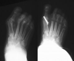
What is this disease and what was done on the left post op?
|
RA - met head arthroplasty
|
|
|
RA
|
|
|
Sausage digit or psoriatic arthritis
|
|
|
swan neck of RA
|