Reading...
![]()
Play button
![]()
Play button
![]()
Use LEFT and RIGHT arrow keys to navigate between flashcards;
Use UP and DOWN arrow keys to flip the card;
H to show hint;
A reads text to speech;
34 Cards in this Set
- Front
- Back
|
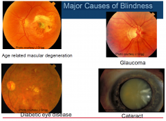
Major Causes of Blindness
|
|
|
|
Age related macular degeneration
STATS |
›30% of Australians will develop AMD over the age of 70
›10% of AMD convert to “wet” AMD requiring 4-12 weekly intravitreal injections of anti VEGF agents ›Results in loss of central vision ›Unable to read or drive |
|
|
Glaucoma
|
›Age 50yrs 1% Australians are affected
›Age 80 yrs 10% Australians are affected ›50% of Australians who have glaucoma currently Do Not Know they have the condition |
|
|
Cataract
|
›18% of Australians have a cataract requiring surgical removal NOW based on visual acuity and symptoms
›If you live to 100 yrs everyone will develop a cataract |
|
|
Diabetes
|
›1.7 million Australians have diabetes (≈ 8% of population)
›Diabetic retinopathy -microvascular complication of diabetes, -affects one-third of people with diabetes -threatens vision in 10 per cent COMMONEST cause of blindness in the working age 20-65 |
|
|
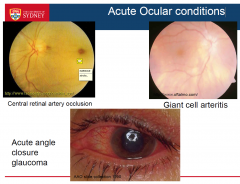
Acute Ocular conditions
|
|
|
|
Visual Acuity
|
A measure of the resolving power of the eye and refers to the spatial limit of visual discrimination
|
|
|
Minimum resolvable (ordinary visual acuity)
|
›Visual acuity as measured clinically also called:
-minimum resolvable or minimum separable. ›Measure of the resolving power of the eye ›A spatial discrimination function representing the smallest visual angle at which two separate objects can be discriminated |
|
|
Snellen Chart
|
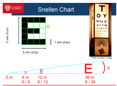
the letters are stanerdised
- numerator is testing distance - denominator is wat the patient sees - young |
|
|
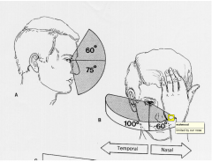
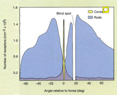
Extent of Visual Field
|
|
|
|
Visual field sensitivity and degree of
eccentricity |
|
|
|
Visual field sensitivity and degree of eccentricity (Large) TESTING
|
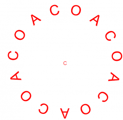
look at this with one eye and you can still ddx between the C & O
|
|
|
Eccentricity further out
|
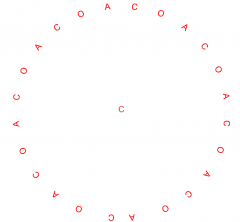
but as you move out into the peripheral vision the detail decreases
|
|
|
Nerve fibre layer arrangement in retina
|
›Optic tract fibres rotate their topographic axis
-Superior retinal fibres rotate laterally -Ventral fibres rotate medially ›Changes horizontal field defects to vertical field defects fibres rotate 90 degrees and so then it becomes vertical fields now |
|
|
Iris Anatomy
|
|
|
|
Pupil Light reflex
PERLA |
›Direct reaction – constriction to light
›Consensual – constriction of other pupil with light to ipsilateral eye ›Accommodation reflex -pupils constrict to near target (& dilate for further distances) ›PERLA -Pupils Equal React Light (direct & consensual) and Accommodation |
|
|
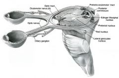
Neural connections pupil light reflex
|
|
|
|
Relative afferent pupillary defect (RAPD)
|
›normal consensual response when illumination of normal side
›diminished pupillary constriction on direct illumination abnormal side ›related to lesions within the anterior visual pathways (retina, optic nerve chiasm and optic tract) ›almost always present in unilateral or asymmetric bilateral optic nerve disease. |
|
|
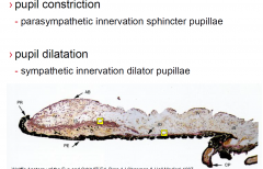
Sympathetic neural supply to eye
|

pupil constriction
parasympathetic innervation sphincter pupillae pupil dilatation sympathetic innervation dilator pupillae |
|
|
Macular Landmarks
|
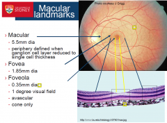
|
|
|
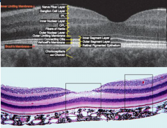
Normal Macular appearance OCT & Histology
|
|
|
|
Central and peripheral retinal connections
|
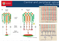
1 to 1 in the central retina
100-200 to 3-4 in the periphery |
|
|
Normal Colour Vision
|
much more red and green
less blue |
|
|
Age related macular degeneration
2 types |
›Two types:
-Dry (non-neovascular) 90% of ARMD cases = slow, progressive decline in central visual function (ATROPHY of cells) -Wet (neovascular) 10% = rapid, more substantial loss of vision (HEALING response in the centre of the fovea, leading to scars and BV leading to viision loss) Tx: Inject Ant-VGEF 4/year $2500 each injection |
|
|
Dry Age-related macular degeneration
|
|
|
Wet ARMD Choroidal neovascularisation
leakage scar |
|
|
Retinal circulation
|
›two sources of blood supply to the mammalian retina:
-central retinal artery -choroidal blood vessels. ›choroid circulation -65-85% of blood flow -vital for maintenance of outer retina (particularly photoreceptors) ›retinal circulation -20-30% central retinal artery from the optic nerve head -supply inner retinal layers. -central retinal artery has 4 main branches in the human retina |
|
|
1. one cherry red spot; because the retina is thin thats the blood here only (but all the connections are loss so no vision)
2. embolism 3. anomylous vessel suppling the fovea from the post. cilliary aa., thus maintain central vision due to anatomically variation |
Retinal artery occlusion
|
|
|
Giant cell arteritis
|
Giant Cell arteritis
- ddx for opthalamic stroke - inflammation of the blood vessel - lots of the medium-large aa. are involved with the lumen is ruined - pale retina without a cherry spot and and the optic nerve is affected so here the choriod and opthalmic aa. are both affected |
|
|
Red Free photo showing nerve fibre defects
|
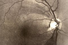
Note the nerve fibre layer arrangement
and horizontal demarcation commonly glaucoma |
|
|
Optic nerve structure
|
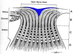
have to turn 90 degrees in bundles
|
|
|
Optic disc assessment
|
›Vertically oval disc is normal.
›Rim width greatest inferior > superior > nasal > temporal (ISNT rule) - If not ? Glaucoma ›Concentric enlargement of cup - if increase over time diagnostic BV in the centre of the optic nerve - due to the 90degree bends, the centre of the nerve is hollow anteriorly thus a divot in the cup |
|
|
Glaucomatous optic disc changes
|
›Asymmetry of cups in both eyes > 0.2
›Focal loss of neuroretinal rim /notch / acquired pit ›Changes in vessels on optic disc: nasalisation, bayoneting, flyover vessels, focal narrowing of vessels, disc haemorrhage |
|
|
Glaucomatous visual field defects
|
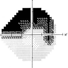
arching shape
- inferior defect will be seen as a superior visual defect |