![]()
![]()
![]()
Use LEFT and RIGHT arrow keys to navigate between flashcards;
Use UP and DOWN arrow keys to flip the card;
H to show hint;
A reads text to speech;
96 Cards in this Set
- Front
- Back
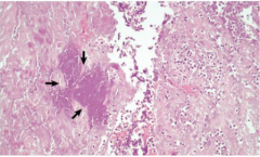
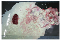
What substance are arrows pointing to
|
Fibrin |
|
|
Define Exudate. What type of inflammation does it belong to? |
Exudate is protein rich (albumin, globulins, fibrinogen) fluid that leaks from vascular walls when permeable. Acute inflammation. |
|
|
What are the inflammatory mediators that cause vasodilation, important in acute phase inflammation? |
Prostaglandins released from mast cells and endothelial cells Bradykinin and C5a from protease cascade/complement cascade Histamine released from mast cells/platelets when degranulating Substance P released by neutrons |
|
|
Define Transudate. What type of inflammation does it belong to? |
Extravasation of fluid due to increase of hydrostatic pressure. Acute inflammation |
|
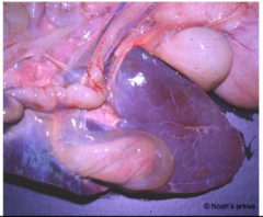
What material is covering the liver/gall bladder? |
Transudate
|
|
|
What is the purpose of the exudative (fluidic) phase of acute inflammation? |
Dilute and localise the inciting agent or substance. |
|
|
what are the cardinal signs of inflammation? |
Rubor (redness) Calor (heat( Tumor (swelling) Dolor (pain) Function laesa (loss of function) |
|
|
What are the two phases involved in acute inflammation? |
Fluidic (exudative) phase (first step) Cellular Phase (second step) |
|
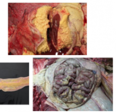
What material are these images portraying? what type of inflammation is associated? |
Fibrin Acute inflammation |
|
|
What is the role of the lymphatic system in acute inflammation? |
Control of exudate- the vessels dilate when tissue pressure rises and drains area Antigen presentation to local lymph nodes clearing of larger particles |
|
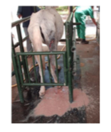
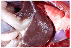
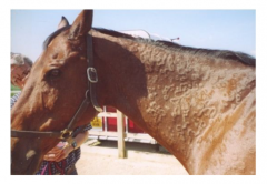
What is this image of a cat depicting and what is causing this? |
Effusive exudative wet form of Feline infectious peritonitis Fibrinous exudate |
|
|
What are the steps involved in the exudative phase? |
vasoconstriction of endothelial cells Vasodilation first at the arteriole (NO, histamine) increased vascular permeability Protein leaks into extravascular space Leukocytes move through endothelial wall |
|
|
What is the purpose of the cellular phase in acute inflammation? |
Leukocyte margination and extravasation (leukocyte adhesion cascade) delivers leukocytes for destruction and removal of stimulus and provides wound healing environment |
|
|
What are the steps of the leukocyte adhesion cascade? What type of inflammation does this occur in and which step? |
1. Margination 2.Tethering 3. Rolling 4. Slow rolling 5. Firm adhesion 6. Emigration 7.Accumulation Acute inflammation; cellular phase |
|
|
What occurs during margination in the leukocyte adhesion cascade? |
Cells which were in the central axial stream approach thevessel wall |
|
|
What occurs during tethering (capture) in the leukocyte adhesion cascade? |
Tumour necrosis factor (TNF) and Interleukin (IL-1)induce expression of cell adhesion molecules E- and P-selectin |
|
|
What occurs during Rolling in the leukocyte adhesion cascade? |
bonds are formed at the leading edge and broken at thetrailing edge |
|
|
What occurs during Slow Rolling in the leukocyte adhesion cascade? |
replacement of selectins by integrin ligands expression on thecell surface |
|
|
What occurs during Firm Adhesion in the leukocyte adhesion cascade? |
firm integrin-mediated binding of leukocytes on endothelialcells |
|
|
What occurs during Emigration (transmigration) in the leukocyte adhesion cascade? |
migration through the inter endothelial spaces attracted bychemokines, through basement membrane |
|
|
What occurs during Accumulation in the leukocyte adhesion cascade? |
Leukocytes adhere to extracellular matrix |
|
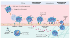
Which cascade is this diagram representing |
Leukocyte adhesion cascade a part of the cellular phase of acute inflammation |
|
What is occurring in these blood vessels |
Leukocytes, under the leukocyte adhesion cascade are exiting blood vessels to an inciting stimulus |
|
|
Which cells leukocytes act first in the cellular phase? How long does it take for them to get to the tissue |
Neutrophils or eosinophils (depending on stimulus). 6-24 hours May be lymphocytes depending on the stimulus. |
|
|
What the the second type of leukocytes to act in the cellular phase? How long does it take for them to get there? Which leukocytes are they replacing? |
Macrophages. 24-48 hours Replace neutrophils as the main cell type |
|
|
What is a disadvantage to the cellular phase? |
By product is tissue damage and maybe prolonged inflammation |
|
|
What is the disadvantage of the exudative phase? |
Edema fluid is the ideal medium for certain bacterial growth In prolonged edema fibroblasts are activated and proliferate |
|
|
What is absent in leukocyte adhesion deficiencies (LAD)? Which animals are they most common in? What is the result? |
Lack of functional expression of integrins on neutrophils Cattle and dogs Leads to animals dying within a few days of birth |
|
|
What is Chediak Higashi Syndrome? |
Neutropenia, defective granulation is a rare autosomal recessive disorder that arises from a mutation of a lysosomal trafficking regulator protein involved in organelle docking and fusion |
|
|
What is Chemotaxis? |
Locomotion oriented along a chemical gradient towards a chemo-attractive substance This is the process by which leukocytes migrate in tissue towards site of injury |
|
|
What are some chemotactic factors? |
Bacterial products Products of tissue, cytokines Products of certain plasma proteins |
|
|
How do leukocytes move in response to chemotaxis? |
By extending filopodia and actin and myosin causing contraction |
|
|
What causes the activation of leukocytes? |
cytokines (including chemotactic factors) microbes, antigen-antibody complexes, products of necrotic cells |
|
|
What is the result of leukocyte activation? |
-- Modulation of adhesion molecules – Degranulation e.g. Neutrophils have cytoplasmic granules containing enzymes and antimicrobial peptides – Secretion e.g. Cytokines – Activation of oxidative burst |
|
|
How do leukocytes recognise their target? |
Receptors for microbial products (TLRs) Receptors for opsonins (FcR, C3b) G protein coupled receptor Mannose receptor, scavenger receptor |
|
|
What is the purpose of opsonisation? What is the process? |
To enhance efficiency of phagocytosis. Coat microbe with opsin (e.g. IgG antibodies, complement protein C3b, fibrinogen, C-reactive protein, carbohydrate-binding proteins) |
|
|
State the events by which a phagocyte "ingests" a microbe. |
Attachment- binding to the surface of a phagocyte Extension of cytoplasm (pseudopods) extend around a microbe until complete closure forms a phagosome. phagosome fuses with lysosome forms a phagolysosome-- within which the membrane oxidises and H2Ox kills the microbe discharge of waste |
|
|
What are the issues of continuous extracellular release of lysosomal enzymes by leukocytes? |
Causes ongoing chronic inflammatory changes -Phagolysosome membrane can become destroyed leading to Asbestos, rate crystals -Phagosome remains open toward extracellular space, when lysosome fusion occurs (regurgitation) -phagocytose material is stuck to membrane-->frustrated phagocytosis |
|
|
What are some microbes that can evade phagocytosis? |
Mycobacteria spp: contained in wax in cellular membrane Toxoplasma, L. monocytogenis: inhibit fusion of phagosome and lysosome Rickettsia, trypanosomes: enter cytoplasm from phagosome. |
|
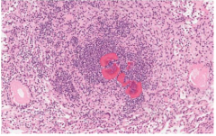
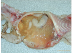
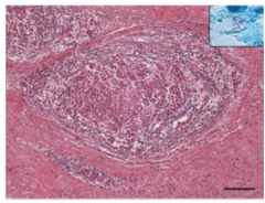
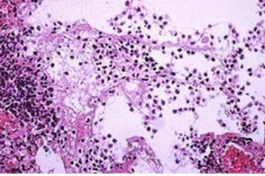
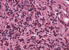
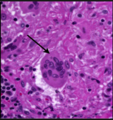
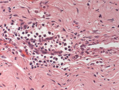
What is this image representing? What causes it? |
Splendore-Hoeppli material formed by fungi, bacteria, or parasites |
|
|
What are the outcomes if acute inflammation is resolved? |
Inflammatory response is completed in correct sequence the inciting stimulus is eliminated macrophages and lymphatic vessels removed exudate stroma is intake |
|
|
What is the outcome if acute inflammation does not occur? |
Progression to chronic inflammation abscess formation healing with scar formation, fibrosis |
|
What is this liquid representing? |
Inflammation due to Bacterial Growth |
|
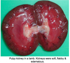
What is the inciting agent causing this? What type of inflammation? |
Clostridium Perfinges Type D Acute Inflammation |
|
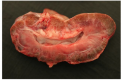
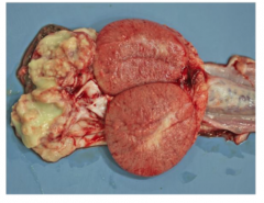
What type of inflammation is occurring in this dogs kidney? how can you tell? |
Chronic inflammation causing nephritis there is no exudate fibrosis is occurring inflammatory response thing to get rid of the stimulus but can't so cells continuously migrate to site. |
|
|
What is the reason for Chronic Inflammation? |
failure of acute inflammation to eliminate the stimulus. repeated episodes of acute inflammation unique virulence factors of microbe/stimulus autoimmunity- chronic tissue damage unidentified mechanisms |
|
|
What are some bacteria that can cause chronic inflammation and how? |
Mycobacteria and R. equi can avoid phagocytosis. (persistence/resistance) Staphylococcus and streptococcus can hide in pus (isolation) |
|
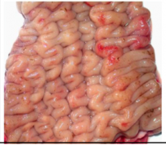
What is causing this mucosal thickening and corrugation in the large intestine of this cow? |
Mycobacterium avian paratuberculosis (Johne's disease) chronic granulomatous inflammation |
|
|
What are the morphological features of chronic inflammation? |
- Infiltration of mononuclear cells: lymphocytes,plasma cells, macrophages - Tissue destruction by persistent offendingagent or by the inflammatory cells - Attempts at healing; replacement of damagedtissue, angiogenesis, fibrosis |
|
|
Outcome of chronic inflammation? |
Encapsulating stimulus with fibroblasts proliferation of fibroblasts can affect organ function extent of debilitation depends on lesion debilitation by continuous release of inflammatory mediators--TNF, IL-1 m-->appetite and temperature |
|
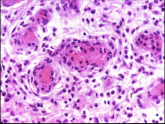
What formation is occurring here? Which cell types are seen? |
Granuloma fibroblasts encapsulating macrophages and epithelioid cells. Lymphocytes surrounding them. |
|
|
Vascular endothelial cells role in inflammation |
Release chemical mediators (NO, prostaglandins) expression of adhesion molecules and receptors for leukocytes contraction |
|
|
Describe Serous Exudate |
Inciting stimulus causes fluid leakage from vessels alongs with low levels of protein can be from virus (FIP) |
|
Where is this abscess formation? |
Abscesses in the epididymis from neutrophils |
|
|
Describe Fibrinous exudate |
More vascular damage so fibrinogen is able to exit vessels along with RBCs and converts to fibrin in the tissue early onset of fibrin deposition causes a granular appearance that can be pulled off chronic fibrin deposition results in fibrous connective tissue that is firmer Can be cause by P. multocida or FIP |
|
What type of exudate is featured here? What is the cause? |
Fibrinous exudate (granular) Feline infectious peritonitis |
|
what substance is featured here? what cell type is commonly associated with it? |
Fibrin Neutrophils Neutrophils in large amounts forming pus with fibrin is called fibrinopurulent inflammation |
|
|
What is diphtheric membrane? |
When fibrin adheres to the underlying mucosal surface. |
|
|
What is the result if fibrinous exudate does not resorb? |
Fibrin is an attractant for fibroblasts to start producing collagen leading to fibrosis (acute to chronic) |
|
|
Describe Cattarhal Exudate and examples |
Leakage of fluid with mucus Canine distemper (from nose) Moniezia (tapeworm-sheep) |
|
|
describe purulent exudate and examples |
Pus Streptococcus equi Corynebacterium pseudotuberculosis-- caseuous llymphadenitis |
|
|
describe hemorrhagic exudate and examples |
Blood vessels damage large enough to release erythrocytes. Mannheimia Haemolytica |
|
|
describe hemorrhagic exudate and examples |
Blood vessels damage large enough to release erythrocytes.Mannheimia Haemolytica |
|
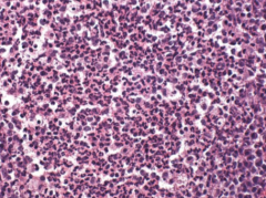
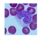
Which type of leukocytes are depicted? How can you tell? |
Neutrophils Segmented Nuclei |
|
What is the pus due to? |
Liquefaction of neutrophils |
|
|
Which antibody receptors do mast cells express on their surface? |
IgE |
|
|
What do the mast cell granules contain? |
Vasoactive amines: Histamine; increases vascularpermeability, causes smooth muscle contraction – Chemotactic factors: Stimulate leukocyteinfiltration especially eosinophils – Proteolytic enzymes: tryptase, chymase; causedegradation of extracellular matrix – Prostaglandin A, Leukotrienes: increase vascularpermeability, vasodilation and contraction ofbronchial smooth muscles |
|
What type of hypersensitivity reaction is occurring and which type of cell is involved? |
Hypersensitivity type I Mast cell |
|
|
What do the eosinophil granules contain? |
• Major basic protein- Toxic to parasites, tumour cells, host tissue • Eosinophil cationic protein- Anti parasitic • Eosinophil peroxidase- Microbicidal • Acid phosphatase, Catalase- Inactivate leukotrienes (regulate mast cell functions) • Hydrolytic lysosomal enzymes- Tissue degradation |
|
What major leukocyte is depicted? What are the attractants involved? |
Histamine, ECFA from mast cells Chemokines released from helminths |
|
What leukocyte is depicted. How can you tell? |
Mast cells basophilic metachromatic granules |
|
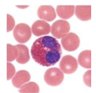
What type of cell is depicted here? How can you tell? |
Eosinophils eosin loving granules; bilobed nucleus |
|
What leukocyte is depicted here? How can you tell? |
Macrophage foamy eosinophilic cytoplasm kidney bean shaped nuclei |
|
|
What are macrophages activated by? |
microbes, endotoxins, chemical mediators |
|
|
what are the main functions of macrophages? |
Eliminate injurious agents – Microbes, tumour cells, foreign material • In acute inflammation – Main role is phagocytosis and cytokine production • Initiate the process of tissue repair • Process and present antigen |
|
What type of cell is this? Why do they arise? |
Epithelioid macrophage response to a persistent stimulus resulting to localisation in granulomas |
|
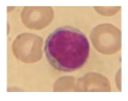
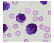
Which type of leukocyte is depicted here? Which form is predominant in the circulation? |
Lymphocyte T lymphocyte predominating in circulation |
|
What are the spindle shaped cells? What is their function? |
Fibroblasts produce collagen and extracellular matrix protein proliferation in chronic inflammatory lesions |
|

What material is forming within the peritoneum? What is the condition called? |
Fibrin Fibrinous pleurisy |
|
|
What is the pivotal step in the compliment cascade? |
Cleavage of C3 to C3b and C3a |
|
|
what are the three pathways that can activate the complement cascade and how do they act? |
Classical: fixation of C1 to antigen- antibody complex on pathogen surface Alternate: triggered by microbial surface molecules (LPS) Lectin: plasma lectin binds to carbohydrates (mannose) on pathogen surface. |
|
|
Important results of the complement cascade? |
C3a and C5a produced to recruit phagocytes C3b produced to bind to complement receptors on phagocytes C3b further reduced to Membrane attack complex, lysis of certain pathogens and cells (C5b, C6,C7, C8, C9) |
|
|
what is the function of the kinin system? |
• Effects: (Short lived) – Increased vascular permeability – Contraction of smooth muscle – Dilation of blood vessels – Pain |
|
|
How is the kinin system activated? |
System is activated by Hageman factor (XII) Generates vasoactive peptides (bradykinin) from plasma proteins (kininogens) by the action of specific proteases (kallikrein) -kallikrein is also a potent activator of Hageman factor |
|
|
What are the two pathways involved in the clotting system and how are they activated? |
Intrinsic pathway: Hageman Factor (XII) Extrinsic pathway: Tissue injury |
|
|
What is a major chemoattractant for neutrophils? |
LTB4 is a potent neutrophil chemoattractant and activator. It stimulates leukocyte adhesion, phagocytosis, oxygen radical production, and degranulation. |
|
|
what is the common pathway between the coagulation system and inflammation? |
thrombin |
|
|
what is the result of binding thrombin to endothelial cells? |
• Mobilisation of P-selectin • Production of chemokine • Expression of endothelial adhesion molecules • Induction of cyclooxygenase-2 • Production of PAF and NO • Changes in endothelial shape |
|
|
What is the important product of the fibrinolytic pathway? What is its function and what does it act on |
plasmin It breaks down fibrin and cleaves C3 to C3a in the complement cascade |
|
|
What does PGH2 cause the formation of and what are the functions of these products and where are they produced? |
PGI2 (prostacyclin): inhibits platelet aggregation; vasodilation; endothelial cells PGD2: vasodilation mast cells PGE2: Inflammation 4_15 HyperalgesicVasodilationIncreased vascularpermeability Fever epithelial cells; fibroblasts; smooth muscle Thromboxane: promotes platelet aggregation; vasoconstriction platelets |
|
|
what is the function of lipoxins? |
Inhibition of neutrophil recruitment, chemotaxis and adherence block superoxideproduction negative regulators of Leukotrienes (vasodilation, decrease vascular permeability, bronchodilation |
|
|
what are the two main groups of cytokines? |
1. Cytokines that mediate and regulate the innate immune response (inflammatory cytokines) – TNF-α and IL-1 are the main actors. 2. Cytokines that mediate and regulate the adaptive immune response – IL-2, IL-4, IL-5, IL-10, IL-12, and IFN-γ. |
|
|
What are chemokines? |
They are a subclass of cytokines involved in chemotaxis |
|
|
What is the function of Platelet Activating Factor? |
Platelet aggregation Vasoconstriction and bronchoconstriction Increases leukocyte adhesion to endothelium Chemotaxis Degranulation Oxidative burst Boosts release of other mediators(e.g.Prostaglandins) |
|
|
What are cytokines and what are they produced by? |
They are proteins that modulate the function of other cells; produced by macrophages and lymphocytes mainly |
|
|
What are the cytokines released by macrophages? |
IL-1, IL6, IL-8, IL12, TNFa |