Reading...
![]()
Play button
![]()
Play button
![]()
Use LEFT and RIGHT arrow keys to navigate between flashcards;
Use UP and DOWN arrow keys to flip the card;
H to show hint;
A reads text to speech;
45 Cards in this Set
- Front
- Back
- 3rd side (hint)
|
Hyperplasia definition
two types of physiologic hyperplasia with examples of each |
increase in cell number
|
Hormonal- female breast tissue
Compensatory- post partial hepatectomy |
|
|
Cellular response to physiologic hyperplasia
|
inc growth factos --> intracellular signaling ptw --> transcriptional regulators --> cell cycle regulation
|
|
|
|
Main cause of physiologic hyperplasia?
|
hormones:
estrogen (from fat/therapy) --> androgens--> chronic infections--> |
- endometrial hyperplasia
- prostatic hyperplasia - follicular lymphoid hyperplasia |
|
|
(T/F) Pathogenic Hyperplasia of the endometrial tissue of the uterus is irreversible.
|
False- all physiologic hyperplasias are reversible as long as the stressor is removed
|
|
|
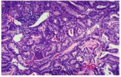
Name this tissue. Normal or abnormal? If abnormal, how so?
endometrial pathogenic hyperplasia |
endometrial pathogenic hyperplasia
|
|
|
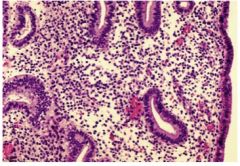
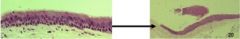
Name this tissue. Normal or abnormal? If abnormal, how so?
|
Normal early secretory phase endometrium
|
|
|
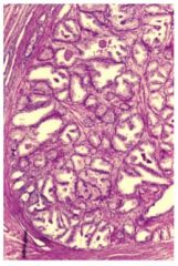
Name this tissue. Normal or abnormal? If abnormal, how so?
|
physiologic hyperplastic prostatic glandular epithelium
|
|
|
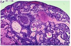
Name this tissue. Normal or abnormal? If abnormal, how so?
|
endometrial pathogenic hyperplasia
|
|
|
|
Define Hypertrophy
Cellular Mechanism? |
increase in cell size
|
increased transcription of structural and functional protein genes
|
|
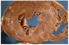
This image is an example of what?
What is the cause? |
pathogenic myocardial hypertrophy
|
Mechanical
- stretch Trophic - growth factors (IGF1) - vasoactive agents angiotensin II & α adrenergics |
|
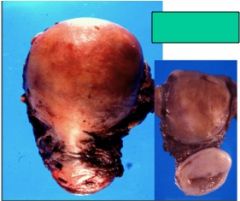
The image on the right is normal. What is represented by the image on the left?
|
Uterine physiologic hypertrophy
|
|
|
|
Which of the following are physiologic atrophy?
Denervation thyroglossal duct notochord Decreased blood supply Inadequate nutrition ductus arteriosus Loss of endocrine stimulation |
thyroglossal duct
notochord ductus arteriosus |
|
|
|
How would you describe cerebral atrophy?
|
narrowed gyri, widened sulci
|
|
|
|
Chemical factors leading to atrophy? (in what type of state are you?)
What chemical opposes atrophy? |
catabolic state such as cachexia:
glucocorticoids thyroid hormone cytokines (TNF) |
insulin
|
|
|
(T/F) Metaplasia is an irreversible process
|
False
|
|
|
|
In what type of tissue can metaplasia occur?
|
connective tissue- epithelium or stromal
|
|
|
|
Vitamin A deficiency and epithelium
|
inability to maintain a highly specialized surface (leads to metaplasia)
-->keratomalacia (thickening of conjunctiva of eye) -->glandular to squamous transition in Lung |
|
|
|
consequence of a sialolith in the bile duct
|
Glandular to squamous metatplasia
|
|
|
classify this transition and tissue
|
Pseudostratified, ciliated bronchial mucosa becomes Stratified squamous metaplastic mucosa
|
|
|
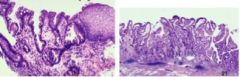
Classify the tissue and the pathogenic process.
increases risk of? |
Barrett’s esophagus (note globlet cells)
Metaplasia - Squamous to columnar Glandular |
Risk of Adenocarcinoma
|
|
|
Two types of stromal metaplasia
|
condroid (cartilage)
osseous (bone) |
|
|
|
(T/F) Metaplasia refers to a morphological change a cell to one more suited to the new microenvironment.
|
False. Metaplasia refers to a morphological change in a TISSUE brought about by changes in the tissue's stem cells
|
|
|
|
what are residual bodies?
|
Undigested remnants (lipid, often) of Phagolysosomes. also called lipofuscin
|
|
|
|
Heterophagy
|
ingested extracellular material
|
|
|
|
Autophagy
|
digestion of cell’s own constituents
|
|
|
|
immotile cilia syndrome may be due to?
|
mitochondrial functional abnormalities
|
|
|
|
What are Mallory Bodies?
|
increased intermediate filament generation in hepatocytes following alcohol injury
|
|
|
Name tissue and pathology.
|
Glycogen storage disease (heart)
|
|
|
Name tissue and pathology
|
Liver- Hepatitis B viral products fill golgi
|
|
|
Name tissue and pathology
|
Hepatocytes- Mallory Bodies
 |
|
|
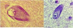
Name tissue and pathology.
|
neurons- Neurofibrillary Tangles
|
|
|
Name tissue and pathology
|
hepatocytes- Alpha-1 antitrypsin mutation leads to impaired secretion of a1antitrypsin
|
|
|
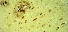
Name tissue and pathology
|
Brain- Plaques
|
|
|
|
Steatosis
|
= Fatty Change = Intracellular Lipid Accumulation
|
|
|
|
xanthomas
|
macroscopic masses of lipid (chl & esters) bloated cells
|
|
|
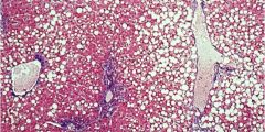
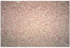
Name the tissue and pathogenesis.
|
Steatosis, liver
|
|
|

Name the tissue and pathogenesis.
|
Steatosis, liver
|
|
|
Name tissue and pathogenesis
|
Cholesterolosis, gallbladder
|
|
|

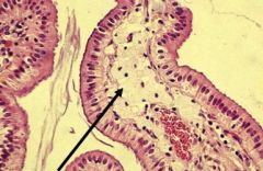
name tissue and pathogenesis
|
Cholesterolosis, gallbladder
|
|
|
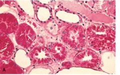
Intensely red material represents what?
|
intracellular accumulation of protein in proximal renal tubule.
|
|
|
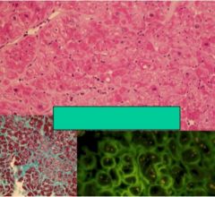
pick,blue, and fluorescent represents what? What type of tissue?
|
Cardiac Amyloidosis- abnormal protein folding
|
|
|
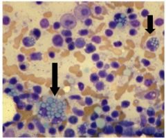
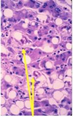
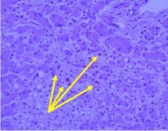
What is represented by the light blue circles? In what type of cells are they located?
|
Russell Bodies (normal Ig's in massive #'s) in Plasma Cells
|
|
|
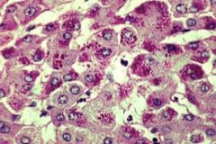
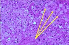
dark pink dots represent? which tissue?
|
Abnormal alpha-1 antitrypsin inclusions (abnormally synthesized protein) in hepatocytes
|
|
|
|
Which of the following describes the calcification of cells/tissues following necrosis?
Metastatic Neoplastic Dystrophic Hypercalcemic |
Dystrophic
|
|
|
|
Causes of Metastatic Calcification?
|
Hypercalcemia due to:
-elevated PTH -caner (inc bone metabolism) -renal failure -vit D disorders |
|