Reading...
![]()
Play button
![]()
Play button
![]()
Use LEFT and RIGHT arrow keys to navigate between flashcards;
Use UP and DOWN arrow keys to flip the card;
H to show hint;
A reads text to speech;
155 Cards in this Set
- Front
- Back
- 3rd side (hint)
|
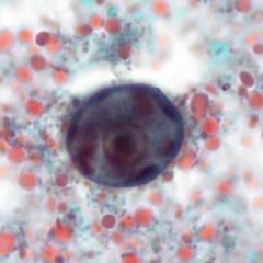
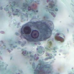
What am I?
List genus, species and stage. |
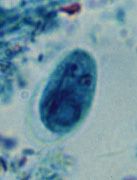
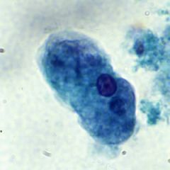
Giardia Lamblia - Trichrome cyst
This is a trichrome stain of the cyst stage of Giardia lamblia. The cysts measure 8 to 19 microns. |
|
|
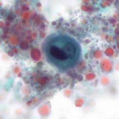
What am I?
List genus, species and stage. |
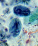
Giardia Lamblia - Trichrome cyst
This is a trichrome stain of the cyst stage of Giardia lamblia. The cysts measure 8 to 19 microns. |
|
|
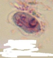
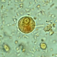
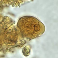
list genus species and stage - what type of slide is this?
|
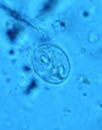
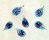
Giardia lamblia (wet mount cycst)
This organism is a pathogenic flagellate. This animated GIF shows the view one would see as they focus up and down through the organism’s cyst stage in a wet mount preparation. |
|
|
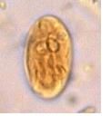
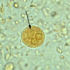
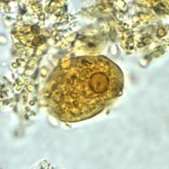
genus species stage, type of stain
|
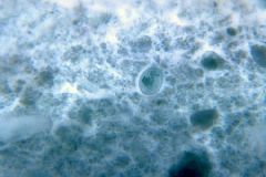
Giardia lamblia cyst. Iodine stain
|
|
|
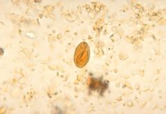
genus species, stage,
|
Giardia lamblia cyst. Iodine stain
|
|
|
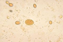
genus species stage, type of stain
|
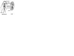
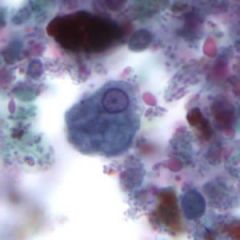
Cysts of Giardia lamblia,stained with iron- hematoxylin (A, B) and in a wet mount (C; from a patient seen in Haiti). Size: 8-12 µm in length. These cysts have two nuclei each (more mature ones will have four).
CDC |
|
|
genus, species, stage, stain
|
Cysts of Giardia lamblia,stained with iron- hematoxylin (A, B) and in a wet mount (C; from a patient seen in Haiti). Size: 8-12 µm in length. These cysts have two nuclei each (more mature ones will have four).
CDC |
|
|
genus, species, stage, stain
|
Cysts of Giardia lamblia,stained with iron- hematoxylin (A, B) and in a wet mount (C; from a patient seen in Haiti). Size: 8-12 µm in length. These cysts have two nuclei each (more mature ones will have four).
CDC |
|
|
genus species stage (not normal stain, but cool pic)
|
Giardia lamblia cyst. Chlorazol black.
CDC/Dr. George R. Healy |
|
|
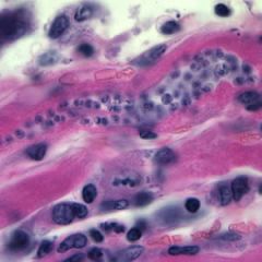
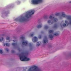
genus, stage, where am I?
|
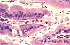
Giardia trophozoites in section of intestine (H&E)
|
|
|
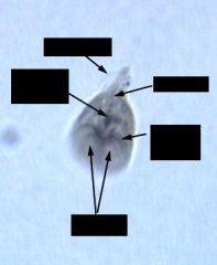
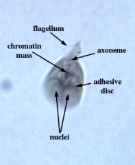
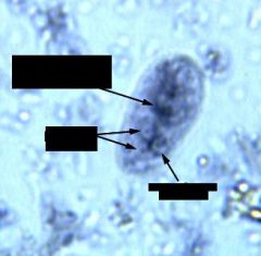
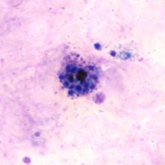
genus, species, stage, name the labeled parts
|
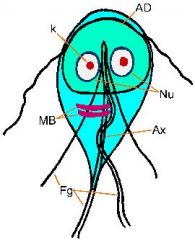
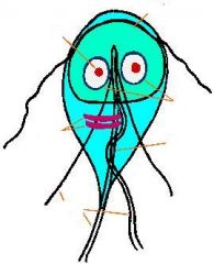
The Giardia trophozoite exhibits a characteristic pear, or tear-drop, shape with bilateral symmetry when viewed from the top (Figure). It is typically 12-15 µm long, 5-10 µm wide, and 2-4 µm thick. Characteristic features of the stained trophozoite include: two nuclei (Nu) with central karyosomes (k), fibrils running the length of the parasite, and median bodies (MB). The large karyosome and lack of peripheral chromatin gives the nuclei a halo appearance. The fibrils are called axonemes (Ax) and are formed from the proximal regions of the flagella (Fg) within the body of the trophozoite. The median bodies are a pair of curved rod-shaped structures which lie posterior to the nuclei. At the ultrastructural level the median bodies contain an array of microtubules. The function of the median bodies is not known, but most believe they are somehow involved with the adhesive disk and its formation. An adhesive disk (AD), not always visible by light microscopy, occupies the ventral side of the anterior end.
|
|
|
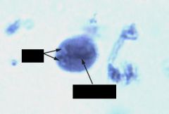
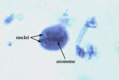
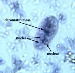
name genus, species, stage and labeled parts
|
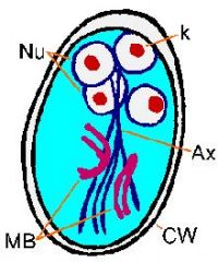
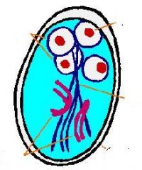
After cyst wall formation the parasite undergoes one round of nuclear division without cytokinesis resulting in four nuclei. These four nuclei (Nu) are usually located at the anterior end of the cyst (Figure). The flagella and adhesive disk are lost as the cyst matures, but the axonemes (Ax) and median bodies (MB) persist. The distinctive fibrils (ie, axonemes), which extend across the length of the cyst, result in Giardia being relatively easy to unambiguously identify. The cysts are oval shaped and typical measure 11-14 µm in length and 6-10 µm wide. Other characteristics of Giardia cysts include a well-defined wall (CW) which is often set apart from the cytoplasm of the parasite.
|
|
|
genus, species, stage and specific parasite part
|
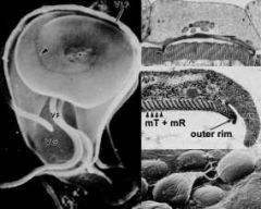
A unique ultrastructural feature of Giardia is the adhesive disk (also called ventral disk, sucking disk, sucker, or striated disk). The adhesive disk is a concave structure which occupies approximately two-thirds of the anterior end of the ventral surface (Figure, left panel). As the names imply, this structure plays a role in the attachment of the trophozoite to the intestinal epithelium and ultrastructural studies reveal close associations between the adhesive disk and the intestinal brush border
|
|
|
Name genus, species, stage.
What are the arrow heads pointing to? These are part of what structure? |
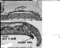
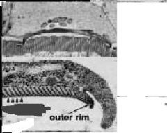
These striations are the result of microtubules (mT) and a unique cytoskeletal element called microribbons (mR). Microribbons are long flattened structures and each microribbon is associated with a microtubule (Figure, middle right panel). The combined microtubule-microribbon structure are arranged in concentric rows that form a flatten spiral with minimal overlap. The outer rim of the adhesive disk, called the lateral crest, contains components of the actin-myosin cytoskeleton.
- adhesive disk |
|
|
name genus species stage and name labeled parts
|
Giardia lambdia trophozoite
|
|
|
name genus species stage and labeled parts
|
giardia labdia early cyst
|
|
|

name genus species stage and labeled parts
|

giardia labdia late trophozoite
|
|
|
name genus species stage and labeled parts
|
giardia labdia late trophozoite
|
|
|
What is my life cycle?
|

Infection occurs after cysts are ingested. This marks the beginning of the life cycle. After ingestion, mature cysts in the small intestine release trophozoites through a process called excystation. Cysts are able to survive exposure to gastric acid; gastric acid may actually trigger excystation.
Trophozoites colonize the small intestine, attaching to the mucosa of the bowel using a ventral sucking disks. The trophozoites then multiply by longitudinal binary fission. As the Giardia trophozoites move toward the colon, they retreat into the cyst stage (known as encystation) and the new cysts are excreted in the feces. Bile salts and intestinal mucus boost trophozoite multiplication and encystations. |
|
|

name genus species stage and labeled parts
|
giardia lamdia trophozoite and cyst
|
|
|
|
name genus species stage and labeled parts
|
giardia lamdia trophozoite and cyst
|
|
|

name genus species stage and labeled parts
|

giardia lambdia trophozoite and cyst
|
|
|

name genus species stage and labeled arrows and arrow heads
|

Giardia cysts stained with trichrome (above). The figure on the right is taken at 1000X magnification (oil immersion) shows the characteristic features including nucleus (Nu), median bodies (mb) and axonemes (Ax). One of the nuclei in the cysts on the left is in focus whereas the other three (arrowheads) are out of focus and barely visible.
|
|
|
name genus species stage
|

Giardia cysts stained with trichrome (above). The figure on the left is taken at 400X magnification (high dry) and shows the distinctive oval cysts (arrowheads).
|
|
|
genus species stage
|
giardia lambdia trophozoites
Trichrome stain is more widely used than iron-hematoxylin. However, the morphology of the trophozoites is the same. The trophozoites in these slides are more evenly distributed in the sample and are easy to find. Be sure to identify the trophozoites in various orientations (ie, top view, side view, etc.). |
|
|

genus, species, stage and labeled parts
|

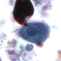
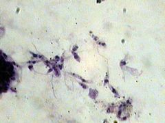
Entamoeba histolytica trophozoites
|
|
|

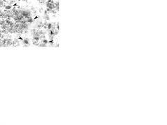
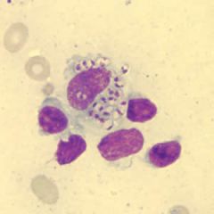
- genus, species, stage
- |

This specimen was fixed and prepared soon after collection as evidenced by the trophozoites (marked with arrowheads) with highly extended pseudopodia which are indicative of highly motile amebas. In addition, a few precysts, containing chromatoid bodies and 1 or 2 nuclei, are present. An ameba which as started to round up and become cyst like is denoted with an asterisk (*). However, it still has a pseudopodium (arrow) which is a feature of trophozoites. Numerous polymorphonuclear cells (PMNs) are also present on this slide. These can be sometimes confused with the cysts. The nuclear morphology is a key feature which helps to distinguish PMNs from cysts.
|
|
|

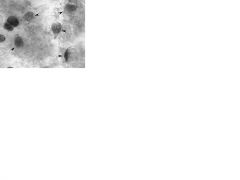
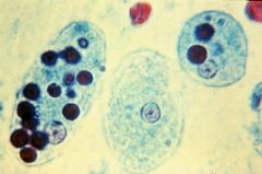
Name genus species and the different stages
|

Various stages of E. histolytica cysts. A) An early precyst with a single nucleus. B and D) A cyst at the 2 nucleus stage. C) A mature cyst with 4 nuclei and no chromatoid bodies. Note how the nuclei decrease in size with each subsequent division. A-C are stained with trichrome and D is stained with iron hematoxylin.
The chromatoid bodies vary from spherical to elongated. Many of the cysts are precysts, containing 1-3 nuclei and chromatoid bodies. The size of nuclei decreases with each nuclear division. |
|
|

- genus species stageS
|

|
|
|

genus, species, stages
- What am I doing? |

Entamoeba histolytica trophozoite and cyst
- I am encysting Encystation of Entamoeba histolytica trophozoite rounds up secretion of cyst wall aggregation of ribosomes (= chromatoid bodies) 2 rounds of nuclear division (14 nuclei) |
|
|

genus, species, stages
- What am I doing? |

Entamoeba histolytica troph and cyst
- I am encystating trophozoite rounds up secretion of cyst wall aggregation of ribosomes (= chromatoid bodies) 2 rounds of nuclear division (14 nuclei) |
|
|
|
What is the life cycle of entamoeba histolytica?
|

Cysts and trophozoites are passed in feces . Cysts are typically found in formed stool, whereas trophozoites are typically found in diarrheal stool. Infection by Entamoeba histolytica occurs by ingestion of mature cysts in fecally contaminated food, water, or hands. Excystation occurs in the small intestine and trophozoites are released, which migrate to the large intestine. The trophozoites multiply by binary fission and produce cysts , and both stages are passed in the feces . Because of the protection conferred by their walls, the cysts can survive days to weeks in the external environment and are responsible for transmission. Trophozoites passed in the stool are rapidly destroyed once outside the body, and if ingested would not survive exposure to the gastric environment. In many cases, the trophozoites remain confined to the intestinal lumen (: noninvasive infection) of individuals who are asymptomatic carriers, passing cysts in their stool. In some patients the trophozoites invade the intestinal mucosa (: intestinal disease), or, through the bloodstream, extraintestinal sites such as the liver, brain, and lungs (: extraintestinal disease), with resultant pathologic manifestations. It has been established that the invasive and noninvasive forms represent two separate species, respectively E. histolytica and E. dispar. These two species are morphologically indistinguishable unless E. histolytica is observed with ingested red blood cells (erythrophagocystosis). Transmission can also occur through exposure to fecal matter during sexual contact (in which case not only cysts, but also trophozoites could prove infective).
Geographic Distribution: Worldwide, with higher incidence of amebiasis in developing countries. In industrialized countries, risk groups include male homosexuals, travelers and recent immigrants, and institutionalized populations. |
|
|

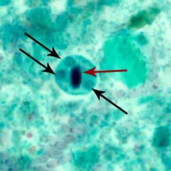
genus, species, stage,
- what is the arrow? |
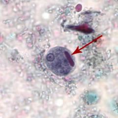
Mature Entamoeba histolytica/Entamoeba dispar cysts have 4 nuclei that characteristically have centrally-located karyosomes and fine, uniformly distributed peripheral chromatin. Cysts usually measure 12 to 15 µm.
Cyst of E. histolytica/E. dispar stained with trichrome. Note the chromatoid body with blunt ends (red arrow). |
|
|

how am i
|
Cyst of E. histolytica/E. dispar stained with trichrome.
|
|
|
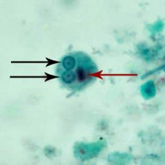
who am i and what are the arrows pointing at?
|

Cysts of E. histolytica/E. dispar stained with trichrome. Two to three nuclei are visible in the focal plane (black arrows), and the cysts contain chromatoid bodies with typically blunted ends (red arrows). The chromatoid body is particularly well demonstrated.
|
|
|

who am i and what are the arrows?
|
Cysts of E. histolytica/E. dispar stained with trichrome. Two to three nuclei are visible in the focal plane (black arrows), and the cysts contain chromatoid bodies with typically blunted ends (red arrows).
|
|
|
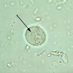
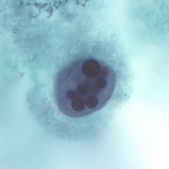
who am i and what is the arrow pointing at?
|

Mature Entamoeba histolytica/Entamoeba dispar cysts have 4 nuclei that characteristically have centrally-located karyosomes and fine, uniformly distributed peripheral chromatin. Cysts usually measure 12 to 15 µm.
Cysts of E. histolytica/E. dispar in an unstained concentrated wet mount of stool. Notice the chromatoid bodies with blunt, rounded ends (arrows). |
|
|
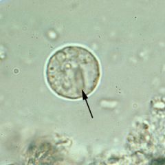
who am I and what is the arrow pointing at?
|

Mature Entamoeba histolytica/Entamoeba dispar cysts have 4 nuclei that characteristically have centrally-located karyosomes and fine, uniformly distributed peripheral chromatin. Cysts usually measure 12 to 15 µm.
Cysts of E. histolytica/E. dispar in an unstained concentrated wet mount of stool. Notice the chromatoid bodies with blunt, rounded ends (arrows). |
|
|
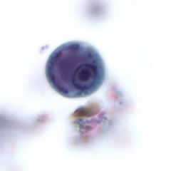
Who am i?
|

Cysts of E. histolytica/E. dispar in a concentrated wet mount stained with iodine.
|
|
|

Who am I and what is the arrow pointing at?
|
Cysts of E. histolytica/E. dispar in a concentrated wet mount stained with iodine. Notice the chromatoid body with blunt, rounded ends
|
|
|

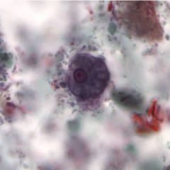
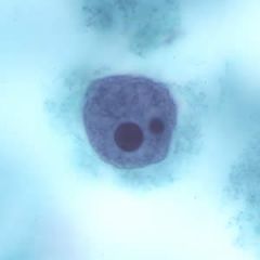
Who am I and at what stage?
|
Immature cyst of E. histolytica. The specimen was preserved in poly-vinyl alcohol (PVA) and stained with trichrome. PCR was performed on this specimen to differentiate between E. histolytica and E. dispar.
|
|
|

who am I and at what stage?
|
Immature cyst of E. histolytica/E. dispar stained with trichrome. Image taken at 1000× oil magnification and contributed by the Kansas Department of Health and Environment
|
|
|

who am i and at what stage?
|
Immature cyst of E. histolytica/E. dispar stained with trichrome. Image taken at 1000× oil magnification and contributed by the Kansas Department of Health and Environment
|
|
|

who am i and at what stage?
|
Immature cyst of E. histolytica/E. dispar stained with trichrome. Image taken at 1000× oil magnification and contributed by the Kansas Department of Health and Environment
|
|
|
who am i and at what stage?
|

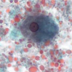
Entamoeba histolytica/Entamoeba dispar trophozoites have a single nucleus and usually measure 15 to 20 µm (range 10 to 60 µm), tending to be more elongated in diarrheal stool.
Trophozoites of E. histolytica/E. dispar in a direct wet mount stained with iodine. |
|
|

who am i and at what stage?
|
Trophozoites of E. histolytica/E. dispar in a direct wet mount stained with iodine.
|
|
|
who am i and at what stage?
|

Trophozoites of E. histolytica/E. dispar stained with trichrome.
Entamoeba histolytica/Entamoeba dispar trophozoites have a single nucleus, which have a centrally placed karyosome and uniformly distributed peripheral chromatin. This typical appearance of the nucleus is not always observed as some trophozoites can have nuclei with an eccentric karyosome and unevenly distributed peripheral chromatin. The cytoplasm has a granular or "ground-glass" appearance. E. histolytica/E. dispar trophozoites usually measure 15 to 20 µm (range 10 to 60 µm), tending to be more elongated in diarrheal stool. Erythrophagocytosis (ingestion of red blood cells by the parasite) is the only morphologic characteristic that can be used to differentiate E. histolytica from the nonpathogenic E. dispar. However, erthrophagocytosis is not typically observed on stained smears of E. histolytica. |
|
|
who am i and at what stage?
|

Trophozoites of E. histolytica. The specimen was preserved in poly-vinyl alcohol (PVA) and stained with trichrome.
|
|
|

who am i and what stage?
|
Trophozoites of E. histolytica. The specimen was preserved in poly-vinyl alcohol (PVA) and stained with trichrome. The vacuolated cytoplasm may be the result of less than optimal preservation. PCR was performed on this specimen to differentiate between E. histolytica and E. dispar.
|
|
|
who am i and at what stage?
|

Trophozoite of E. histolytica/E. dispar, measuring approximately 16.7 µm, stained with trichrome. The image was taken at 1000× magnification and contributed by the Kansas Department of Health and Environment.
|
|
|
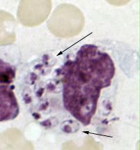
who am i and at what stage?
|

Erythrophagocytosis (ingestion of red blood cells by the parasite) is the only morphologic characteristic that can be used to differentiate E. histolytica from the nonpathogenic E. dispar. However, erthrophagocytosis is not typically observed on stained smears of E. histolytica.
Trophozoites of E. histolytica with ingested erythrocytes stained with trichrome. The ingested erythrocytes appear as dark inclusions. The parasites above show nuclei that have the typical small, centrally located karyosome, and thin, uniform peripheral chromatin. |
|
|

who am i and at what stage?
|
Trophozoites of E. histolytica with ingested erythrocytes stained with trichrome.
|
|
|
who am i and at what stage?
|

Trophozoites of E. histolytica with ingested erythrocytes stained with trichrome.
|
|
|

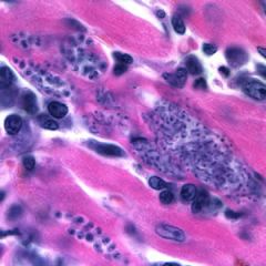
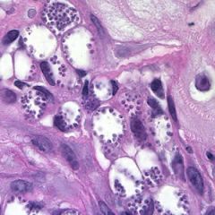
who am I, what stage and where am I???
|
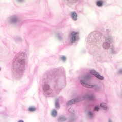
Entamoeba histolytica trophozoites in colon tissue stained with H&E.
|
|
|
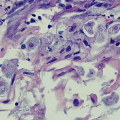
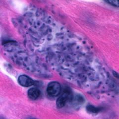
who am I, what stage and where am I?
|

Entamoeba histolytica trophozoites in a perianal biopsy, stained with H&E.
|
|
|

who am I, what stage and where am I??
|
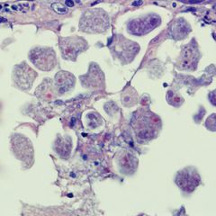
Entamoeba histolytica trophozoites in a perianal biopsy, stained with H&E.
|
|
|

who am I?
|

Cryptosporidium
4-5 mm oocysts 4 sporozoites no sporocysts |
|
|
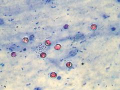
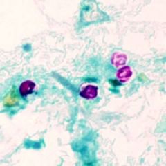
name genus species stage
|

Cryptosporidium hominis (or parvum), human feces, acid fast stain.
Cryptosporidium oocysts will appear as small bright pink spherical objects against a blue background. Their small size makes it difficult to distinguish the internal structures seen in some of the organisms. |
|
|
|
What is the preferred method of staining crypto spp. fecal smears to detect oocysts?
|
The more common stains used on fecal smears such as trichrome and iron hematoxylin are not useful for the identification and diagnosis of Cryptosporidium. The preferred method is a modified acid-fast stain to detect the oocysts in fecal smears.
|
|
|

what is my life cycle?
|
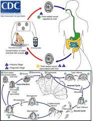
Life cycle of Cryptosporidium parvum and C. hominis.
Sporulated oocysts, containing 4 sporozoites, are excreted by the infected host through feces and possibly other routes such as respiratory secretions . Transmission of Cryptosporidium parvum and C. hominis occurs mainly through contact with contaminated water (e.g., drinking or recreational water). Occasionally food sources, such as chicken salad, may serve as vehicles for transmission. Many outbreaks in the United States have occurred in waterparks, community swimming pools, and day care centers. Zoonotic and anthroponotic transmission of C. parvum and anthroponotic transmission of C. hominis occur through exposure to infected animals or exposure to water contaminated by feces of infected animals . Following ingestion (and possibly inhalation) by a suitable host , excystation occurs. The sporozoites are released and parasitize epithelial cells (, ) of the gastrointestinal tract or other tissues such as the respiratory tract. In these cells, the parasites undergo asexual multiplication (schizogony or merogony) (, , ) and then sexual multiplication (gametogony) producing microgamonts (male) and macrogamonts (female) . Upon fertilization of the macrogamonts by the microgametes (), oocysts (, ) develop that sporulate in the infected host. Two different types of oocysts are produced, the thick-walled, which is commonly excreted from the host , and the thin-walled oocyst , which is primarily involved in autoinfection. Oocysts are infective upon excretion, thus permitting direct and immediate fecal-oral transmission. Note that oocysts of Cyclospora cayetanensis, another important coccidian parasite, are unsporulated at the time of excretion and do not become infective until sporulation is completed. Refer to the life cycle of Cyclospora cayentanensis for further details. |
|
|
genus species stage and how big am I?
|
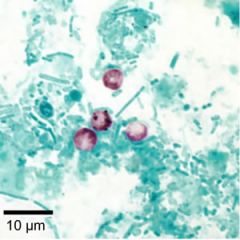
Cryptosporidium parvum oocysts stained with modified acid-fast. Against a blue-green background, the oocysts stand out in a bright red stain. Sporozoites are visible inside the two oocysts to the right.
Oocyst - 4-6um |
|
|
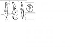
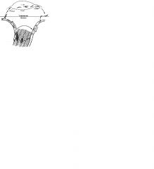
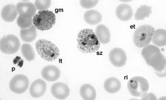
What group of parasites am I and what are my different morphologies?
What are the items at the lables? |
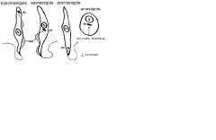
KINETOPLASTIDS
The different forms are dis¬tinguished by the position of the kinetoplast in relation to the nucleus, the presence or absence of an undulating membrane, and the presence or absence of a flagella. The undulating mem¬brane is a structure formed from a flagellum that is attached to cell body via interactions between the cell membrane and the membrane surrounding the axoneme of the flagellum. The undulating membrane becomes a 'fin-like' structure that results in increased mobility. |
|
|
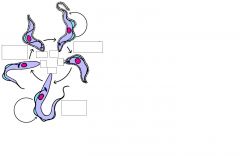
Name genus, species, stages.
Which stages are in the tsetse midgut? Which stages are in the tsetse salvilary gland? Which stages are in the tsetse human blood? |
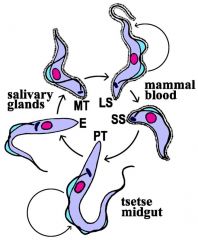
The infection is initiated in humans when metacyclic trypomastigotes are introduced during tse-tse (Glossina) feeding. The parasites replicate as trypomastigotes within the blood, lymph nodes and spleen. If the bloodstream forms are taken up by a tse-tse, they convert to procyclic trypomastigotes which undergoes replication within the gut of the fly. The procyclic trypomastigotes migrate to the salivary glands of the tse-tse and convert to epimastigotes. The epimastigotes are non-infective for the mammalian host and mature into an infective metacyclic trypomastigote.
|
|
|
Who am I?
What is my life cycle? |
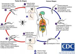
During a blood meal on the mammalian host, an infected tsetse fly (genus Glossina) injects metacyclic trypomastigotes into skin tissue. The parasites enter the lymphatic system and pass into the bloodstream . Inside the host, they transform into bloodstream trypomastigotes , are carried to other sites throughout the body, reach other blood fluids (e.g., lymph, spinal fluid), and continue the replication by binary fission . The entire life cycle of African Trypanosomes is represented by extracellular stages. The tsetse fly becomes infected with bloodstream trypomastigotes when taking a blood meal on an infected mammalian host (, ). In the fly’s midgut, the parasites transform into procyclic trypomastigotes, multiply by binary fission , leave the midgut, and transform into epimastigotes . The epimastigotes reach the fly’s salivary glands and continue multiplication by binary fission . The cycle in the fly takes approximately 3 weeks. Humans are the main reservoir for Trypanosoma brucei gambiense, but this species can also be found in animals. Wild game animals are the main reservoir of T. b. rhodesiense.
|
|
|
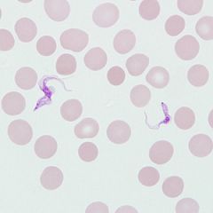
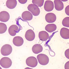
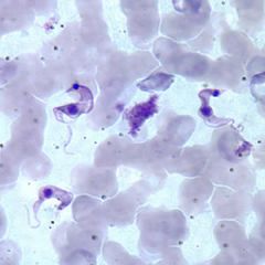
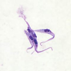
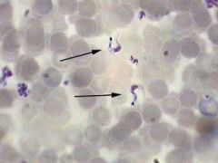
- Genus Species Stage(s)
- name labeled sections - What is the difference between the white and black thick arrows? |
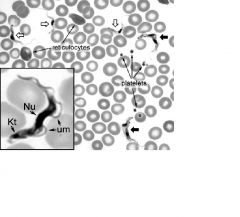
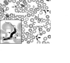
Giemsa-stained blood smear of African trypanosomes viewed under the 100X objective lens. The block arrows denote trypomastigote forms of the African trypanosomes found within the blood and tissue of the vertebrate host. The filled block arrows are replicating forms which are in the process of cell division. (Note the two nuclei, two kinetoplasts, and two undulating membranes.) Also indicated are reticulocytes (immature erythro¬cytes) and platelets. The inset in the lower left shows an enlarged trypomastigote with the kinetoplast (Kt), nucleus (Nu), and undulating membrane (um) indicated.
|
|
|

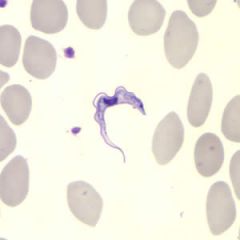
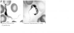
Name genus, species, stage
- Can you name the subspecies?? |
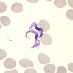
The two Trypanosoma brucei subspecies that cause African trypanosomiasis, T. b. gambiense and T. b. rhodesiense, are indistinguishable morphologically. A typical trypomastigote has a small kinetoplast located at the posterior end, a centrally located nucleus, an undulating membrane, and a flagellum running along the undulating membrane, leaving the body at the anterior end. Trypomastigotes are the only stage found in patients. Trypanosomes range in length from 14 to 33 µm.
|
|
|

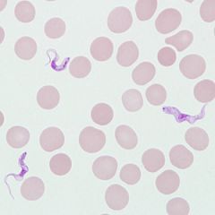
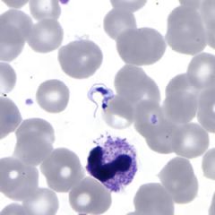
name genus species, stage
|
Trypomastigotes of T. brucei ssp. in a blood smear stained with Giemsa.
The two Trypanosoma brucei subspecies that cause African trypanosomiasis, T. b. gambiense and T. b. rhodesiense, are indistinguishable morphologically. A typical trypomastigote has a small kinetoplast located at the posterior end, a centrally located nucleus, an undulating membrane, and a flagellum running along the undulating membrane, leaving the body at the anterior end. Trypomastigotes are the only stage found in patients. Trypanosomes range in length from 14 to 33 µm. |
|
|

name genus, species, stage
|
Trypomastigotes of T. brucei ssp. in a blood smear stained with Giemsa.
The two Trypanosoma brucei subspecies that cause African trypanosomiasis, T. b. gambiense and T. b. rhodesiense, are indistinguishable morphologically. A typical trypomastigote has a small kinetoplast located at the posterior end, a centrally located nucleus, an undulating membrane, and a flagellum running along the undulating membrane, leaving the body at the anterior end. Trypomastigotes are the only stage found in patients. Trypanosomes range in length from 14 to 33 µm. |
|
|
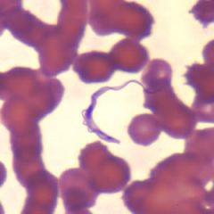
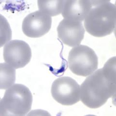
name genus species stage
|

Trypanosoma brucei ssp. in thin blood smears stained with Wright-Giemsa.
The two Trypanosoma brucei subspecies that cause African trypanosomiasis, T. b. gambiense and T. b. rhodesiense, are indistinguishable morphologically. A typical trypomastigote has a small kinetoplast located at the posterior end, a centrally located nucleus, an undulating membrane, and a flagellum running along the undulating membrane, leaving the body at the anterior end. Trypomastigotes are the only stage found in patients. Trypanosomes range in length from 14 to 33 µm. |
|
|

name genus species stage
|
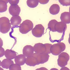
Trypanosoma brucei ssp. in thin blood smears stained with Wright-Giemsa.
The two Trypanosoma brucei subspecies that cause African trypanosomiasis, T. b. gambiense and T. b. rhodesiense, are indistinguishable morphologically. A typical trypomastigote has a small kinetoplast located at the posterior end, a centrally located nucleus, an undulating membrane, and a flagellum running along the undulating membrane, leaving the body at the anterior end. Trypomastigotes are the only stage found in patients. Trypanosomes range in length from 14 to 33 µm. |
|
|
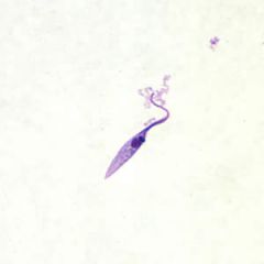
name genus species and stage
|

Trypanosoma brucei ssp. in thin blood smears stained with Wright-Giemsa.
The two Trypanosoma brucei subspecies that cause African trypanosomiasis, T. b. gambiense and T. b. rhodesiense, are indistinguishable morphologically. A typical trypomastigote has a small kinetoplast located at the posterior end, a centrally located nucleus, an undulating membrane, and a flagellum running along the undulating membrane, leaving the body at the anterior end. Trypomastigotes are the only stage found in patients. Trypanosomes range in length from 14 to 33 µm. |
|
|
|
List the important steps of the American Trypanosome Life Cycle
|
Humans: metacyclic trypomastigotes, amasitgotes in muscle (replication),
trypomastigote. Triatomine: trypomastigote, converts to epimastigote in midgut, matures to metacyclic trypomastigote in hindgut |
|
|
what is the american trypanosome life cycle
|

Humans: metacyclic trypomastigotes - convert to amastigote in muscle and replicates, converts to trypomastigote and released to blood
Triatomine: trypomastigote, converts to epi in midgut and replicates, goes to hindgut to mature to metacyclic trypomastigote - excreted in feces |
|
|
Who am I?
Name my labeled parts |
Two examples of T. cruzi trypomas¬ti-gotes from Giemsa-stained blood smears in which the kinetoplast (kt), nucleus (nu), undulating membrane (um), and free flagellum (fl) are denoted.
|
|
|

name genus species stage? How do you know this?
|
T. cruzi in the blood exhibits typical trypomastigote morphology (see figure below). Note the lightly staining cytoplasm, spherical or slightly oval nucleus, and the large spherical kinetoplast near the posterior end. The kinetoplast sometimes appears to be larger than the diameter of the organism at its pointed end.
|
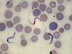
Three T. cruzi trypomastigotes in a thin blood smear stained with Giemsa.
|
|

name genus species stage
|
T. cruzi trypomastigote in a thin blood smear stained with Giemsa.
Motile circulating trypomastigotes are readily seen on slides of fresh anticoagulated blood in acute infection but are rarely detectable by microscopy in chronic T. cruzi infection. A typical trypomastigote has a large, subterminal or terminal kinetoplast, a centrally located nucleus, an undulating membrane, and a flagellum running along the undulating membrane, leaving the body at the anterior end. Trypanosomes measure from 12 to 30 µm in length. Trypomastigotes may be seen in cerebrospinal fluid (CSF) in central nervous system infections; also the amastigote stage parasite may be seen in histopathology specimens from affected organs. |
|
|

name genus species stage
|
T. cruzi trypomastigote in a thin blood smear stained with Giemsa. Note the kinetoplast is subterminal.
Motile circulating trypomastigotes are readily seen on slides of fresh anticoagulated blood in acute infection but are rarely detectable by microscopy in chronic T. cruzi infection. A typical trypomastigote has a large, subterminal or terminal kinetoplast, a centrally located nucleus, an undulating membrane, and a flagellum running along the undulating membrane, leaving the body at the anterior end. Trypanosomes measure from 12 to 30 µm in length. Trypomastigotes may be seen in cerebrospinal fluid (CSF) in central nervous system infections; also the amastigote stage parasite may be seen in histopathology specimens from affected organs. |
|
|
name genus species stage
|

The epimastigote stage is not seen in humans but can be found in the midgut of triatomines that have ingested trypomastigotes from an infected host.
Trypanosoma cruzi epimastigotes from culture. Note the location of the kinetoplast anterior to the nucleus. |
|
|

name genus species stage
|
The epimastigote stage is not seen in humans but can be found in the midgut of triatomines that have ingested trypomastigotes from an infected host.
Trypanosoma cruzi epimastigotes from culture. Note the location of the kinetoplast anterior to the nucleus. |
|
|
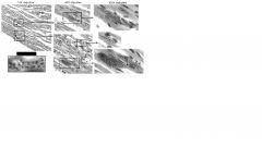
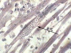
name genus species, stage and where am I?
|
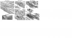
T. cruzi in heart muscle. The successive enlargements of the boxed areas are shown at the indicated magnifications. Infected myocytes (i.e., muscle cells) appear as a collection of small dots using the 10X objective as indicated by the arrowheads. The infected host cells are sometimes called pseudocysts. These dots become more evident using the 40X objective. To clearly see the kinetoplasts it is necessary to use the 100X objective lens (i.e., oil immersion). It is often difficult to see the individual amastigotes in the larger pseudocysts which are most evident at the lower magnifications. Distinct amastigotes (indicated by arrowheads) are usually easier to see in the pseudocysts which contain fewer amastigotes. The inset at the lower left is a further enlargement of amastigotes in which the nuclei and kinetoplasts are more evident.
|
|
|
who am I and what stage?
|

Epimastigotes of T. cruzi. Shown is a Giemsa-stained smear from in vitro cultures of T. cruzi. T. cruzi epimastigotes have a prominent bar-shaped kinetoplast just anterior to the nucleus and an undulating membrane that extends from the kinetoplast to the anterior end. The undulating membrane is sometimes difficult to see in epimastigotes and it often appears that the anterior end of the parasite is twisted. The open block arrows denote epimastigotes in which the features (i.e., nucleus, kinetoplast, undulating membrane) are relatively apparent. The filled block arrows denote dividing forms.
|
|
|
name genus species stage and where it is
|

Trypanosoma cruzi amastigotes in heart tissue. The section is stained with hematoxylin and eosin (H&E).
|
|
|

name genus species stage and place
|
Trypanosoma cruzi amastigotes in heart tissue. The section is stained with hematoxylin and eosin (H&E).
|
|
|
name genus species and stage
|

Trypanosoma cruzi amastigotes in heart tissue. The section is stained with H&E.
The amastigotes in this image appear to be transforming into trypomastigotes. |
|
|

name genus species stage, place, and what is going on here?
|
Trypanosoma cruzi amastigotes in heart tissue. The section is stained with H&E.
The amastigotes in this image appear to be transforming into trypomastigotes. |
|
|

what is the life cycle of leishmaniasis?
|
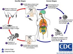
Leishmaniasis is transmitted by the bite of female phlebotomine sandflies. The sandflies inject the infective stage, promastigotes, during blood meals . Promastigotes that reach the puncture wound are phagocytized by macrophages and transform into amastigotes . Amastigotes multiply in infected cells and affect different tissues, depending in part on the Leishmania species . This originates the clinical manifestations of leishmaniasis. Sandflies become infected during blood meals on an infected host when they ingest macrophages infected with amastigotes (, ). In the sandfly's midgut, the parasites differentiate into promastigotes , which multiply and migrate to the proboscis .
|
|
|

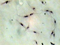
name genus species stage - where am I?
|
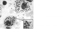
Giemsa-stained impression smear of Leishmania amastigotes. Shown are two infected macrophages. The macrophage nuclei are marked with *. The inset shows and enlarge-ment of some of the amastigotes. The kinetoplasts are denoted with arrowheads and the arrows denote nuclei. The morphology of amastigotes is generally better in impression smears that in tissue sections
|
|
|

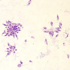
name genus species stage
|
Leishmaniasis promastigotes in vitro culture
|
|
|

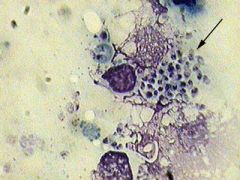
name genus speices stage and labeled parts
|
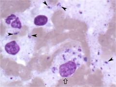
Bone marrow smear of patient with visceral leishmaniasis. Free amastigotes (arrowheads) and an infected macrophage (block arrow) can be seen. Many of the amastigotes are out of focus.
|
|
|

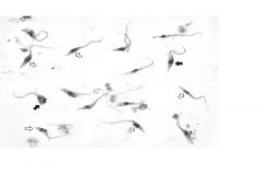
name genus species stage
|

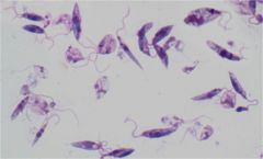
Promastigotes of Leishmania derived from in vitro culture. Promastigotes are found within the gut of the sandfly vector. Distinct kinetoplasts and nuclei are evident in many of the promastigotes. Dividing forms are also evident. Note the range of morpho¬logies. Different morphological forms representing different developmental stages are also present within the gut of the vector. It can be difficult to distinguish promastigotes from epimastigotes especially in cases where the kinetoplast of the promastigote is just anterior to the nucleus. The distinguishing feature is the presence or absence of an undulating membrane and not the position of the kinetoplast. Be sure that you are able to recognize the undulating membrane and that you are able to distinguish epimasti¬gotes from promastigotes (see figure of T. cruzi epimastigotes).
|
|
|
name genus species stage
|

Leishmania spp. amastigotes in a Giemsa-stained tissue scraping.
Amastigotes of Leishmania are spherical to ovoid and measure 1-5 µm long by 1-2 µm wide. They possess a large nucleus, a prominent kinetoplast, and a short axoneme, the last of which is rarely visible by light microscopy. The organisms reside in macrophages of the host and can be found throughout the body. |
|
|

name genus species stage and identify arrows
|
Leishmania tropica amastigotes from an impression smear of a biopsy specimen from a skin lesion. An intact macrophage is practically filled with amastigotes (arrows), several of which have a clearly visible nucleus and kinetoplast;
|
|
|
name genus species stage
|

Amastigotes of Leishmania sp. in a biopsy specimen from a skin lesion, stained with hematoxylin and eosin (H&E).
Amastigotes of Leishmania are spherical to ovoid and measure 1-5 µm long by 1-2 µm wide. They possess a large nucleus, a prominent kinetoplast, and a short axoneme, the last of which is rarely visible by light microscopy. The organisms reside in macrophages of the host and can be found throughout the body. |
|
|
name genus species stage
|

Leishmania sp. promastigotes from culture.
Promastigotes are not found in human tissue; this stage occurs in the mid-gut of the sand fly (genera Phlebotomus and Lutzomyia) intermediate hosts. Promastigotes are elongate, slender and measure about 10-12 µm in length. They have a large central nucleus and a kinetoplast located near the anterior end. A flagellum arises at the anterior end, that may be longer than the rest of the promastigote. |
|
|
name genus species stage
|

Trypanosoma cruzi epimastigotes in culture, 900x.
This is what the protozoan looks like in the insect gut. |
|
|
name genus species stage
|

Trypanosoma cruzi amastigotes in a heart smear, 450x. The large mass is the pseudocyst containing the amastigotes.
|
|
|

name genus species stage
|
Trypanosoma cruzi trypanomastigote in blood, 900x.
Note the large kinetosome at the "head". |
|
|

name genus species stage
|
Leishmania donovani amastigote in spleen smear, 900x
|
|
|

name genus species stage
|
Leishmania donovani promastigotes in culture, 900x.
These represent the insect gut stage. |
|
|
name genus species stage
|

Trypanosoma lewisi trypanomastigote in rat blood, 900x.
|
|
|

life cycle of t cruzi
|

Trypanosoma cruzi causes Chagas disease in Central and South America, occasionally appearing in Mexico and the Rio Grande border of the US. Bugs of the family Reduviidae such as Panstrongylus megistus and Triatoma spp. serve as vectors. These bloodsucking bugs, known as assassin or kissing bugs inhabit roof thatching and as well as crevices in the house and feed nocturnally. The bug commonly bites in the orbits of the eyes or on and around the lips, thus the name kissing bug. The bite is very painful, hence another monicker... assassin bug. The bug often defecates as it feeds and the feces containing the infective metacyclic trypomastigotes, may be rubbed into the wound by the victim. The trypomastigotes are found in large numbers in the bloodstream shortly after inoculation, though they soon enter cells of the spleen, liver, heart and others tissues where they assume the amastigote stage. It is in the amastigote stage that they quickly divide, producing such numbers that they kill the host cell, escape and attack other cells. Promastigotes and epimastigotes can be seen in interstitial spaces and some fully develop into trypomastigotes which enter the bloodstream to continue the life cycle should they be ingested by a biting assassin bug. They multiply in the bug's midgut as epimastigotes, transforming into short metacyclic trypomastigotes in about 8-10 days post-infection. A number of domestic and wild mammals serve as reservoir hosts, principally, wood rats, armadillos, opossums, cats and dogs. Hosts may be infected by eating infected insects and in some villages of Mexico, bugs are eaten as aphrodisiacs, leading to infections. Transplacental transmission occurs resulting in newborns with advanced Chagas disease. Blood transfusions serve as potential risk factors. Coitus and mother's milk are being investigated as a possible source of transmission.
|
|
|
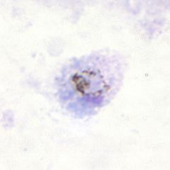
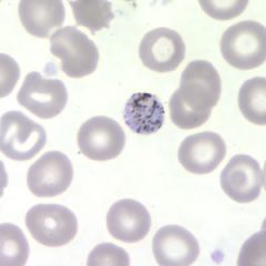
name genus species stage
|

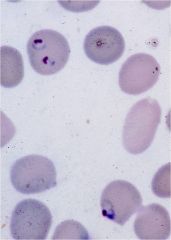
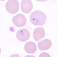
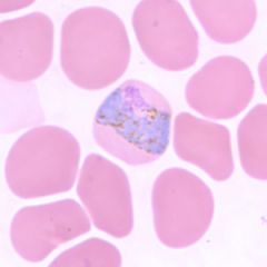
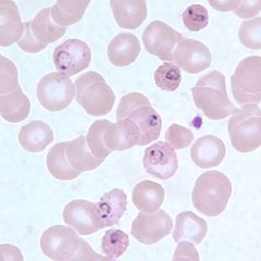
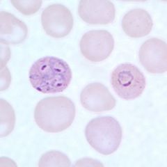
Plasmodium falciparum
numerous rings without mature forms slightly smaller marginal forms |
|
|

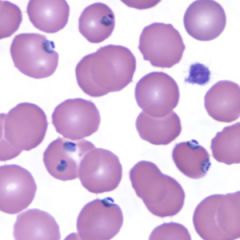
name genus species and stage
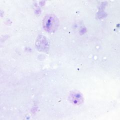
|

. All of the parasites are young trophozoites and are almost always inside an erythrocyte. The early tropho¬zoite is also referred to as a ring form (or sometimes as a 'signet ring'). Parasites are recognized by their blue staining cytoplasm, often appearing in the shape of a ring, and the purple or reddish-purple nucleus. Note that some of the ring forms appear to have two nuclei. This is an optical phenomenon due to an elongated and 'dumbbell-shaped' nucleus. In addition, some erythrocytes are infected with 2-3 ring forms. Multiple infected erythrocytes is more common in P. falciparum than the other species.
|
|
|
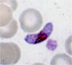
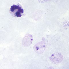
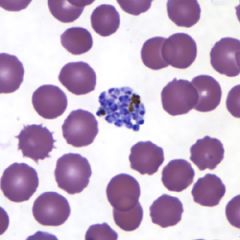
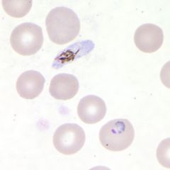
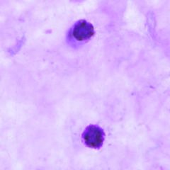
name genus species stage
|

the sausage shaped (or crescent shaped) gametocytes. The gametocytes appear to be extracellular. However, they are within the erythrocytes and occasionally the edge of the erythrocyte can be seen (arrows). This is known as Laveran's bib. The cytoplasm of the macrogametocyte usually stains more blue, has more pointed ends, and has a denser, more compact nucleus which is frequently obscured by malarial pigment in the central part of the parasite. The cytoplasm of the microgametocyte tends to stain lighter (lavender or pink), the ends are more blunt, and the nuclear material consists of what often appears as a loose aggregate of reddish or purplish granules partially obscured by pigment. These crescent shaped gametocytes are unique to P. falciparum.
|
|
|
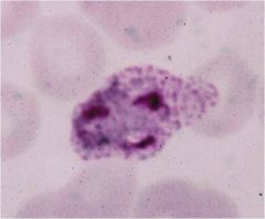
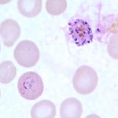
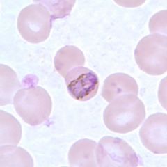
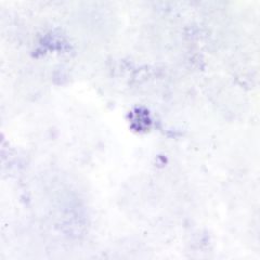
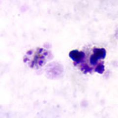
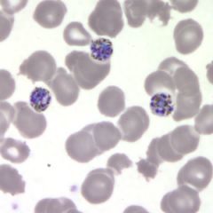
genus species stage
|

Plasmodium falciparum trophozoites and schizonts
not normally in peripheral circulation severe disease compact parasite |
|
|
genus species stage
|

the sausage shaped (or crescent shaped) gametocytes. The gametocytes appear to be extracellular. However, they are within the erythrocytes and occasionally the edge of the erythrocyte can be seen (arrows). This is known as Laveran's bib. The cytoplasm of the macrogametocyte usually stains more blue, has more pointed ends, and has a denser, more compact nucleus which is frequently obscured by malarial pigment in the central part of the parasite. The cytoplasm of the microgametocyte tends to stain lighter (lavender or pink), the ends are more blunt, and the nuclear material consists of what often appears as a loose aggregate of reddish or purplish granules partially obscured by pigment. These crescent shaped gametocytes are unique to P. falciparum.
|
|
|

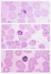
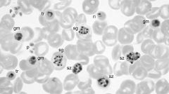
genus species stages
|
Composite showing various morphological forms of P. vivax. ri = ring; et = early trophozoite; lt = late trophozoite; sz = schizont; gm= gametocyte; p = platelets
The young trophozoites, or ring forms, will have the same appearance as other Plasmodium species. As the trophozoites mature they may be in the form of a larger ring or may have an irregular ameboid form. The cytoplasm of erythrocytes infected with trophozoite or schizont stages usually contain reddish Schüffner granules (or dots). These are in the erythrocyte and not in the parasite itself. Parasites with two or more nuclei are referred to as schizonts. Mature gametocytes of P. vivax are large parasites which nearly fill the erythrocyte but still have a single nucleus. In addition, the pigment tends to be dispersed in small granules in the cytoplasm of the gametocytes rather in a large granule as is found in trophozoites destined to become schizonts. The male form, or microgametocyte, stains a lighter blue and has a large nucleus composed of a loose aggregate of nuclear particles. The female, or macrogametocyte, stains a deeper blue and has a smaller denser nucleus. These are usually more numerous than the microgametocytes. The gametocyte stages develop from merozoites just as do the trophozoites; however, it is a dead end unless these stages are picked up by a susceptible mosquito. |
|
|

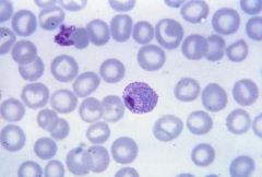
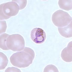
genus species stage
|
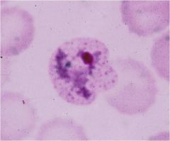
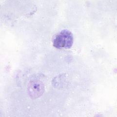
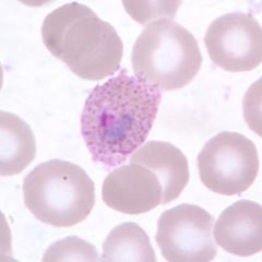
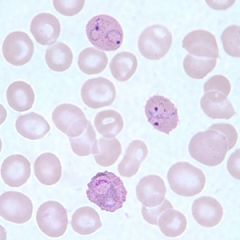
Plasmodium vivax
enlarged erythrocyte Schüffner’s dots ‘ameboid’ trophozoite many merozoites in mature segmenter |
|
|
genus species stage
|

Plasmodium vivax
enlarged erythrocyte Schüffner’s dots ‘ameboid’ trophozoite many merozoites in mature segmenter |
|
|
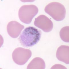
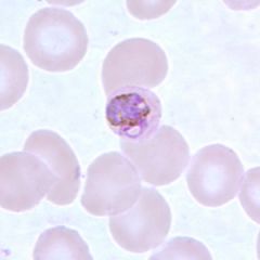
genus speces stage, what is special about me?
|

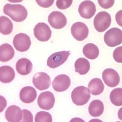
Schüffner’s dots due to caveola-vesicle complexes on erythrocyte surface
Plasmodium vivax, thin blood smear showing large trophozoite in an erythrocyte. Note the numerous pigment dots in the infected erythrocyte (= Schüffner's dots) characteristic of P. vivax and P.ovale infections. From CDC PHIL 5863 |
|
|
|
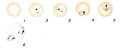
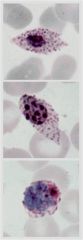
genus species and stages
|
Stages of P. vivax in thin smears. Fig. 1: Normal red cell; Figs. 2-6: Young trophozoites (ring stage parasites); Figs. 7-18: Trophozoites; Figs. 19-27: Schizonts; Figs. 28 and 29: Macrogametocytes (female); Fig. 30: Microgametocyte (male).
|
|
|
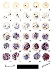
genus species and stages
|
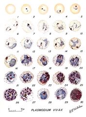
Stages of P. vivax in thin smears. Fig. 1: Normal red cell; Figs. 2-6: Young trophozoites (ring stage parasites); Figs. 7-18: Trophozoites; Figs. 19-27: Schizonts; Figs. 28 and 29: Macrogametocytes (female); Fig. 30: Microgametocyte (male).
|
|
|
name genus species stages
|

Ring-form trophozoites of P. vivax. Fig. 1: Normal red blood cell; Figs. 2-6: Developing ring-form trophozoites.
|
|
|

genus species and stage
|
Ring-form trophozoites of P. vivax in a thick blood smear.
Ring-form trophozoites of P. vivax usually have a thick cytoplasm with a single, large chromatin dot. Rings may be difficult to distinguish from those of P. ovale. The cytoplasm becomes amoeboid and Schüffner's dots may appear as the trophozoites mature. Infected RBCs are often larger than uninfected RBCs. Multiply-infected RBCs are not uncommon. |
|
|

genus species stage
|
Ring-form trophozoites of P. vivax in a thick blood smear.
Ring-form trophozoites of P. vivax usually have a thick cytoplasm with a single, large chromatin dot. Rings may be difficult to distinguish from those of P. ovale. The cytoplasm becomes amoeboid and Schüffner's dots may appear as the trophozoites mature. Infected RBCs are often larger than uninfected RBCs. Multiply-infected RBCs are not uncommon. |
|
|
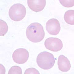
genus species stage
|

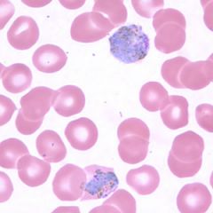
Ring-form trophozoites of P. vivax in thin blood smears.
Ring-form trophozoites of P. vivax usually have a thick cytoplasm with a single, large chromatin dot. Rings may be difficult to distinguish from those of P. ovale. The cytoplasm becomes amoeboid and Schüffner's dots may appear as the trophozoites mature. Infected RBCs are often larger than uninfected RBCs. Multiply-infected RBCs are not uncommon. |
|
|
genus species stage
|

Ring-form trophozoites of P. vivax in thin blood smears.
Ring-form trophozoites of P. vivax usually have a thick cytoplasm with a single, large chromatin dot. Rings may be difficult to distinguish from those of P. ovale. The cytoplasm becomes amoeboid and Schüffner's dots may appear as the trophozoites mature. Infected RBCs are often larger than uninfected RBCs. Multiply-infected RBCs are not uncommon. |
|
|
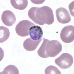
Genus species stage
|

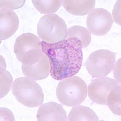
Developing trophozoites of P. vivax become increasingly amoeboid, with tenuous pseudopodial processes and large vacuoles. Schüffner's dots are visible with proper staining. Pigment tends to be fine and brown. Infected RBCs are usually noticeably larger than uninfected RBCs.
|
|
|

genus species stage
|
Trophozoites of P. vivax in thin blood smears. Note the amoeboid appearance, Schüffner's dots and enlarged infected RBCs.
|
|
|

genus species stage
|
Trophozoites of P. vivax in a thin blood smear. Note however the fine, light brown pigment that is distributed throughout the cytoplasm (pigment in P. malariae is usually darker and coarser and distributed on the periphery of the cytoplasm). The infected RBCs are also noticeably larger than the uninfected RBCs.
|
|
|
genus species stage
|

Trophozoites of P. vivax in a thin blood smear. Note the band-like appearance of the trophozoite in Figure F that may be mistaken for a band-form trophozoite of P. malariae. Note however the fine, light brown pigment that is distributed throughout the cytoplasm (pigment in P. malariae is usually darker and coarser and distributed on the periphery of the cytoplasm). The infected RBCs are also noticeably larger than the uninfected RBCs.
|
|
|

genus species stage
|
Gametocyte (upper) and trophozoite (lower) of P. vivax in a thick blood smear.
Macrogametocytes of P. vivax are round to oval and usually fill the host cell. The infected RBC is usually noticeably larger than uninfected RBCs. The cytoplasm is usually a darker blue and contains fine brown pigment throughout. Schüffner's dots may be seen with proper staining. Microgametocytes are usually the size of an uninfected RBC and have a paler blue, pink or gray cytoplasm. |
|
|
genus species stage
|

Macrogametocytes of P. vivax in thin blood smears. Note the enlargement of the gametocytes compared to uninfected RBCs.
Macrogametocytes of P. vivax are round to oval and usually fill the host cell. The infected RBC is usually noticeably larger than uninfected RBCs. The cytoplasm is usually a darker blue and contains fine brown pigment throughout. Schüffner's dots may be seen with proper staining. Microgametocytes are usually the size of an uninfected RBC and have a paler blue, pink or gray cytoplasm. |
|
|
genus species stage
|

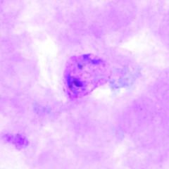
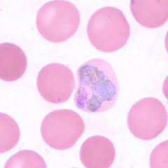
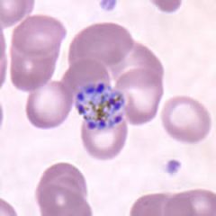
Schizonts of P. vivax in thick blood smears.
Developing schizonts of P. vivax are large and amoeboid. Chromatin is arranged in two or more masses; pigment is also usually arranged in more than one mass. Mature schizonts contain 12-24 merozoites, each of which contains a dot of chromatin and a mass of cytoplasm. Pigment is usually organized in one or two clumps. Like other stages, infected RBCs are usually larger than uninfected RBCs. |
|
|
genus species stage
|

Schizonts of P. vivax in thick blood smears.
Developing schizonts of P. vivax are large and amoeboid. Chromatin is arranged in two or more masses; pigment is also usually arranged in more than one mass. Mature schizonts contain 12-24 merozoites, each of which contains a dot of chromatin and a mass of cytoplasm. Pigment is usually organized in one or two clumps. Like other stages, infected RBCs are usually larger than uninfected RBCs. |
|
|

genus species stage
|
Schizonts of P. vivax in thin blood smears.
Developing schizonts of P. vivax are large and amoeboid. Chromatin is arranged in two or more masses; pigment is also usually arranged in more than one mass. Mature schizonts contain 12-24 merozoites, each of which contains a dot of chromatin and a mass of cytoplasm. Pigment is usually organized in one or two clumps. Like other stages, infected RBCs are usually larger than uninfected RBCs. |
|
|

genus species stage
|
Trophozoites of P. falciparum in thin blood smears. In Figure D, a gametocyte can also be seen in the upper half of the image.
Developing trophozoites of P. falciparum tend to remain in ring form, but may become thicker and more compact. The amount of pigment and chromatin may also increase. Compact or amoeboid forms may be seen in smears where there was a delay in processing the blood. |
|
|

genus species stage
|
Trophozoites of P. falciparum in thin blood smears.
Developing trophozoites of P. falciparum tend to remain in ring form, but may become thicker and more compact. The amount of pigment and chromatin may also increase. Compact or amoeboid forms may be seen in smears where there was a delay in processing the blood. |
|
|

genus species stage
|
Ring-form trophozoites of P. falciparum in a thin blood smear, exhibiting Maurer's clefts.
Maurer's clefts can be seen in P. falciparum infections containing older ring-form trophozoites and asexual stages. Maurer's clefts resemble the Schüffner's dots seen in P. vivax and P. ovale, but are usually larger and more coarse. Visualization of these structures is dependent on the quality of the smear preparation and the pH of the Giemsa stain. Like Schüffner's dots, Maurer's clefts appear to play a role in the metabolic pathways of the infected RBCs. |
|
|
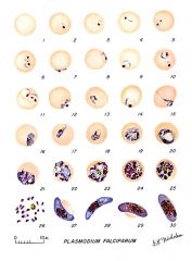
Pf life stages
|

The stages of P. falciparum. 1: Normal red cell; Figs. 2-18: Trophozoites (among these, Figs. 2-10 correspond to ring-stage trophozoites); Figs. 19-26: Schizonts (Fig. 26 is a ruptured schizont); Figs. 27, 28: Mature macrogametocytes (female); Figs. 29, 30: Mature microgametocytes (male).
|
|
|
genus species stags
|
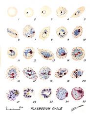
Stages of P. ovale in thin blood smears. Fig. 1: Normal red cell; Figs. 2-5: Young trophozoites (Rings); Figs. 6-15: Trophozoites; Figs. 16-23: Schizonts; Fig. 24: Macrogametocytes (female); Fig. 25: Microgametocyte (male).
|
|
|
genus species stages
|

Ring-form trophozoites of P. ovale in thin blood smears. Note the multiply-infected RBC
Ring-form trophozoites usually contain a single chromatin dot, but may contain double-chromatin dots. Multiply-infected RBCs may be seen, making the rings difficult to differentiate from P. falciparum. The single rings may be difficult to differentiate from P. vivax, as the cytoplasm is usually thick with a large chromatin dot. As the trophozoites mature, they are less amoeboid than P. vivax and may exhibit fimbriation and Schüffner's dots. Infected RBCs are not usually enlarged as in P. vivax infections. |
|
|
genus species stage
|

Trophozoite of P. ovale in a thin blood smear. Note the fimbriation
Developing trophozoites of P. ovale are compact with little vacuolation. Infected RBCs are often slightly enlarged and may exhibit fimbriation and Schüffner's dots. Pigment is less-coarse and diffuse. |
|
|

genus species stage
|
Trophozoite of P. ovale in a thin blood smear. Note the fimbriation and Schüffner's dots.
|
|
|
genus species stage
|

Infected RBCs showing developing (lower) and ring-form (upper two) trophozoites of P. ovale in a thin blood smear.
Developing trophozoites of P. ovale are compact with little vacuolation. Infected RBCs are often slightly enlarged and may exhibit fimbriation and Schüffner's dots. Pigment is less-coarse and diffuse. |
|
|

genus species stage
|
Gametocytes of P. ovale can be difficult to distinguish from those of P. vivax, although there is generally less enlargement of the infected RBC. The mature macrogametocyte fills the host RBC; the microgametocyte is smaller. Schüffner's dots may be seen with proper staining and fimbriation may occur.
|
|
|

genus species stage
|

Microgametocyte of P. ovale in a thin blood smear. Note the elongated, oval shape and the Schüffner's dots.
Gametocytes of P. ovale can be difficult to distinguish from those of P. vivax, although there is generally less enlargement of the infected RBC. The mature macrogametocyte fills the host RBC; the microgametocyte is smaller. Schüffner's dots may be seen with proper staining and fimbriation may occur |
|
|
genus species stage
|

Macrogametocyte of P. ovale in a thin blood smear. Note the fimbriation.
|
|
|
genus species stage
|

Schizonts of P. ovale in thick blood smears.
Schizonts of P. ovale can be similar to P. vivax, although tend to be smaller and contain fewer merozoites (4-16, on average 8). Elongation to an oval shape and fimbriation are common. Schüffner's dots can be observed with proper staining. Pigment is lighter and less coarse, similar to P. vivax. |
|
|
genus species stage
|

Schizont (upper right) and ring-form trophozoite (lower left) of P. ovale in a thin blood smear.
Schizonts of P. ovale can be similar to P. vivax, although tend to be smaller and contain fewer merozoites (4-16, on average 8). Elongation to an oval shape and fimbriation are common. Schüffner's dots can be observed with proper staining. Pigment is lighter and less coarse, similar to P. vivax. |
|
|
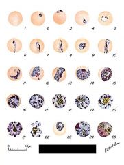
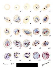
genus species stages
|
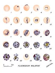
Stages of P. malariae in thin blood smears. Fig. 1: Normal red cell; Figs. 2-5: Young trophozoites (rings); Figs. 6-13: Trophozoites; Figs. 14-22: Schizonts; Fig. 23: Developing gametocyte; Fig. 24: Macrogametocyte (female); Fig. 25: Microgametocyte (male).
|
|
|
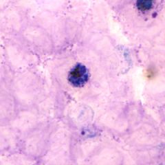
genus species stage
|

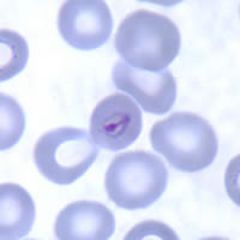
Ring-form (lower right) and developing (upper left) trophozoites of P. malariae in a thick blood smear.
Ring-form trophozoites have one (rarely two) chromatin dots and a cytoplasm ring that tends to be thicker than P. falciparum. 'Bird's-eye' forms may appear. There is no enlargement of infected RBCs. |
|
|
genus species stage
|

"Birds-eye" trophozoite of P. malariae in a thin blood smear.
|
|
|

genus species stage
|
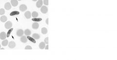
Band-form trophozoites of P. malariae in a thin blood smear.
In developing trophozoites of P. malariae, chromatin is rounded or streaky and the cytoplasm is usually compact with no vacuole. Pigment may be coarse and peripheral. As the trophozoites mature, the cytoplasm may elongate across the host RBC, forming a 'band-form', or may be oval with a vacuole forming a 'basket-form'. Chromatin is usually in a single mass, less definite in outline. Pigment granules become larger and tend to have a more peripheral arrangement. |
|
|
genus species stage
|

Basket-form trophozoites of P. malariae in a thin blood smear.
In developing trophozoites of P. malariae, chromatin is rounded or streaky and the cytoplasm is usually compact with no vacuole. Pigment may be coarse and peripheral. As the trophozoites mature, the cytoplasm may elongate across the host RBC, forming a 'band-form', or may be oval with a vacuole forming a 'basket-form'. Chromatin is usually in a single mass, less definite in outline. Pigment granules become larger and tend to have a more peripheral arrangement. |
|
|
genus species stage
|

Gametocytes of P. malariae in thin blood smears.
Gametocytes of P. malariae are compact and tend to fill the host RBC. There is no enlargement of the infected RBC and sometimes there is a reduction in size. The cytoplasm stains blue and the chromatin pink to red. Abundant dark pigment may be scattered throughout the cytoplasm. |
|
|

genus species stage
|
Gametocytes of P. malariae in thin blood smears.
Gametocytes of P. malariae are compact and tend to fill the host RBC. There is no enlargement of the infected RBC and sometimes there is a reduction in size. The cytoplasm stains blue and the chromatin pink to red. Abundant dark pigment may be scattered throughout the cytoplasm. |
|
|
genus species stage
|

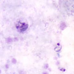
Schizonts of P. malariae in thick blood smears.
Schizonts of P. malariae have 6-12 (usually 8-10) merozoites, often arranged in a rosette or irregular cluster. Mature schizonts nearly fill the normal-sized host RBC. Pigment is course and often peripheral. Schizonts can be common in peripheral blood circulation. |
|
|

genus species stage
|
Schizonts of P. malariae in thick blood smears.
Schizonts of P. malariae have 6-12 (usually 8-10) merozoites, often arranged in a rosette or irregular cluster. Mature schizonts nearly fill the normal-sized host RBC. Pigment is course and often peripheral. Schizonts can be common in peripheral blood circulation. |
|
|
genus species stage
|

Schizonts of P. malariae in thin blood smears.
Schizonts of P. malariae have 6-12 (usually 8-10) merozoites, often arranged in a rosette or irregular cluster. Mature schizonts nearly fill the normal-sized host RBC. Pigment if course and often peripheral. Schizonts can be common in peripheral blood circulation. |
|
|

genus species stage
|
Schizonts of P. malariae in thin blood smears.
Schizonts of P. malariae have 6-12 (usually 8-10) merozoites, often arranged in a rosette or irregular cluster. Mature schizonts nearly fill the normal-sized host RBC. Pigment if course and often peripheral. Schizonts can be common in peripheral blood circulation. |
|
|
genus species stage
|

Like P. vivax, P. ovale also exhibits Schüffner granules and tends to enlarge the host erythrocyte. P. ovale tends to be more compact and less ameboid and stains a richer blue than P. vivax. Also, the number of merozoites in the mature schizont is much smaller for P. ovale. Note that the shape of the parasitized erythrocytes is not elongated for most of the infected cells except for occasional cells in the thinnest part of the smear. P. ovale infected erythrocytes are often elongated, but this does not always occur and can be misleading; hence other characteristics must be employed in making a species identification. Note malaria pigment in the cytoplasm of the older parasites; this is an end product of hemoglobin metabolism by the parasite.
|
|
|
genus species stages
|
Composite showing various morphological forms of P. malariae. ri = ring; tr = trophozoites; sz = schizont; sg = segmenter; gm= gametocyte; p = platelets
|
|