Reading...
![]()
Play button
![]()
Play button
![]()
Use LEFT and RIGHT arrow keys to navigate between flashcards;
Use UP and DOWN arrow keys to flip the card;
H to show hint;
A reads text to speech;
182 Cards in this Set
- Front
- Back
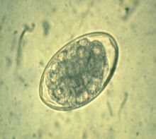
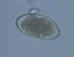
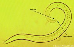
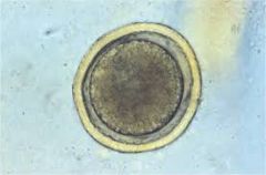
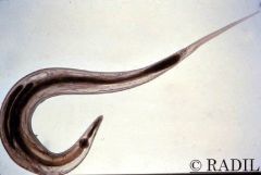
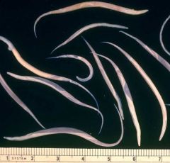
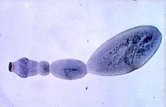
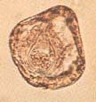
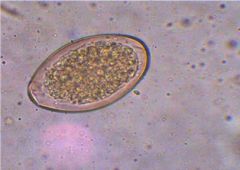
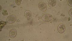
Ruminants
|
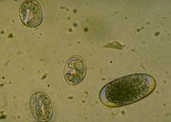
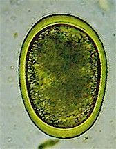
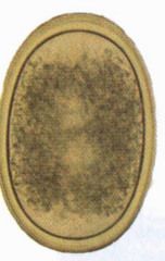
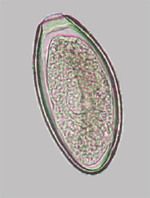
typical strongylid egg - oval, thin shelled
Ruminants - Ostertagia, Haemonchus, Cooperia, Chabertia, Bunostomum (hookworm) |
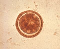
|
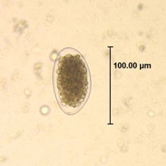
cattle, sheep, goats, horses
|
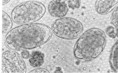
Trichostrongylus axei
typical strongylid egg - oval, thin walled |
|
sheep, goats, cattle
|
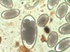
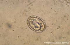
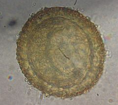
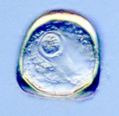
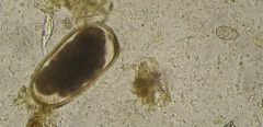
Nematodirus (large egg)
Much larger than typical strongylid eggs |
|
Cattle, Sheep, Goats, Deer
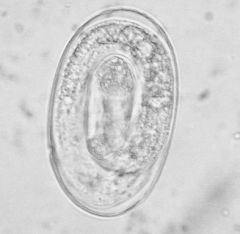
|
Dictyocaulus
thin shell, L1 in egg passed in faeces D. viviparus - cattle D. filaria - sheep, goats D. eckerti - deer |
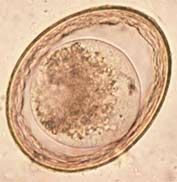
|

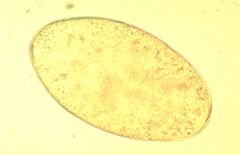
Horses
|
typical strongylid egg - oval, thin shelled
Horses - Strongylus spp. Triodontophorus, Oesophagodontus, Cyathostominae |
|

Pigs
|
typical strongylid egg - oval, thin shelled
Pigs - Hyostrongylus rubidus, Oesophagostomum |
|

Cats
|
typical strongylid egg - oval, thin shelled
Cats - Ollulanus tricuspis |
|

|
typical strongylid egg - oval, thin shelled
|
|
Poultry and other birds
|
Syngamus trachea
bipolar opercula (plugs) at each end otherwise resemble typical strongylid eggs |
|
Dog (rarely cat)
|
Larger eggs - Uncinaria stenocephala
Smaller eggs - Ancylostoma caninum Both hookworms typical strongylid eggs, size differentiation |
|
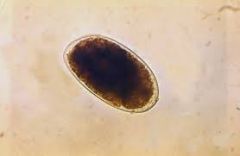
Pig, passed in urine
|
Stephanurus dentatus
|
|
pigs
|
Metastrongylus
lungworm of pigs L1 in egg |
|
sheep, goats
|
Muellerius capillaris
Lungworm eggs hatch in lungs, L1 passed in faeces. |
|

deer
|
Elephostrongylus cervi
adults in IM connective tissue resembles M. capillaris L1 passed in faeces |
|

cat
|
Aelurostrongylus abstrusus
L1 with spine in faeces lungworm |
|
Sheep, goat, pig, horse
|
Strongyloides spp smaller eggs (large egg typical stronylid.
S. papillosus - sheep, goat S. ransomi - pig S. westeri - horse L1 laid and passed in faeces |
|
Pig
|
Ascaris suum
egg containing single zygote when passed in faeces thick shelled, outer proteinaceous coat (sticky) |
|
man
|
Ascaris lumbricoides
egg containing single zygote when passed in faeces thick shelled, outer proteinaceous coat (sticky) |
|
Horse
|
Parascaris equorum
typical ascarid egg |
|
Cat
|
Toxocara cati
characteristic pitted shell, not sticky |
|
Dog
|
Toxocara canis
characteristic pitted shell, not sticky |
|
Cat (dog)
|
Toxascaris leonina
egg similar to Toxocara egg but with smooth surface, also more space around zygote within egg. |
|
Chicken and other birds
|
Ascaridia galli
egg oval and smooth |
|
Poultry
|
Heterakis gallinarum
eggs smooth, thick shelled, oval |
|
Horse
|
Oxyuris equi
Eggs flattened on one side and with an operculum, sticky Female partly emerges from anus to plaster eggs around anus and perineal region |
|
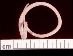
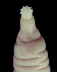
Horse colon/rectum
|
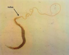
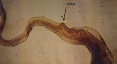
Oxyuris equi
anterior vulva long tapered tali oesophageal bulb |
|
Horse colon/rectum
|
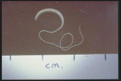
Oxyuris equi
anterior vulva long tapered tali oesophageal bulb females up to 10cm |
|
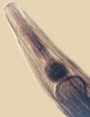
Horse colon/rectum
|
Oxyuris equi
anterior vulva long tapered tali oesophageal bulb females up to 10cm |
|

Horse faeces
|
Habronema microstoma, H. muscae
adult in stomach of horse in lumen or nodules 2 - 5cm fly IH - L1 in egg ingested by fly larvae in faeces, L3 breaks out of fly proboscis in response to moisture no migration Cutaneous habronemiasis |
|
(Ruminants) Pig, Dog, Man
|
Trichuris egg
thick walled, lemon-shaped, two opercula contain single-celled zygote in faeces |
|
Caecum. (Ruminants) Pig, Dog, Man
|
Trichuris - Whipworm
Anterior end much narrower than posterior - diagnostic |
|
Caecum. (Ruminants) Pig, Dog, Man
|
Trichuris spp
Vulva near narrowing of body |
|
|
Oxyuroid found in man (pinworm)
|
Enterobius vermicularis
|
|
Name the Species of Trichuris in pigs, dogs, and one in ruminants
|
pigs - T. suis
dogs - T. vulpis ruminants - T. ovis |
|
|
Capillaria egg
barrel shaped eggs with two opercula opercula often not in same plane |
|
|
Capillaria characteristics
|
slender
approximately even diameter along length narrow oesophagus barrel shaped, double operculate eggs |
|
|
Capillaria species occurring in chickens, stomach of dogs and cats and respiratory tract of dogs and cats
|
Chickens - C. contorta
Stomach (dog, cat) - C. erinacei (Normally hedgehogs) Respiratory tract (dog, cat) - C. aerophila |
|
muscle of pig
|
Trichinella spiralis
infective stage encysted in muscle |
|
removed from muscle of pig
|
Trichinella spiralis
infective L1 encysts in muscle fibres to be ingested by next host |
|
Faecal float, horse
|
Habronema
L1 in egg |
|
Stomach of horse
|
Habronema muscae
rare in NZ Muscoid fly IH L3 deposited on wounds can cause habronemiasis No bursa in male, caudal alae usually present usually two lips around mouth |
|
Stomach of horse
|
Draschia megastoma
similar to Habronema |
|
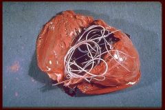
Heart, pulmonary artery, Dog
|
Dirofilaria immitis
females larviparous producing L1 (microfilariae) up to 30cm long |
|

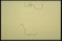
blood smear, dog
|
microfilariae, Dirofilaria immitis
Microfilariae circulate in blood, picked up by mosquito IH, L3 enter connective tissue through mosquito bite |
|
_____________ may be mistaken for _____________
|
Dipetalonema reconditum microfilariae
Dirofilaria immitis microfilariae |
|
|
Principal morphological features of a taeniid cestode
--Scolex |
Adults in SI
Scolex has 4 suckers (no hooks) and a fixed rostellum armed with 2 rows of hooks |
|
|
Principal morphological features of a taeniid cestode
-- Segments |
One set of genitalia per proglottid
Genital pores alternate irregularly between sides of the segment gravid segments longer than wide eggs in uterus which persists as a sac with many lateral pockets |
|
|
Principal morphological features of a taeniid cestode
-- Larva |
Depending on species may be cysticercus, coenurus or hydatid cyst
NEVER a cysticercoid |
|
|
Cysticercus
|
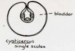
fluid filled bladder like cyst
one protoscolex invaginated vertebrate IH only |
|
|
Cysticercoid
|
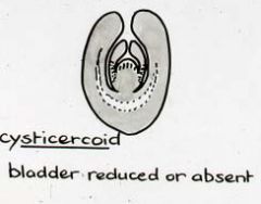
cyst contains no fluid and closely enfolds one protoscolex
invertebrate IH |
|
|
Coenurus
|
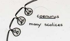
fluid filled cyst lined by germinal membrane from which are budded many protoscolices - multiplication occurs
protoscolices remain attached to germinal layer and are not grouped in capsules vertebrate IH |
|
|
Hydatid Cyst
|
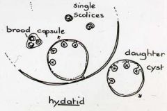
fluid filled cyst lined by germinal membrane, bounded by a thick laminated layer
Germinal membrane produces many protoscolices usually in small, thin walled sacs of germinal epithelium - Brood capsules Hydatid sand - sediment of scolices, brood capsules and daughter cysts |
|
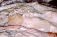
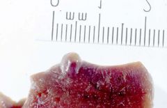
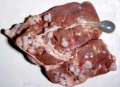
abdominal cavity, sheep/goat/cattle
|
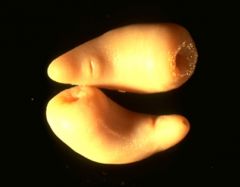
Cysticercus tenuicollis
(Taenia hydatigena) - DH dog |
|
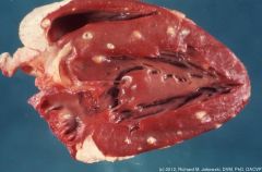
Heart/diaphragm, Sheep, goats
|
Cysticercus ovis
(Taenia ovis) - DH dog |
|
Peritoneal cavity, rabbit/hare
|
Cysticercus pisiformis
(Taenia pisiformis) - DH dog |
|
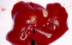
Liver, mice/rats
|
Cysticercus fasciolaris
(Taenia taeniaeformis) - DH cat |
|
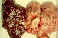
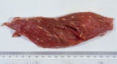
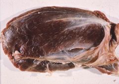
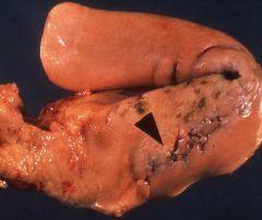
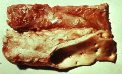
IM connective tissue (tongue, heart, masseter), cattle
|
Cysticercus bovis
(Taenia saginata) - DH human |
|
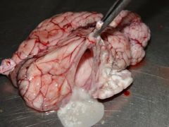
CNS, commonly cerebrum, sheep/goat/cattle
|
Coenurus cerebralis
(Taenia multiceps) - DH dog |
|
|
Coenurus found in IM connective tissue, rabbit/hare
|
Coenurus serialis
(taenia serialis) - DH dog |
|
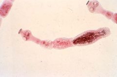
Dog, SI
|
Taeniid cestode
-scolex has 4 suckers without hooks, hooked rostellum -gravid proglottids wider than are long -one set of genitalia per segment -genital pores alternate irregularly between sides -eggs persist in uterus |
|
Dog, SI
|
Taeniid cestode
-scolex has 4 suckers without hooks, hooked rostellum -gravid proglottids wider than are long -one set of genitalia per segment -genital pores alternate irregularly between sides -eggs persist in uterus |
|
Dog, SI
|
Echinococcus granulosus
-3-4 segments, length of gravid segment more than half total length -otherwise similar to other taeniids |
|
Dog, SI
|
Echinococcus granulosus
-3-4 segments, length of gravid segment more than half total length -otherwise similar to other taeniids |
|
Fox, Dog, SI. NOT in NZ
|
Echinococcus multilocularis
Northern Hemisphere IH rodents Humans may be infected with larval stage, multilocular hydatid cyst in liver, lungs. Often fatal |
|
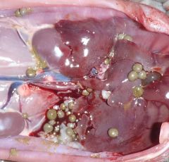
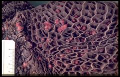
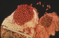
Cyst found in liver/lung of sheep
|
Hydatid cyst - E. granulosus
Hydatid sand in bottom of cyst - brood capsules, protoscolices and daughter cysts Cyst wall has three layers: --inner germinal layer --middle laminated layer --outer adventitious layer derived from host |
|
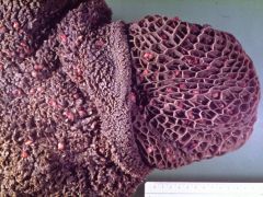
Sheep liver
|
Hydatid cysts - E. granulosus
-hydatid sand -three layers in wall -most common in liver and lungs |
|
Sheep liver, lungs
|
Hydatid cysts - E. granulosus
-hydatid sand -three layers in wall -most common in liver and lungs |
|
SI dog/cat
|
Dipylidium caninum
Scolex: -retractible rostellum with 4-6 rows thorn-like hooks |
|
SI dog/cat
|
Dipylidium caninum
Immature proglottids -two sets of male and female organs -genital pore at each lateral border |
|
SI dog/cat
|
Dipylidium caninum
Mature proglottids -two sets of male and female organs -genital pore at each lateral border |
|
SI dog/cat
|
Dipylidium caninum
Gravid proglottid -eggs contained in groups called egg capsules -each capsule holds up to 20 or so eggs |
|
From gravid segment of cestode in SI of Dog/cat
|
Egg capsules of Dipylidium caninum
eggs contained within these in the gravid proglottid up to about 20 eggs per capsule |
|
Faecal float, Dog/Cat
|
Hexacanth egg, Dipylidium caninum
-three pairs of hooks on hexacanth embryo -embryophore -outer egg membrane |
|
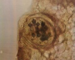
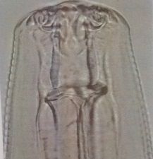
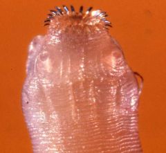
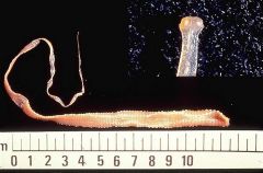
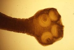
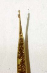
Near ileo-caeco-colic junction of intestines of Horse
|
Anoplocephala perfoliata
Strobila broadens rapidly behind scolex Scolex has no rostellum or hooks, beneath each sucker is a lappet Segments much much wider than are long. |
|
Ileo-caeco-colic junction of Horse
|
Anoplocephala perfoliata
very common in horse sometimes cause severe inflammation of this area |
|
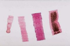
Termminal SI/beginning of LI, Horse
|
Anoplocephala perfoliata
Scolex showing: -suckers -lappets -no rostellum or hooks |
|
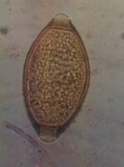
Faecal float, Horse
|
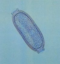
Anoplocephala perfoliata egg
-Hexacanth embryo -pyriform apparatus extending from embryo -egg shell |
|
Faecal float, Horse
|
Anoplocephala perfoliata
-Hexacanth embryo -pyriform apparatus extending from embryo -egg shell -two angles |
|
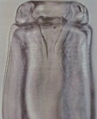
SI, Sheep/Goat
|
Moniezia expansa
Scolex showing: -4 suckers -no rostellum or hooks |
|
SI, Sheep/Goat
|
Moniezia expansa
Mature proglottids Two sets of reproductive organs per segment |
|
SI Sheep/Goat
|
Moniezia expansa
Gravid proglottids Two sets of organs per segment |
|
Faecal float, Sheep/Goat
|
Moniezia egg
Hexacanth Three angles pyriform apparatus |
|
Pig, SI
|
Macracanthorhynchus hirudinaceus
male 10cm, female 35cm proboscis - "thorny headed worm" rare in NZ |
|
Pig, SI
|
Macracanthorhynchus hirudinaceus
male 10cm, female 35cm proboscis - "thorny headed worm" rare in NZ |
|
|
Features of a Digenean Trematode
|
Unsegmented
May be flattened dorso-ventrally, or round and fleshy Complex tegument armed with small spines No body cavity - organs supported by cellular parenchyma Oral and ventral suckers Pharynx, Oesophagus, blind ending Caeca Flame cells Hermaphrodite |
|
|
Sexual features of a Digenean Trematode
|
Male:
-two testis -two vas deferens join and open at the genital pore -cirrus Female: -Ovary, oviduct, ootype, uterus, genital pore -Vitelline glands and Mehlis gland |
|
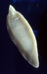
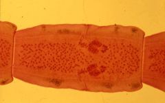
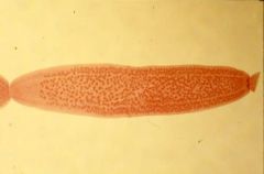
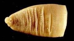
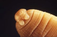
Bile duct, Ruminant
|
Fasciola hepatica
up to 3cm long flattened, ovate body fawn to mottled grey or black IH - lymnaeid snails (freshwater) |
|
Bile duct, Ruminants
|
Fasciola hepatica
up to 3cm long flattened, ovate body fawn to mottled grey or black IH - lymnaeid snails (freshwater) |
|
Bile duct, Ruminant
|
Fasciola hepatica
Cirrus protruding between suckers up to 3cm long flattened, ovate body fawn to mottled grey or black IH - lymnaeid snails (freshwater) |
|
Bile duct, Ruminant
|
Fasciola hepatica - suckers
up to 3cm long flattened, ovate body fawn to mottled grey or black IH - lymnaeid snails (freshwater) |
|
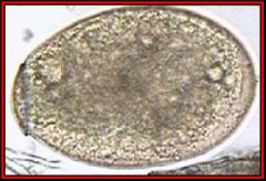
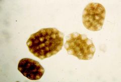
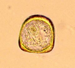
Ruminant faecal float
|
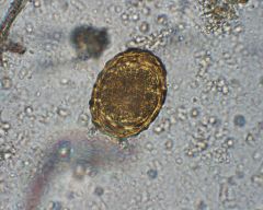
Fasciola hepatica
yellow-brown in colour (from bile fluids) Single operculum Contain morula when passed |
|
Ruminant
|
Fasciola hepatica
yellow-brown in colour (from bile fluids) Single operculum Contain morula when passed |
|
Ruminant
|
Fasciola hepatica
yellow-brown in colour (from bile fluids) Single operculum Contain morula when passed |
|
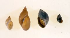
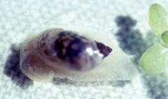
Which freshwater snail species may be a host for Fasciola hepatica?
|
The two on the left
Left-most: Lymnaea tomentosa (native) - IH Left-middle: Lymnaea columella (introduced) - IH Right-middle: Physa/Physastra Right-most: Potamopyrgus |
|
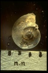
Which Freshwater snail is this?
|
Lymnaea columella
L for Lymnaea basal whorl large but longer relative to width than L. tomentosa Spire of shell considerably longer - conical shape Fine striations perpendicular to growth lines up to 15mm |
|
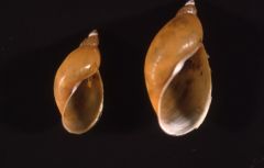
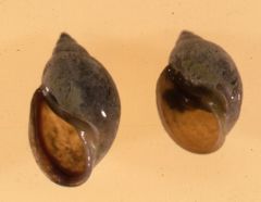
Which freshwater snail is this?
|
Lymnaea columella
L for Lymnaea Usually black when alive basal whorl large but longer relative to width than L. tomentosa Spire of shell considerably longer - conical shape Fine striations perpendicular to growth lines up to 15mm Short, flattened, triangular tentacles, no operculum (opening closed only by foot), characteristic of Lymnaeids |
|
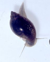
Which freshwater snail is this?
|
Lymnaea tomentosa
Mottled golden-brown and black when alive Basal whorl large and spire of shell short in comparison No striations in shell up to 8mm long Tentacles short, flattened, triangular tenticles, no operculum (opening closed only by foot), characteristic of Lymnaeids |
|
Which freshwater snail are these?
|
Lymnaea tomentosa
Mottled golden-brown and black when alive Basal whorl large and spire of shell short in comparison No striations in shell up to 8mm long Tentacles short, flattened, triangular tenticles, no operculum (opening closed only by foot), characteristic of Lymnaeids |
|
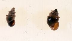
What freshwater snail is this?
|
Lymnaea tomentosa
Mottled golden-brown and black when alive Basal whorl large and spire of shell short in comparison No striations in shell up to 8mm long Tentacles short, flattened, triangular tenticles, no operculum (opening closed only by foot), characteristic of Lymnaeids |
|
Which freshwater snail is this?
|
Lymnaea tomentosa
Mottled golden-brown and black when alive Basal whorl large and spire of shell short in comparison No striations in shell up to 8mm long |
|
Which freshwater snail do these belong to?
|
Physa/Physastra
Opens to the left |
|
Which freshwater snail is this?
|
Physa/Physastra
Shell opens to left Tentacles long and thin |
|
Which freshwater snail genera do these belong to?
|
Potamopyrgus
Small, less than 4mm, black when alive Shell steeply conical with small, almost circular opening closed by an operculum Shell often bears minute spines on each whorl |
|
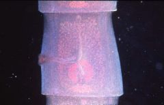
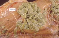
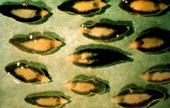
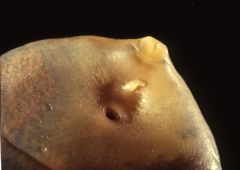
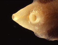
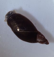
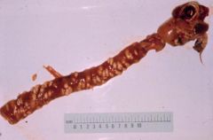
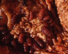
Reticulum/Rumen of Ruminants
|
Calicophoron calicophorum
1-1.5cm long bright pink when alive Firmly attach via acetabulum (ventral sucker) which is at the posterior end of the body and is very large. Plug of epithelium drawn into it to ingest. |
|
Reticulum/Rumen, Ruminant
|
Calicophoron calicophorum
1-1.5cm long bright pink when alive Firmly attach via acetabulum (ventral sucker) which is at the posterior end of the body and is very large. Plug of epithelium drawn into it to ingest. |
|
Reticulum/Rumen, Ruminant
|
Calicophoron calicophorum
1-1.5cm long bright pink when alive Firmly attach via acetabulum (ventral sucker) which is at the posterior end of the body and is very large. Plug of epithelium drawn into it to ingest. |
|
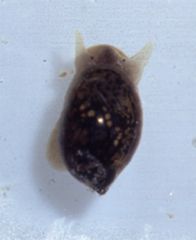
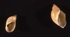
What is this snail and what is it an intermediate host for?
|
Gyraulus corinna
Calicophoron calicophorum Flat-spiralled, freshwater snail Found mainly in streams, sometimes ponds. Common in NZ |
|
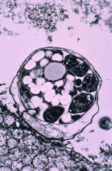
Ruminants, Birds
|
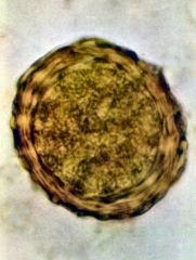
Eimerian oocyst
4 sporocysts per oocyst 2 sporozoites per sporocyst |
|
Cats, Dogs
|
Isosporan oocyst
2 sporocysts per oocyst 4 sporozoites per sporocyst |
|
Small intestine, calf dead from diarrhoea
|
Cryptosporidium spp
invasion of brush border, looks like is sitting on it but is actually in the cell. |
|
Brush border of SI of calf with diarrhoea
|
Cryptosporidium gametocytes
|
|
|
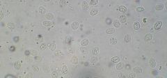
Coccidia oocysts
one partially sporulated in corner |
|
Ruminant faecal sample
|
Nematodirus and coccidia oocyst
note size difference |
|
|
Cryptosporidium gametocyte
immediately infective contains 4 free sporozoites |
|
What sort of faecal smear is this and what does it show?
|
Modified Ziehl-Nielsen technique
Cryptosporidium oocysts |
|
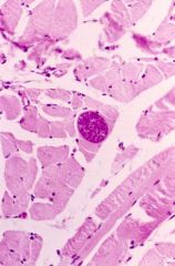
Pig muscle
|
Macrocysts
Sarcocystis spp |
|
sheep oesophagus
|
Macrocysts
Sarcocystis spp |
|
beef muscle
-little white lines |
Macrocysts
Sarcocystis spp |
|
Mouse
|
Macrocysts
Sarcocystis spp |
|
Duck
|
Macrocysts
Sarcocystis spp |
|
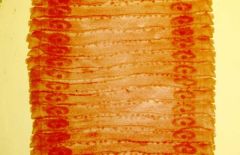
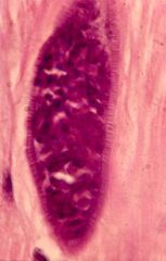
Muscle fibre, sheep
|
Microcysts, thin walled
Sarcocystis spp |
|
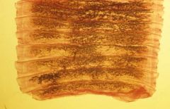
Muscle fibre, sheep
|
Microcyst, thick walled
Sarcocystis spp |
|
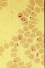
Blood smear, Ruminant erythrocytes
|
Babesia intracellularly
|
|
Blood smear, ruminant erythrocytes
|
Babesia intracellularly.
|
|
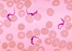
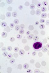
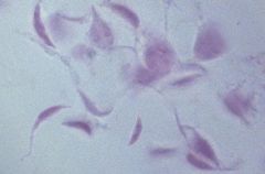
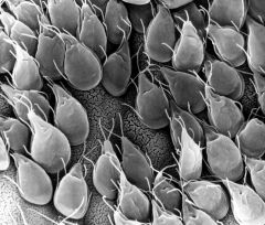
Blood smear
|
Trypanonsome
note single flagellum attached by undulating membrane |
|
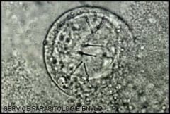
SI Humans, dogs, cats, ruminants
|
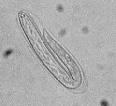
Giardia
pear/leaf/tear-drop shaped |
|
SI Humans, dogs, cats, ruminants
|
Giardia
pear/leaf/tear-drop shaped large ventral disc two nuclei spit by axostyle looks like face |
|
SI Humans, dogs, cats, ruminants
|
Giardia
large ventral disc in contact with mucosa (both pink) |
|
faecal sample
|
Giardia cysts
oval, 8-14 x 5-10microns partial binary fission so 4 nuclei visible hard to detect |
|
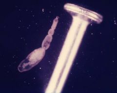
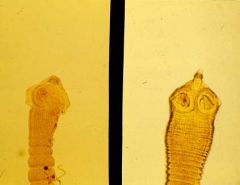
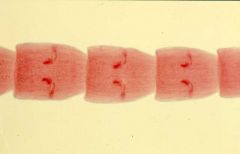
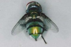
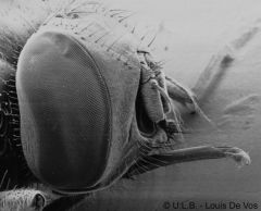
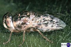
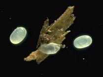
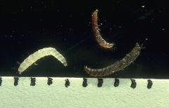
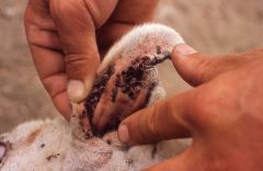
Which species of fly is this?
|
Calliphora stygia
large, golden brown common primary strike fly |
|
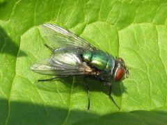
Which genus of fly is this?
|
Lucilia
L. sericata, L. cuprina smaller than Calliphora stygia coppery green in colour Primary strike flies |
|
Which species of fly is this?
|
Chrysomya rufifacies
coppery green, superficially resembles Lucilia but has dark bands on abdominal segmetns. Main secondary strike fly |
|
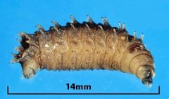
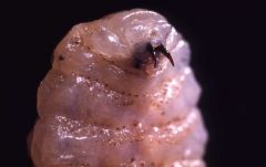
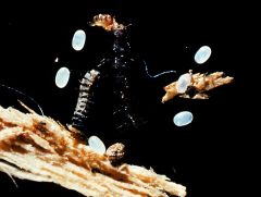
Which species of fly larva is this?
|
Chrysomya rufifacies
main Secondary strike fly pronounced bristles on larva (are 'hairy') |
|
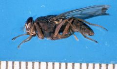
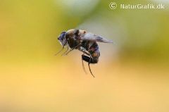
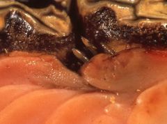
Which species of fly is this?
How is it distinguished from other flies? |
Stomoxys calcitrans
Bites Rigid black proboscis projects forward from mouth, which it projects vertically when feeding. |
|
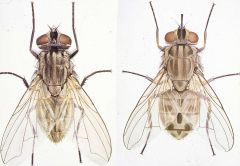
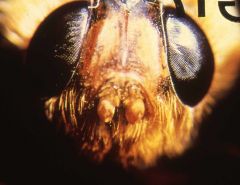
Which fly species are these?
How may they be distinguished from each other? |
Musca domestica - left
--sponging mouthparts, ventrally projecting --does not bite Stomoxys calcitrans - right --piercing mouthparts --proboscis projects forward --bites, sucks blood |
|
Which species of fly is this?
How is it distinguished from other flies? |
Stomoxys calcitrans
forward projecting proboscis bites |
|
Which genera of fly is this?
Found flying around legs of horses |
Gasterophilus
G. intestinalis, G. nasalis Adults bee-like Females cement eggs on hairs of horse's coat |
|
Which genera of fly is this?
Found flying around legs of horses |
Gasterophilus spp.
Rudimentary mouthparts, adults do not feed |
|
Which genera of fly is this?
Mouth of Horse |
Gasterophilus spp
2nd instar larva |
|
Which genera of fly is this?
Horse tongue |
Gasterophilus (intestinalis)
1st larval instar in tongues for 3-4 weeks |
|
Which genera of fly is this?
Stomach of Horse |
Gasterphilus spp
3rd larval instar in aglandular part of stomach (G. intestinalis) or duodenal antrum (G. nasalis) |
|
Which genera of fly is this?
Stomach of horse |
Gasterphilus spp
3rd larval instar in aglandular part of stomach (G. intestinalis) or duodenal antrum (G. nasalis) |
|
Which genera of fly is this?
Found flying around sheep head |
Oestrus ovis
Lays first instar larva on external nares of sheep |
|
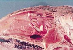
Which genera of fly is this?
Sheep nasal cavity and sinus |
Oestrus ovis
second larval instar in nasal cavity of sheep third larval instar in frontal sinus |
|
Which genera of fly is this?
Found in Sheep nasal cavity and sinus |
Oestrus ovis
Fly lays first instar larva on external nares of sheep second larval instar in nasal cavity third larval instar in frontal sinus |
|
What genera of Diptera this?
Back of Cattle/Deer |
Hypoderma spp.
H. bovis, H. lineatum, H. diana third larval instar lives under skin of animal, usually on back. Cuts hole in skin to breathe Damaging to hides |
|
What genera of Diptera this?
Back of Cattle/Deer |
Hypoderma spp.
H. bovis, H. lineatum, H. diana third larval instar lives under skin of animal, usually on back. Cuts hole in skin to breathe Damaging to hides |
|
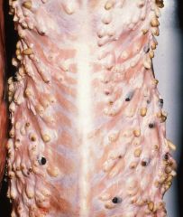
What genera of Diptera this?
Spinal canal of Cattle/Deer |
Hypoderma spp.
H. bovis, H. lineatum, H. diana larvae migrate through body to overwintering areas: --spinal canal --submucosa of oesophagus |
|
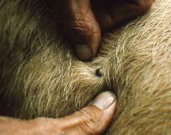
What species is this?
|
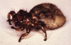
Melophagus ovinus -- sheep ked
Wingless/reduced wings Adults suck blood 4-6mm live in fleece at skin level |
|
What species is this?
|
Melophagus ovinus -- sheep ked
Wingless/reduced wings Adults suck blood 4-6mm live in fleece at skin level Larviparous, third larval instar laid 1-2cm from skin in wool pupation immediately, lasts 3-5 weeks |
|
What species is this?
Found on wool fibre of sheep |
Melophagus ovinus -- sheep ked
Wingless/reduced wings Adults suck blood 4-6mm live in fleece at skin level Larviparous, third larval instar laid 1-2cm from skin in wool pupation immediately, lasts 3-5 weeks |
|
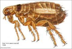
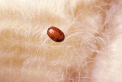
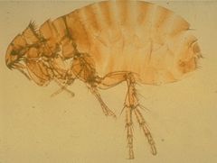
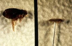
What is this?
|
Flea
Laterally flattened Wingless Suck blood Six long legs adapted for jumping |
|
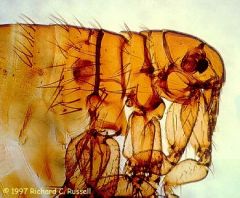
What genera is this?
Found on cats and dogs |
Ctenocephalides
C. felis (more common) or C. canis Both genal and pronotal ctenidia |
|
What species is this?
found on pig and human |
Pulex irritans
No ctenidia Mainly seen around free range pigs these days |
|
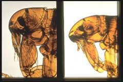
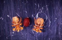
Which species of flea are these?
|
Left: Ctenocephalides canis
Right: Ctenocephalides felis |
|
What are these?
Found in environment of dog/cat |
Flea eggs
White, shiny, subspherical 0.5mm diameter laid on or off the host but if laid on, fall of shortly after. Widely distributed |
|
What are these?
Found in environment of dog/cat |
Flea eggs:
White, shiny, subspherical, 0.5mm diameter laid on or off the host but if laid on, fall of shortly after and are widely distributed Flea larva: resembles a bristly fly maggot three larval instars |
|
What are these?
Found in environment of cat/dog |
Flea larva:
resembles a bristly fly maggot three larval instars |
|
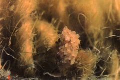
What is this?
Found in carpet of house with a dog |
Flea cocoon
silk, binds to grit/dirt pupation takes >10 days |
|
What are these insects?
|
Left: flea
--laterally flattened Right: louse --dorsoventrally flattened |
|
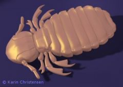
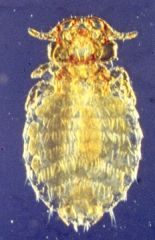
What sort of louse is this?
|
Biting louse:
--head wider than thorax --mouthparts ventral --chew skin, hair, feathers |
|
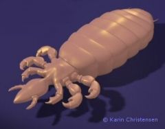
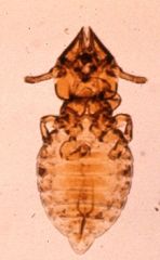
What sort of louse is this?
|
Sucking louse:
--head narrower than thorax --mouthparts project anteriorly --suck blood/lymph |
|
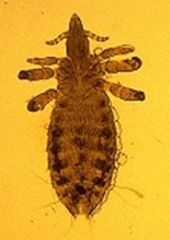
Found on Horse
|
Bovicola equi
Biting louse - head wider than thorax Common |
|

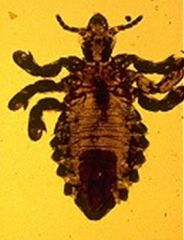
Found on horse
|
Haematopinus asini
Sucking louse - head narrower than thorax Common |
|
Found on cattle
|
Linognathus vituli
sucking louse - head narrower than thorax Very common, especially on dairy breeds |
|

Found on cattle
|
Bovicola bovis
Biting louse, head wider than thorax common |
|

Found on Sheep
|
Bovicola ovis
Biting louse - head wider than thorax common |
|
Found on pig
|
Haematopinus suis
sucking louse - head narrower than thorax very common |
|

Found on dog
|
Linognathus setosus
sucking louse - head narrower than thorax common |
|
Found on dog
|
Trichodectes canis
Biting louse - head wider than thorax common |
|
Found on cat
|
Felicola subrostratus
biting louse - head wider than thorax common |
|
What is this?
cattle, sheep, deer |
Haemaphysalis longicornis
No males, females are parthenogenic Egg - larvae (jan-mar) - nymph (aug-sep) - adult (nov-jan) lifecycle takes about a year most damaging to young |
|
What are these?
|
Haemaphysalis longicornis
unfed larvae/nymphs/adults larvae - jan-mar nymphs - aug-sept adults - nov-jan |
|
What is this?
cattle, sheep, deer |
Haemaphysalis longicornis
adults found on host mid november to mid january |
|
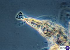
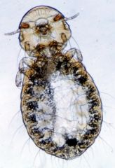
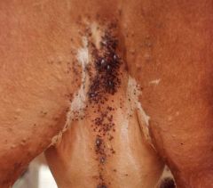
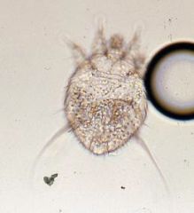
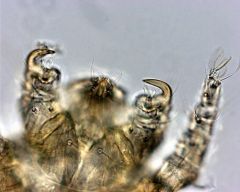
Is this a Chorioptes or Psoroptes mite?
|
Chorioptes
--pedicels short and unjointed (the sucker on the end) --non-burrowing --long posterior legs that extend well beyond the body margin --occur on herbivores --scrotal mange in rams --mange in feathered draught horses |
|
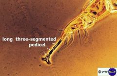
Is this a Chorioptes or Psoroptes mite?
|
Psoroptes
--long, three-segmented pedicel (sucker on end of leg) --non-burrowing --long posterior legs that extend well beyond the body margin --occur on herbivores --otitis externa |
|
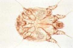
Found in stratum corneum of skin
Dogs, pigs, man in NZ |
Sarcoptes
--approximately spherical, ~0.5mm diameter --tunnelling and feeding activities can be very irritating, causing sarcoptic mange |
|
Found in stratum corneum of skin
Dogs, pigs, man in NZ |
Sarcoptes
--approximately spherical, ~0.5mm diameter --tunnelling and feeding activities can be very irritating, causing sarcoptic mange |
|
Guinea Pigs
|
Trixacarus caviae
Similar to sarcoptes |
|
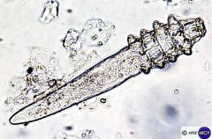
Hair follicle
Virtually any animal |
Demodex spp
--cigar shaped, small --majority of infestations cause not detectable lesions --in dogs especially, progressive demodectic mange may occur which can be hard to cure if neglected --bacteria may be involved in mange |
|
Skin of rabbits, cats, dogs
|
Cheyletiella spp
--easily identifiable by large palpal hooks and feathered bristles --feed on superficial epidermal cells and debris --can cause severe irritation and inflammation although often cause no clinical signs |
|
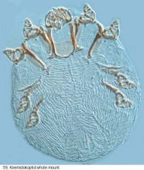
Burrowed in keratinised skin
Birds |
Cnemidocoptes
--morphologically and ecologically resemble sarcoptes |