![]()
![]()
![]()
Use LEFT and RIGHT arrow keys to navigate between flashcards;
Use UP and DOWN arrow keys to flip the card;
H to show hint;
A reads text to speech;
55 Cards in this Set
- Front
- Back
|
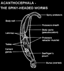
Phylum Acanthocephala -Leptorhynchoides thecatus |
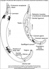
spiny headed worms -live in the gut of vertebrates & earlier in their life cycle -within invertebrates. |
|
|
Leptorhynchoides thecatus life cycle |
1. Arthropods ingests egg with algae 2. Cystacanth develop in haemocoel 3. Fish (paratenic host) eats arthropods 4. Amphipod or paratenic host gets eaten 5. Juvenile released from amphipod. Adult acanthocephalan attached to gastric cecum. 6. Eggs released into water. 7. Egg coat fibers entagled with algea. 8. Cycle starts all over again! |
|
|
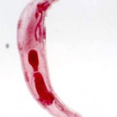
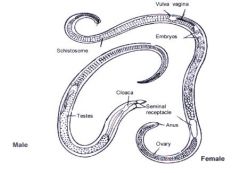
Leptorhynchoides thecatus male -proboscis -testes -bursa -saefftigen's pouch -cement gland -cement reservoir |
|
|
|
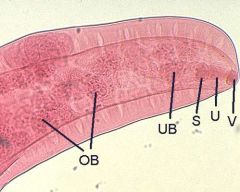
Leptorhynchoides thecatus female -ovarian balls -uterine bell -uterus -vulva |
|
|
|
Leptorhynchoides thecatus male |
|
|
|
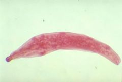
Neoechinorhynchus rutili male |
|
|
|
Neoechinorhynchus rutili male |
|
|
|
Neoechinorhynchus rutili female |
|
|
|
Neoechinorhynchus rutili female |
|
|
|
Nematomorpha (horsehair worms) life cycle |
Intermediate host: Aquatic invertebrate Definitive host: Cricket, crab, shrimp *Infects insects* ^ 1. Larvae in bottom of water, find aquatic invertebrate to encyst. (Paratenic host) 2. Cricket eats aquatic animal, worm grows into adult worm in gut. |
|
|
Leeches, what are they? |
They are annelids -many species are not parasitic (blood sucking) leeches -they are detritivores or predators |
|
|
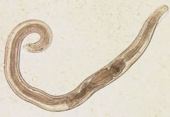
Ascaris lumbricoides life cycle -host organism? |
Host: Humans 1.Female worms produces eggs-passed in feces. (fertilized eggs embryonate & become infective) 2. Infective eggs are swallowed - larvae hatch, mature in lungs, ascend bronchial tree to the throat, & are swallowed. 3. Develop into adult worms in small intestine. 4. Adult worms lives in small intestine |
|
|
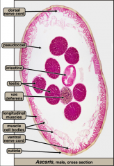
Ascaris lumbricoides male -cuticle -pseudocoel -longitudinal muscles -intestine -testes |
-has spicule at end |
|
|
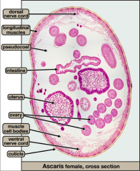
Ascaris lumbricoides female -pseudocoel -longitudinal muscles -intestine -uterus -ovaries |
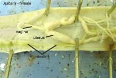
-has obvious genital pore and vagine -longer than male |
|
|
Ascaris lumbricoides cross section of male |
|
|
|
Ascaris lumbricoides cross section of female |
|
|
|
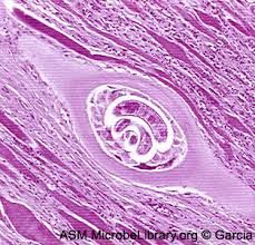
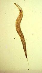
Trichinella spiralis what are they? -characteristics |
-Roundworm -males: 1.5mm females: 3mm -short esophagus -have stichosome, pseudobursa, papilla -no copulatory spicule -1 gonad -uterus contains fully developed eggs |
|
|
Trichinella spiralis (roundworm) life cycle |
-Acquired by ingesting meat containing cysts of Trichinella. 1. Larvae are released from cysts & invade small intestine where they develop into adult worms. 2. After 1 week, females release larvae that migrate to muscles where they encyst. |
|
|
Trichinella spiralis (roundworm) encysted larvae |
|
|
|
Trichinella spiralis (roundworm) female |
|
|
|
Trichinella spiralis (roundworm) male & female |
|
|
|
Nematode: -Trichuris trichiura what are they? |
whipworms! -complete digestive system -mature eggs (footballs) -conspicous walls w/ abopercular knob -stichosome!! |
|
|
Trichuris trichuria (whipworm) life cycle |
Host: Humans 1. Unembryonated eggs passed in feces. 2. 2-Cell Stage., Advanced cleavage. 3. Embryonated eggs are ingested by self-contamination of hands/food. 4. Eggs hatch in small intestine & release larvae that mature & become adults in colon. 4. Adult worms live in cecum. 4. Females oviposit and cycle starts all over again. |
|
|
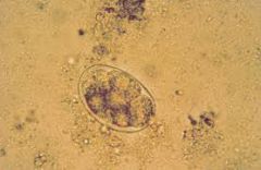
Trichuris trichiuria eggs |
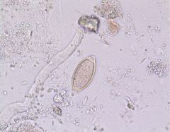
50-54 um X 22-23 um conspicuous wall abopercular knobs at ends |
|
|
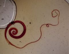
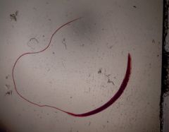
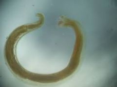
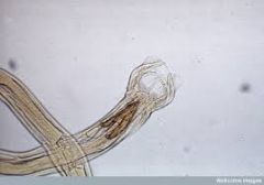
Trichuris trichiura (whipworm) |
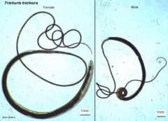
30-50 mm long esophagus surrounded by stichocytes (glands) single gonad |
|
|
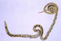
Trichuris trichiura (whipworm) male |
single spicule |
|
|
Trichuris trichiura female |
|
|
|
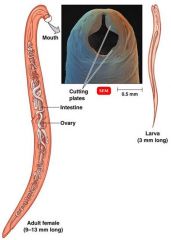
Hookworms life cycle (Necator americanus & Ancylostoma braziliensis) |
Host: Humans ---- Cause blood loss 1. Eggs in feces 2. Rhabditiform larva hatches & develops into filariform larva that are infective. 4. On contact w/ human host, larvae penetrate skin and travel to heart & lungs thru blood vessels. 5. Larvae reach small intestine & mature into adults. 6. Adult worms live in small intestine. |
|
|
Necator americanus (hookworm) male |
|
|
|
Necator americanus (hookworm) female |
|
|
|
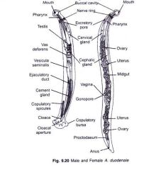
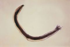
Ancylostoma duodenale (hookworm) male & female |
|
|
|
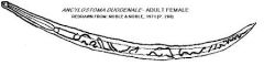
Ancylostoma duodenale female |
|
|
|
Ancylostoma duodenale (hookworm) male |
|
|
|
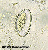
Ancylostoma caninum (hookworm) egg |
-thin egg shell |
|
|
Oswaldocruzia (trichostrongyle) male |
have copulatory bursa & spicules |
|
|
Oswaldocruzia (trichostrongyle) female |
ovijector |
|
|
Enterobius vermicularis (pinworm) egg |
-very tiny on 4x |
|
|
Enterobius vermicularis (pinworm) male |
-large esophageal bulb -sharp, pointed tail -alae at anterior end |
|
|
Enterobius vermicularis female |
-prominent esophageal bulb |
|
|
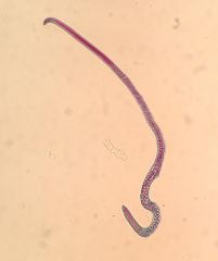
Dirofilaria immitis microfilariae |
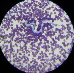
-no sheath -cephalic end pointed -tail straight w/ the end pointed |
|
|
Dirofilaria immitis (microfilariae) life cycle |
1. Mosquito takes a blood meal (larvae enters bite wound) into dog. 2. Adults in pulmonary arteries. they produce microfilaria. 3. Mosquito takes a blood meal, ingests microfilaria. |
|
|
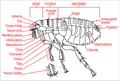
ARTHROPOD: Cimex lectularius (bed bug) Flea: head, thorax, abdomen, pronatal comb, genal comb, femur, tibia, tarsus |
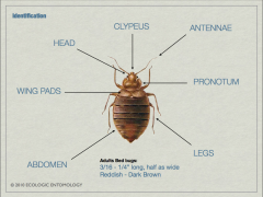
-Antennae has 4 segments each -Reddish, brown segmented body -wingless! -(female contains eggs in abdomen-very obvious under microscope) -traumatic insemination |
|
|
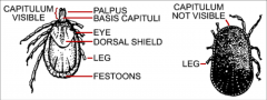
ARACHNID: Dermacentor variabilis TICK: gnathosoma, idiosoma, scutum, festoon, hypostome, pedipalp, hallers organ |
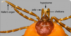
-Body composed of 18 somites -6 unit prosoma & 12 unit opisthosoma -chelicerae of 2/3 podomeres -Pedipalps of six podomeres -Haller's organ: present on 1st tarsi -contains hair like sensory structures |
|
|
ARTHROPOD: Amblyomma americanum |
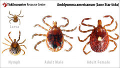
-longer gnathostome!! |
|
|
ARTHROPOD: Ctenocephalides canis Flea: head, thorax, abdomen, pronatal comb, genal comb, femur, tibia, tarsus |
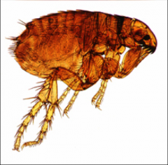
-dog flea |
|
|
Cimex lectularius (bedbug) |
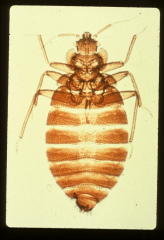
-pronotum about 2 1/2 times broader than long. -wide pronotum! -wingless |
|
|
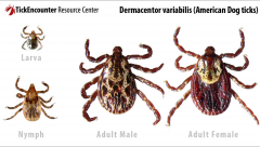
Dermacentor variabilis |
|
|
|
Dermacentor variabilis (American Dog Tick) Male & Female |
|
|
|
Cnetocephalides canis (dog flea) Flea: head, thorax, abdomen, pronotal comb, genal comb, femur, tibia, tarsus |
|
|
|
Prevalence |
-# of hosts infected w/ 1 or more individuals of a particular parasite species divided by # of hosts examined for that parasite species. -requires only detection of the presence of parasite -percent infected ** # of infected hosts of a particular parasite / # of examined hosts for that particular parasitic species |
|
|
Abundance |
-# of individuals of a particular parasite in/on a single host regardless of whether or not the host is infected "# of parasites in a host that is/isnt infected" |
|
|
Intensity (of infection) Mean Intensity |
-# of individuals of a particular parasite species in a single infected host -Syn: "parasite load" Mean Intensity= -AVG intensity of a particular species of parasite among the infected members of a particular host species -total # of parasites of a particular species found in a sample divided by # of hosts infected with that parasite |
|
|
Flotation technique: Simple Flotation |
-Sheather's Solution, centrifuged for 5 min -used to concentrate helminth ova Adv: detection of nematode & non-operculate cestode eggs Dis: Doesn't concentrate more operculate eggs or schistosomes. |
|
|
Flotation Technique: The Fecalyzer |
-Fecasol flotation medium, green insert Adv: higher densities of flotation solution (FECASOL) is capable of floating heavier parasite eggs Dis: since Fecasol is a higher density of flotation solution, it allows other fecal particles to float to the top, which makes it difficult to examine under microscope. May obscure thin shelled eggs. |
|
|
Flotation Technique: Sedimentation |
Tap water, gravity, long test tube -Top, bottom layer of sediment Adv: Can detect trematode eggs, which are larger and heavier than cestode eggs. |