Reading...
![]()
Play button
![]()
Play button
![]()
Use LEFT and RIGHT arrow keys to navigate between flashcards;
Use UP and DOWN arrow keys to flip the card;
H to show hint;
A reads text to speech;
44 Cards in this Set
- Front
- Back
|
Benign conditions with a follicular pattern
|
Follicular hyperplasia
Castleman disease PTGC HIV related lymphadenopathy Rheumatoid lymphadenopathy Syphilitic lymphadenopathy |
|
|
Benign conditions with a paracortical pattern
|
Viral infections NOS
Post vaccinial lymphadenitis EBV Drug induced Dermatopathic SLE Histiocytic necrotizing lymphadenitis |
|
|
Benign conditions with a sinus pattern
|
Sinus histiocytosis
LCH Rosai Dorfman Monocytoid B cell hyperplasia Whipple disease |
|
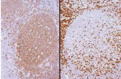
Stains?
|
CD20 left
CD3 right |
|
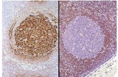
Stains?
|
CD21 left, BCL2 right
|
|
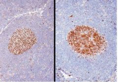
Stains?
|
BCL6 right, CD10 left
|
|
|
What are some features that distinguish follicular hyperplasia from follicular lymphoma?
|
Hyperplasia: increased mitosis, BCL2 negative, T14:18 negative, architecture preserved, GC varied, no back to back follicles
Lymphoma: Decreased mitosis, decreased macrophages, BCL2+ t15:18 positive Architecture effaced, little variation in GC, back to back follicles |
|
|
What form of castleman disease has a worse prognosis?
|
Multicentric
|
|
|
What is POEMS?
|
Syndrome associated with castleman
Polyneuropathy, organomegaly, endocrinopathy, monoclonal gammopathy, and skin abnormalities |
|
|
What are the three stages of HIV lymphadenopathy
|
Follicular hyperplasia
Follicular involution Lymphocyte depletion |
|
|
What are the histologic features of toxoplasma?
|
Follicular pattern hyperlasia
Epithelioid histiocytes near GC Monocytoid B cells in sinuses Serology confirms diagnosis |
|
|
Histologic features of infectious mononucleosis?
|
Paracortical proliferation of immunoblasts
Sinuses distended by monocytoid B cells or immunoblasts Focal necrosis/apoptosis LMP+ EBER+ DDx: DLBCL |
|
|
What markers do you see in acute EBV (serology)?
|
IGM+ IgG VCA
|
|
|
What serology do you see with remote EBV infection?
|
IgG EBNA, IgG VCA
|
|
|
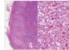
Histologic features of dermatopathic lymphadenopathy?
|
Paracortical expansion with many pale, sometimes melanin filled histiocytes
(Mycosis fungoides must be considered if confluence is present) |
|
|
Dermatopathic lymphadenitis
|
|
|
Clinical presentation of kikuchi?
|
Young asian women, cervical nodes, serology negative
|
|
|
Architecture/cytology of Kikuchi?
|
Necrosis with karyorrhectic dust
ABSENT acute inflammation Plasmacytoid monocytes, crescentic histiocytes Absent plasma cells |
|
|
How can you tell Kikuchi from SLE?
|
Absence of plasma cells in Kikuchi
|
|
|
Architecture/cytology of cat scratch disease?
|
Suppurative granolumas (Stellate abcesses)
Neutrophils and monocytoid B cell hyperplasia Granulomas can be seen outside of the LN Warthy starry demonstrates organisms (B.henselae) |
|
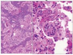
Diagosis?process?
|
Rosai Dorfman
|
|
Diagnosis
|
Rosai dorfman
|
|
|
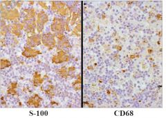
Architecture/cytology of rosai dorfman?
|
Distended sinuses with foamy histiocytes
Emperipoiesis No erythroid phagocytosis S100+ CD68+ Lysozyme + |
|
|
Architecture/cytology of Bacillary angiomatosis?
|
Vascular nodal parenchymal proliferation
Amphophilic and eosinophilic material Vascular spaces are lined by plump endothelium Warthy starry shows organisms |
|
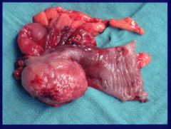
Diagnosis?
|
Bacillary angiomatosis
|
|
|
How can you distinguish bacillary angiomatosis from kaposi sarcoma?
|
Bacillary: Plump endothelial cells, little atypia, HHV8-, pos for organisms
Kaposi: Spindle cels, slit shaped vessels, more atypic, more mitosis, negative for organisms, HHV8+ |
|
|
What is a pseudofollicle?
|
Pale staining spherical structures not surrounded by a mantle zone and seen in CLL
|
|
|
Phenotype of CLL?
|
CD20 weak, CD79a, CD5, CD23, CD43, CD11c dim
|
|
|
Good prognosis for CLL?
|
CD38 and Zap70 negative, means hypermutated
|
|
|
Good prognosis genetics for CLL?
|
Del 13q
|
|
|
Bad prognosis for CLL?
|
Trisomy 12, Del 11q, Del 17p
|
|
|
Bone marrow findings in HCL?
|
Subtle infiltrate, dispersed B cells with pale cytoplasm (fried egg)
Increased reticulin fibrosis |
|
|
Immunophenotype for HCL?
|
Bright: CD11c, CD25, CD103
Annexin A1, TRAP, DBA.44 |
|
|
Immunophenotype for Follicular lymphoma?
|
CD10+, BCL-2 + , T(14:18) IgH-Bcl-2
|
|
|
Immunophenotype of Mantle Cell Lymphoma?
|
CD10 -
CD 5 + BCL2 + CD23 - CD20 - Bright CD43 + |
|
|
Immunophenotype of Marginal Zone Lymphoma?
|
CD10 -
CD5 - No specific Marker |
|
|
Genetics for Mantle Cell?
|
t(11;14) CCND1-IgH
|
|
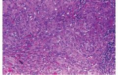
Diagnosis?
|
Mantle cell lymphoma
|
|
|
Histologic features of marginal zone lymphoma?
|
Monocytoid cells, plasmacytoid cells, dutcher bodies, confluent marginal zones, lymphoepithelial lesions
|
|
|
Genetics for marginal zone lymphoma?
|
t(11;18) MLT and API2
|
|
|
How does t(11;18) vary amongst other lymphomas
|
Not seen in primary nodal MZL
Not in MALT with DLBCL It is resistant to antibiotic therapy |
|
|
What are some precursor lesions for MZL?
|
H Pylori (90% for gastric)
Sjogren Hashimoto HepC Borrelia burgdorferi for skin Campylobacter jejuni for eye lymphoma |
|
|
What is the WHO definitions of Waldenstrom macroglobulinemia?
|
LPL with bone marrow involvement + IgM monoclonal gammopathy
|
|
|
Clinical findings in waldenstrom?
|
Hyperviscosity, neuropathy, cryoglobulinemia
|