![]()
![]()
![]()
Use LEFT and RIGHT arrow keys to navigate between flashcards;
Use UP and DOWN arrow keys to flip the card;
H to show hint;
A reads text to speech;
61 Cards in this Set
- Front
- Back
- 3rd side (hint)
|
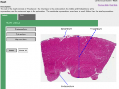
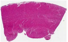
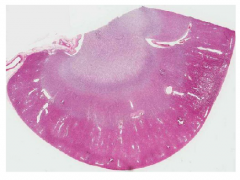
Heart |
Cardio |
|
|
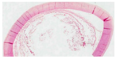
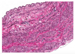
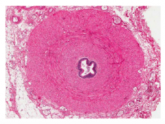
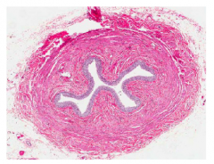
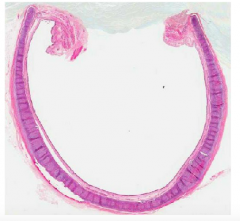
Aorta |
Cardio |
|
|
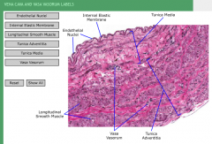
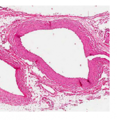
Large Vein |
Cardio |
|
|
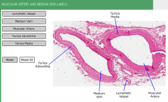
Muscular Artery |
Cardio |
|
|
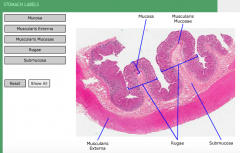
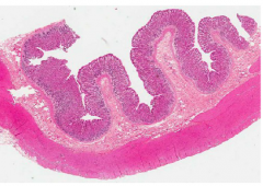
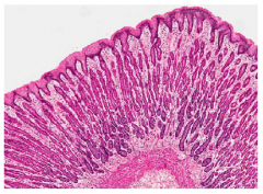
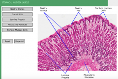
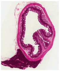
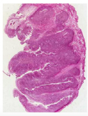
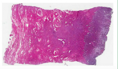
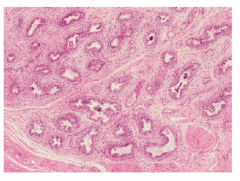
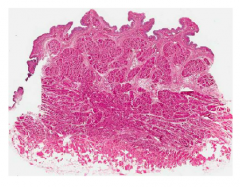
Stomach |
GI Tract |
|
|
Stomach |
GI Tract |
|
|
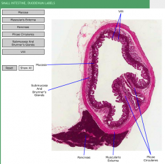
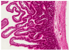
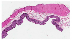
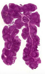
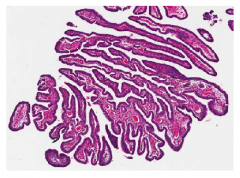
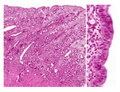
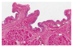
Duodenum |
GI Tract |
|
|
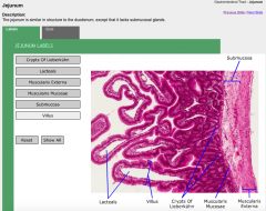
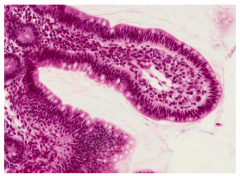
Jejunum |
No submucosal glands |
|
|
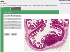
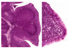
Ileum |
will have increased galt |
|
|
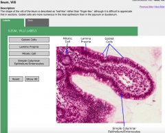
Ileum |
villi are "leaflike" and have increased number of goblet cells |
|
|
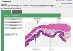
Colon |
|
|
|
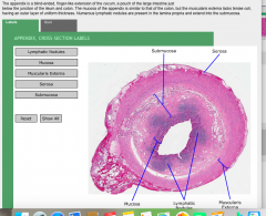
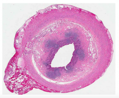
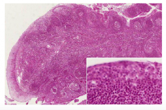
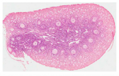
Appendix |
|
|
|
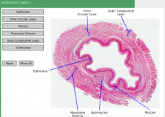
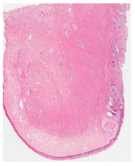
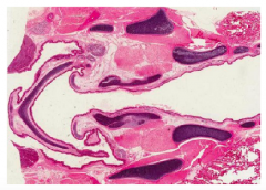
Esophagus |
|
|
|
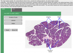
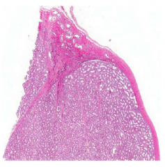
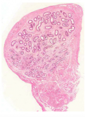
SubmandibularGland |
|
|
|
|
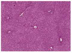
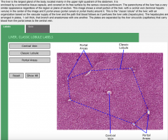
Portal Area? |
|
|
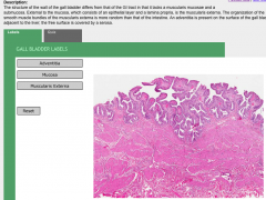
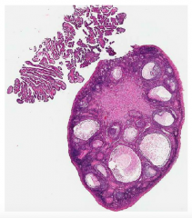
GallBladder |
|
|
|
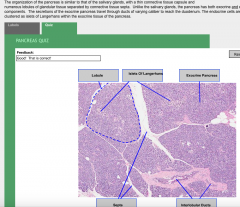
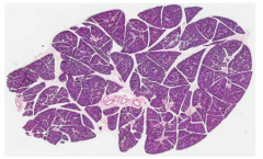
Pancreas |
|
|
|
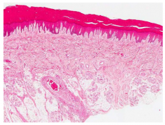
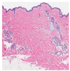
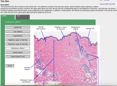
Thick Skin |
|
|
|
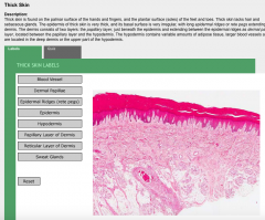
Thick Skin |
|
|
|
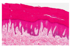
Thin Skin |
|
|
|
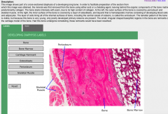
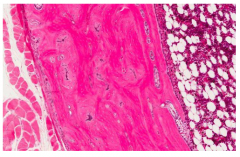
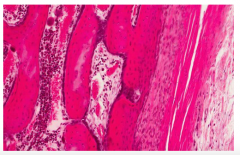
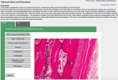
Bone |
|
|
|
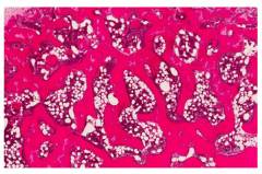
Bone (trabecular) |
|
|
|
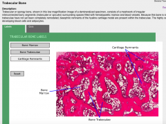
Bone (Trabecular) |
|
|
|
cartilage |
|
|
|
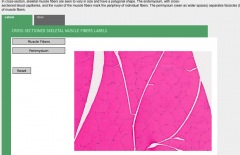
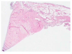
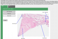
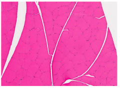
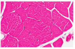
Skeletal Muscle |
|
|
|
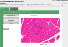
Skeletal Muscle |
|
|
|
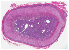
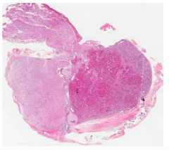
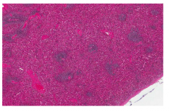
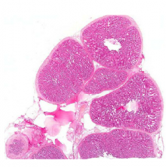
Adrenal Gland |
|
|
|
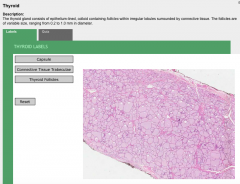
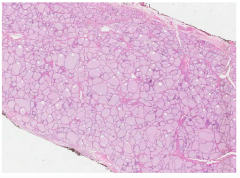
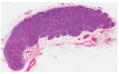
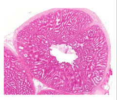
Thyroid Gland |
|
|
|
Pituitary Gland |
|
|
|
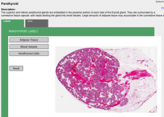
Parathyroid Gland |
|
|
|
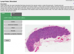
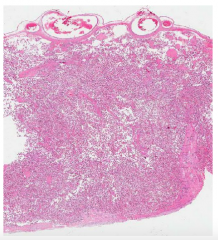
Lymph Node |
|
|
|
Spleen |
|
|
|
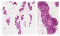
Thymus (infant) |
|
|
|
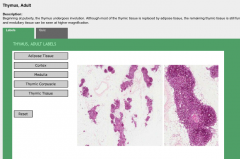
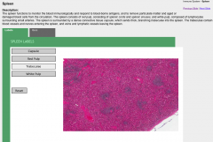
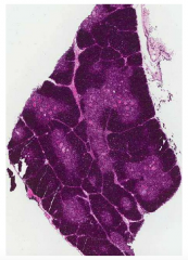
Adult Thymus |
|
|
|
Palatine Tonsil |
|
|
|
Pharyngeal Tonsil |
|
|
|
Cervix |
|
|
|
Ovary (adult) |
|
|
|
ovary (infant) |
|
|
|
Oviduct |
|
|
|
uterus; proliferative |
|
|
|
uterus, secretory |
|
|
|
Placenta |
|
|
|
Mammary Gland; Nonlactating |
|
|
|
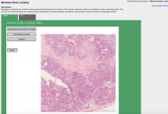
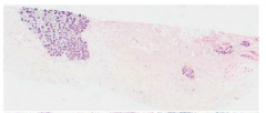
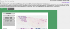
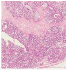
Mammary Gland - Lactating |
|
|
|
Testis |
|
|
|
Rete testis |
|
|
|
Epididymis |
|
|
|
Vas Deferens |
|
|
|
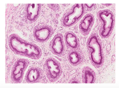
Efferent Dutules |
bout 20 highly coiled efferent ductules connect the rete testis to the ductus epididymis. The luminal surfaces of the efferent ductules have irregular, scalloped outlines. |
|
|
Prostate |
|
|
|
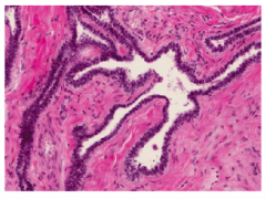
Seminal Vesicle |
tortuous tubular structure with a single lumen and a highly folded mucosa. A layer of smooth muscle surrounds each of the tubular segments. |
|
|
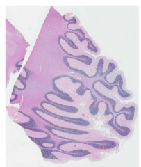
Cerebellum |
|
|
|
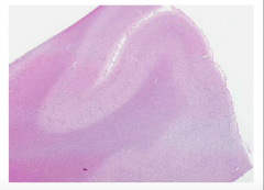
Cerebral Cortex (Cerebrum) |
|
|
|
Kidney |
|
|
|
Ureter |
|
|
|
Bladder |
Like the ureter, the wall of the bladder consists of three layers, a mucosa, a muscularis and an adventitia (or serosa on the superior surface and upper part of the base). The muscularis, also called the detrussor muscle, consists of large bundles of smooth muscle, separated by connective tissue containing numerous blood vessels and nerves. The bladder is innervated by sympathetic nerves that regulate contraction of the smooth muscle of the internal sphincter, parasympathetic nerves that regulate contraction of the muscularis, and sensory nerves that reflexively activate the parasympathetic efferent nerves and inhibit the sympathetic efferent nerves. The muscle layers are less regular than in the ureter, allowing compression of the entire bladder upon contraction. |
|
|
Trachea |
|
|
|
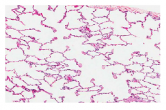
Lung |
|
|
|
Larynx |
|
|

|
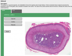
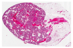
Adrenal |
|