Reading...
![]()
Play button
![]()
Play button
![]()
Use LEFT and RIGHT arrow keys to navigate between flashcards;
Use UP and DOWN arrow keys to flip the card;
H to show hint;
A reads text to speech;
28 Cards in this Set
- Front
- Back
|
Describe the features, location, food, and hosts for Superfamily Ancylostomatoidea
|
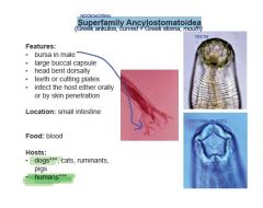
♦FEATURES
-bursa in male -large buccal capsule -head bent dorsally -teeth or cutting plates -infect the host either orally or by skin penetration ♦LOCATION: Small intestine ♦FOOD: BLOOD ♦HOSTS: Mainly dogs***, cats, ruminants, pigs & CAN INFECT HUMAN |
|
|
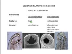
How do Ancylostomatinae differ from Bunostominae?
|
|
|
|
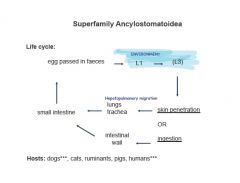
Describe the lifecycle of Ancylostomatoidea
|
|
|
|
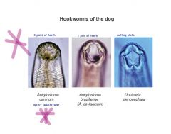
What are the different hookworms in dogs?
|
|
|
|
Describe the lifecycle of ANCYLOSTOMA CANINUM
|
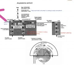
♦LIFECYCLE
PPP***IMPORTANT -skin penetration is important -transmammary infection is important -L1 to L3 develop in faeces, L3 emerges from faecal mass -oral or percutaneous infection possible larvae penetrating skin secrete collagenase which breaks down basement membrane of skin, depolymerises proteins and dermal ground substance -larvae reach blood vessel or lymphatic duct -reach venous system, then pass to lungs -tracheal migration -prepatent period - 2 weeks -ingested larvae may undertake tracheal migration, most penetrate mucosal glands, return to lumen and moult to L4, then adult -prenatal infection and transcolostral infection routes significant -larval inhibition (hypobiosis) can occur, especially in A. duodenale |
|
|
Describe the effect on the host, epidemiology, and diagnosis of ANCYLOSTOMA CANINUM
|
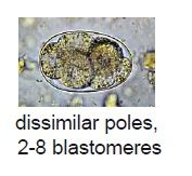
♦EFFECT ON HOST
-use blood for food & respiration -black tarry feces -microcytic, hypochromic anaemia -heavy infections (pups) cause death -strong immunity following exposure -can cause eosinophilic enteritis in man -5000-10,000 eggs can kill pups ♦EPIDEMIOLOGY – important in pups 2-3 months -common in warm climaes -less common in Southern Australia -sandy or loam areas for larvae ♦DIAGNOSIS -eggs in fees -egg counts high (>5000) = pathogenic -eggs small (< 65 μm) |
|
|
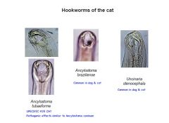
Describe ANCYLOSTOMA BRAZILIENSE
|
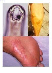
ANCYLOSTOMA BRAZILIENSE
-less pathogenic than previous -no transmammary transmission -ingestion or through skin -common in tropical & subtropical resions -can cause hypoproteinemia and diarrhea -primarily cause of “CUTANEOUS LARVA MIGRANS” or “creeping eruption” in man -tortuous erythematous inflammatory tracts within dermis associated with severe pruritus |
|
|
Describe UNCINARIA STENOCEPHALA
|
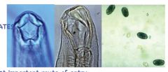
-cutting plates
-common in cooler areas -L3 are ingested -no extra-intestinal migration -less pathogenic -eggs > 65 μm -hypoprotenemia, diarrhea |
|
|
What worms can infect sea lions?
|
♦U. lucasi
- serious pathogen in northern hemisphere seals - disease of pups; adult seals do not harbour adult nematodes, only larvae - transmammary transmission most important route of infection - larvae penetrate skin ♦U. hamiltoni - causes anaemia in Australian sea-lions |
|
|
What hookworms can infect cats?
|
|
|
|
What are the hookworms of ruminants?
|
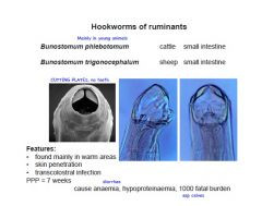
❤Genus Bunostomum
♦Life cycle -L1-L3 develop in dung -L3 penetrate skin of host -reaches lung, moults to L4, undertakes tracheal migration -final mount in small intestine - oral infection occurs but not important -prepatent period 7 weeks -transcolostral infection occurs ♦Pathogenesis -adult nematodes feed on blood, lacerate mucosa -anaemia -hypoproteinaemia -diarrhoea -infection with 1000 nematodes in young animals fatal ❤Genus Gaigeria (named after Gaiger) G. pachyscelis - occurs in ruminants in Africa - feeds on blood - infections with 25 nematodes fatal |
|
|
What are the hookworms of pigs?
|
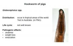
❤Genus Globocephalus
Species occur in pigs in many parts of the world except Australia. Life cycles and pathogenesis not known; blood feeders. |
|
|
What are the hook worms found in humans?
|

|
|
|
Describe the features, lifecycle, and diagnosis of MEGASTRONGYLOIDEA – LUNGWORMS
|
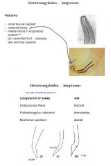
MEGASTRONGYLOIDEA – LUNGWORMS
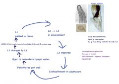
♦FEATURES -small buccal capsule -reduced bursa -mainly found in respiratory system (or connected to it – vascular & nervous system) ♦LIFECYCLES -L1 passed in feces*** -viviparous ✶Simple direct lifecycle: -L1-L3 in feces, L3 ingested -after exsheathment, penetrate intestine, pass to mesenteric lymph nodes, moult & then go to lung -migrates to lungs via lymphatic system HOST: ruminants & horse GENUS: Dictyocaulus ✶Intermediate host: -L1-L3 in mollusks or earthworms, L3 ingested -migrates via lymphatics to lung -HOST: ruminants, pig & cat GENERA: Protosrongylus, Muellerius, Metastrongylus, Aelurostrongylus ✶Direct Lifecycles -L1 infective vs. L3 for others -L1 infects definitive host -migration not worked out, all moults in lungs -HOST: dogs -GENERA: Oslerus, Filaroides ♦DIAGNOSIS: -Baermann technique -larvae with distinctive morphology |
|
|
Describe the features, lifecycle, effect on host, clinical signs, and diagnosis of DICTYOCAULUS FILARIA
|
♦FEATURES
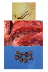
-long thin worm (10 cm) -small buccal capsule -“spongy” spicules ♦LIFECYCLE -L1 passed in faeces; eggs coughed up and swallowed, hatch in intestine -recognised by dome on anterior end or short tail spike -L3 ensheathed -D. viviparus larvae spread up to 10' by faecal fungus Pilobolus -exsheath in abomasum -penetrate small intestine -migrate via lymphatics to venous circulation then lungs -prepatent period 3 weeks ♦EFFECT ON HOST -block airways -excess mucous production -damage epithelium -loss of mucocilliary escalator -loss of epithelium = loss of protection -interstitial pneumonia -alveoli fill with fluid -detachment of alveolar macrophages -consolidation of lung lobes ♦CLINICAL SIGNS: -chronic cough -laboured breathing -secondary bacterial infection -eosinophilia -death ♦DIAGNOSIS: -larvae in feces (L1) |
|
|
Describe the features, lifecycle, and effect on the host of PROTOSTRONGYLUS RUFESCENS
|
♦FEATURES:
-up to 65 mm long -slender, red worm -in small bronchi & bronchioles ♦LIFECYCLE: INDIRECT -L1 in feces -penetrates foot of snail (Cernuella) -L1-L3 in snail -sheep ingests snail -migrates via lymphatics to lung -L3 released when ingested PPP= 2-3 months ♦EFFECT ON HOST -mild pneumonia -most infection subclinical |
|
|
Describe P. stilesi
|
P. stilesi
-major pathogen in Rocky Mountain sheep -REALLY PATHOGENIC -transcolostral transmission |
|
|
Describe the features, lifecycle, and effect on the host of MUELLERIUS CAPILLARIS
|
♦FEATURES
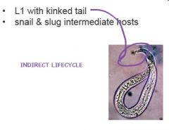
-tiny coiled worms in alveoli ♦LIFECYCLE: Indirect -L1 with kinked tail -snail & slug are intermediate hosts ♦EFFECT ON HOST -high prevalence -virtually non-pathogenic in sheep -can be highly pathogenic in goats -lots of inflammatory cells, granulomatous response |
|
|
Describe the recognition, lifecycle, pathogenesis, immunity, and epidemiology of Dictyocaulus viviparus
|
♦Recognition
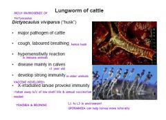
-location, large size -round, poorly lobed bursa -short, spongiform spicules ♦Life cycle -L1 passed in faeces; eggs coughed up and swallowed, hatch in intestine -recognised by dome on anterior end or short tail spike -L3 ensheathed -D. viviparus larvae spread up to 10' by faecal fungus Pilobolus -exsheath in abomasum -penetrate small intestine -migrate via lymphatics to venous circulation then lungs -prepatent period 3 weeks ♦Pathogenesis -block airways -leads to excess mucus production -chronic cough, "husk", laboured breathing -predispose to secondary bacterial infection -see pneumonia involving entire lobules, particularly diaphragmatic lobes -provoke pronounced eosinophilia -disease due to D. viviparus a major problem in cooler regions of Europe -in Australia, has very limited distribution ♦Immunity -strong immunity develops following exposure -X-irradiated vaccine was successful against D. viviparus but was withdrawn because of short shelf-life and need to vaccinate annually ♦Epidemiology -restricted to cool, moist areas -larvae do not survive hot conditions |
|
|
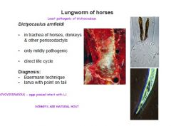
Describe Dictyocaulus arnfieldi
|
|
|
|
Describe the host & lifecycle of PARELAPHOSTRONGYLUS TENUIS
|
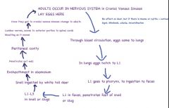
-white tailed deer (North America)
-deer → NO CLINICAL SIGNS In abnormal host: -(moose, cattle, sheep) -produces fatal neurological disease ♦LIFECYCLE: -white-tailed deer are inhabitants of deciduous forests -moose occur in coniferous wet lands -agricultural practices or forestry alter the successional stages of growth in coniferous forests, provide suitable conditions for deer to move into areas inhabited by moose -moose become infected with Pneumostrongylus, larvae in brain cause blindness, circling, ataxia, death -moose are eliminated from areas into which deer move -will also cause disease in cattle which become infected |
|
|
Describe ELAPHOSTRONGYLUS CERVI
|
-parasites of Red Deer (New Zealand)
-occur in connective tissues and meninges -L1 enters blood and reaches lungs -snail intermediate host -no clinical signs in normal host -neurological signs in other ruminants |
|
|
Describe the features, lifecycle, effect on host & diagnosis of AELUROSTRONGYLUS ABSTRUSUS
|
♦FEATURES
-tiny nematode, in alveoli -very common parasite ♦LIFECYCLE -lays eggs -eggs hatch, L1 in feces -L1-L3 in slug or snail -cat eats mollusks or rodent, birds, reptiles → PARATENIC HOSTS -larvae enter bloodstream to reach lungs PPP = 6 weeks ♦EFFECT ON HOST: -generally subclinical if low infection If infection is high: -mild cough -weightloss -eosinophilia ♦DIAGNOSIS: -Baermann for larvae -differentiate from Strongyloides cati by shape of tail |
|
|
Describe the Metastrongylus worms in pigs and their lifecycle & pathogenesis
|
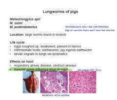
♦Life cycle
-eggs coughed up and swallowed, eggs in faeces (L1 in egg) -eaten by earthworm, moult to L2 and L3, accumulate around heart -pigs ingest earthworms, larvae pass via lymphatics to lungs ♦Pathogenesis -obstruct airways -adults feed on inflammatory exudate in airways -carry swine influenza virus in eggs -controlled effectively in concrete sties |
|
|
Describe the lifecycle of Oslerus osleri in DOGS
|
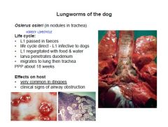
♦Life cycle
-DIRECT -L1 passed in faeces -regurgitation in food and water (in dingoes) is most likely mode of spread -penetrate duodenum, reach lungs -adults migrate from lung parenchyma to trachea -present after 10 weeks -gravid females present by 18 weeks -migratory phase through lung parenchyma can cause severe pneumonia -common in dingoes, uncommon in domestic dogs |
|
|
Describe the lifecycle of Filaroides hirthi in DOGS
|
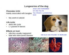
-L1 passed in faeces
-direct life cycle -infection usually inapparent -can be fatal with intercurrent disease -mainly associated with beagle colonies -uncommon in Australia |
|
|
Describe the lifecycle and clinical signs for Angiostrongylus vasorum
|
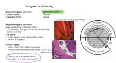
-(right ventricle or pulmonary artery)
•Common in Europe; uncommon in Australia ♦Life cycle -INDIRECT LIFECYCLE -L1 in faeces, snail intermediate hosts •PPP = 4-8 weeks ♦Clinical signs: •mild, cough, interstitial pneumonia; •severe cases develop heart failure -May get damage to arterial walls and formation of thrombi. -Impairment of blood flow may lead to cardiac hypertrophy, congestion, ascites and dyspnoea |
|
|
Describe the lifecycle and pathogenesis for Angiostrongylus cantonensis
|
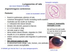
♦Life cycle (A. cantonensis)
-eggs released into pulmonary artery -lodge in lungs -L1 migrates through alveoli, coughed up, swallowed, passed in faeces -Penetrate foot of snail, develop to L3 -ingested by definitive host or L3 shed in mucus on feed plants -larvae penetrate wall of ileum, enter blood vessels -carried to CNS -congregate and moult to L4 in anterior cerebrum -emerge onto surface of brain, remain in subarachnoid space -then enter venous system and pass via heart to pulmonary artery -transport hosts - prawns, land crabs (how they become infected not known) ♦Pathogenesis -non-pathogenic in rats -can infect dogs and less commonly other mammals -causes severe encephalitis in abnormal hosts -cause of eosinophilic meningitis in man -man eats intermediate hosts or vegetables they have been on |